
Stenohelia venusta, Cairns, Stephen D. & Zibrowius, Helmut, 2013
|
publication ID |
https://doi.org/ 10.11646/zootaxa.3691.1.1 |
|
publication LSID |
lsid:zoobank.org:pub:E98CE6DF-AF3B-4AAA-95CB-8ACD615C9FCC |
|
DOI |
https://doi.org/10.5281/zenodo.5619773 |
|
persistent identifier |
https://treatment.plazi.org/id/955B87C9-A156-DD02-FF22-FCDDF2A62CFB |
|
treatment provided by |
Plazi |
|
scientific name |
Stenohelia venusta |
| status |
sp. nov. |
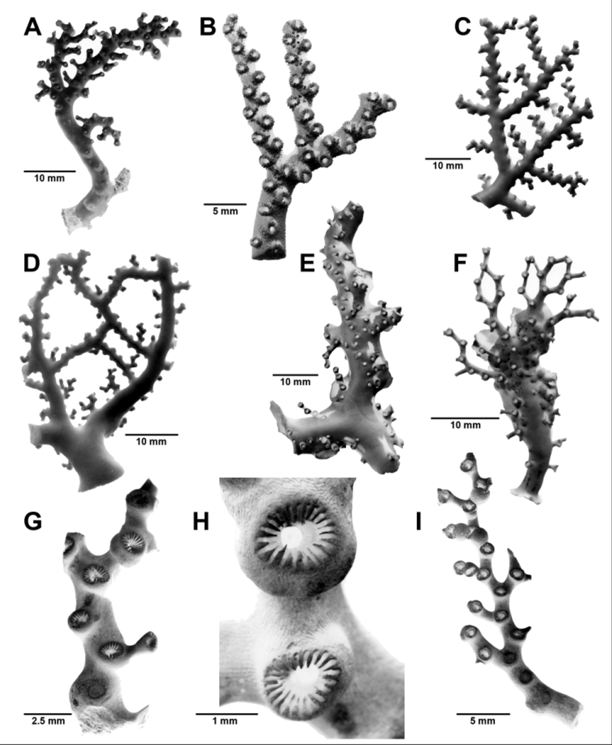
Stenohelia venusta View in CoL sp. nov.
Figs. 3 View FIGURE 3 A, 17A–K, 30
? Stylaster minimus, Hickson & England, 1909: 346 ( Mauritius).
Etymology. From the Latin venustus, meaning beautiful, elegant, and graceful, a name that could be given to virtually any stylasterid.
Types and Type Locality. Holotype: MN SM232, female colony (in alcohol), SAM, and SEM stubs 1682–83 (USNM). Paratypes: MN SM226, 1 male colony, SAM; MN SM232, 3 female and 3 male colonies, SAM, and SEM stub 1681 (USNM); PF 1915, 1 male colony, SAM H1463; PF 13395, 2 branches, SAM H2820; PF 13476, 3 male branches, SAM H1228; Anton Bruun 8—420A, 1 male colony (dry) and 1 male colony (in alcohol), USNM 76773. Type Locality: 32°14.9’S, 29°10.4’E, 620–650 m (off Umtata, northern Eastern Cape Province).
Material Examined. Types. Reference Material, fragment of male syntype of S. tiliatus , Siboga 105, USNM 77280.
Description. Colonies are uniplanar and relatively small, the largest specimen (the holotype, Fig. 3 View FIGURE 3 A) only 4.6 cm in height and 2.6 cm in width, with a basal branch diameter of 4.5 mm; branch anastomosis occurs occasionally. The coenosteum is covered with reticulate strips 50–60 µm in width, each strip covered with small irregularlyshaped granules ( Figs. 17 View FIGURE 17 D, E). The coenosteum is white.
Cyclosystems are unifacial in arrangement, unilinearly positioned on the anterior face ( Fig. 17 View FIGURE 17 A). They are elliptical to irregular in shape, up to 1.3 mm in greater diameter and about 0.8–0.9 mm in lesser diameter, the greater diameter usually oriented perpendicular to the branch axis ( Figs. 17 View FIGURE 17 B, C). Based on 50 cyclosystems, the range of dactylopores per cyclosystem is 13–20; the average is16.46 (ơ = 1.74); and the mode is 16.
Gastropores are also elliptical in shape, up to 0.35 mm in greater diameter and about 0.3 mm in lesser diameter. The gastropore tube is long (up to 1.6 mm) and invariably bent about 90° just beneath the gastropore. The gastrostyle occupies only the lower 30% of the tube, and is composed of a lower section about 0.21 mm in diameter that supports a cylindrical distal portion, which is approximately 0.10 mm in diameter. A solid inner ring (the sphincter) constricts the tube at the transition point of the basal to distal portion of the style ( Figs. 17 View FIGURE 17 H, I). The illustrated style is 0.45 mm in height, and is covered with small spines. The dactylotomes are fairly consistent in width (60–70 µm), whereas the pseudosepta are somewhat irregular in width, ranging from 64–145 µm wide ( Fig. 17 View FIGURE 17 F). The tops of the pseudosepta range from slightly convex to slightly concave. Each dactylopore contains 1–3 dactyloglossae, the uppermost being at the level of the coenosteal surface, and thus most easily seen in damaged cyclosystems ( Fig. 17 View FIGURE 17 J). The dactyloglossae are tongue-shaped, about 60–70 µm in surface dimensions, and about 8–10 µm thick, each blocking approximately 70–80% of the dactylopore tube.
Female ampullae are superficial hemispheres 0.6–0.8 mm in diameter, arranged in close proximity on the posterior faces of terminal branches ( Fig. 17 View FIGURE 17 G); efferent pores are inconspicuous but are lateral in position, not opening within the gastropore tube. The male ampullae are smaller (0.35–0.50 mm diameter) and conical in shape, with a small (about 35 µm diameter), irregularly-shaped apical efferent pore. The male ampullae tend to cluster on the posterior side of the branches ( Fig. 17 View FIGURE 17 K).
Comparisons. Among the 11 other species in the genus, Stenohelia venusta is most similar to S. tiliata (Hickson & England, 1905), originally described and still known only from the Sulu Sea at 275 m. Examination of the syntype of S. tiliata shows it to have15–16 dactylopores per cyclosystem, irregularly-shaped cyclosystems, a rough reticulate coenosteal texture, lacking a ring palisade, and male ampullae about 0.5 mm in diameter. The only difference detectable between the two species is that S. venusta has dactyloglossae, whereas S. tiliata has typical dactylostyles composed of aligned pillars. One must keep in mind, however, that S. tiliata is known only from one specimen, and the taxonomic value of dactyloglossae is yet to be determined. Regardless, these two species would appear to be sister species, if not conspecific.
Stenohelia venusta also bears a resemblance to S. conferta Boschma, 1968 , known only from the Antipodes Islands at 1335 m. They are similar in coenosteal texture, cyclosystem shape, and in lacking a typical ring palisade, but S. venusta differs in having a higher number of dactylopores per cyclosystem, and in having dactyloglossae.
Hickson & England (1909) reported Stylaster minimus from Mauritius, which heretofore was the only record of this genus in the Indian Ocean, the type locality for Stylaster (= Stenohelia ) minimus being the Philippines at 1089 m. They did not supply any substantive description or illustrations for this specimen.
Distribution. Known from the continental shelf and slope off South Africa from Cape Blaize to northern Eastern Cape Province (Fig. 30) (159–710 m); off Kenya (140 m),? Mauritius (Hickson & England, 1909), 140– 710 m.

No known copyright restrictions apply. See Agosti, D., Egloff, W., 2009. Taxonomic information exchange and copyright: the Plazi approach. BMC Research Notes 2009, 2:53 for further explanation.