
Theonoe longiseta Simon 1881: 132–133
|
publication ID |
https://doi.org/ 10.1206/0003-0082(2007)3575[1:OTSSCL]2.0.CO;2 |
|
persistent identifier |
https://treatment.plazi.org/id/03908786-191E-FFF6-3FD4-FAC440EFFB41 |
|
treatment provided by |
Carolina |
|
scientific name |
Theonoe longiseta Simon 1881: 132–133 |
| status |
|
Theonoe longiseta Simon 1881: 132–133 , table 26, fig. 1 View Figs .
Theonoe longiseta, Bertkau, 1890: 10 .
Cepheia longiseta, Simon, 1894: 589 View in CoL ; Simon, 1926: 313– 315; Denis, 1933a: 564; Denis, 1933b: 93; Levi and Levi, 1962: 18, 64, figs. 309–310; Brignoli, 1970: 1410– 1412, figs. 11–14; Wunderlich, 1980: 267, figs. 17 View Figs , 42– 43; Thaler and Noflatscher, 1990: 173–174, figs. 25–29 View Figs View Figs ; Heimer and Nentwig, 1991: 306, fig. 823; Marusik and Lehtinen, 2003: 151; Lopardo et al., 2007:9–11 View Cited Treatment .
TYPES: One male lectotype and 14♀ 173 and 3 juvs paralectotypes from FRANCE (‘‘Gallia’’) coll. Simon 4538, b.849 (in MNHN-AR 1059 , examined) . The label, rewritten by P.M. Brignoli, also includes ‘‘1969, PM Brignoli leg.’’, which should be read as ‘‘det. P.M. Brignoli 1969’’ .
TYPE LOCALITY: ‘‘ France: Dept. du Var, Vallée de Dardennes near Toulon; pierrefeu dans la forèt de Maures’ ’ ( Simon, 1881:133) .
DIAGNOSIS: See generic diagnosis.
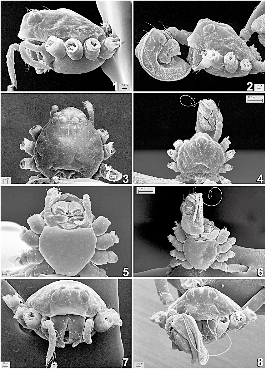
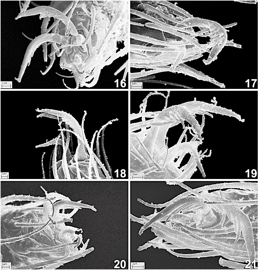
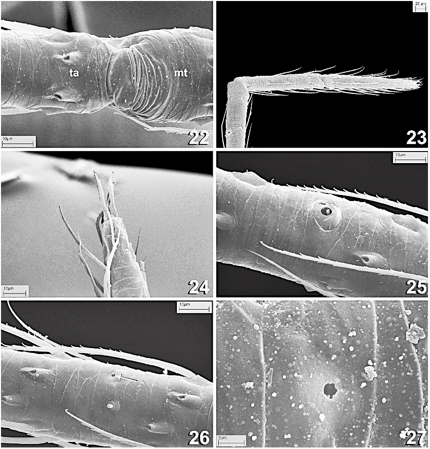
DESCRIPTION: Dorsal carapace with three setae along midline and four laterally, two on each side ( figs. 3, 4, 7, 8 View Figs ). Midline setae slightly posterior to PME (one), and on dorsalmost carapace surface (two). Lateral setae located behind ALE (one pair) and PLE (one pair). Carapace rounded (as long as wide), with clypeal area protruding in dorsal view ( figs. 3, 4 View Figs , 9). Chelicerae with median keel ending in single strong promarginal tooth ( figs. 10, 11, 13, 15); retromarginal teeth absent. Maxillary setae scarce, distal maxillary setae clavate (arrow in fig. 13). Clypeus slightly convex. Sternum cuticle squamate, posterior margin truncated, wide, about twice width of coxa IV ( figs. 5, 6 View Figs ). Legs: Femoral spot absent. Setae on legs with large elevated, striated bases ( figs. 22, 25, 26 View Figs ), weaker on chelicerae ( fig. 10). Leg tarsi without pseudosegmentation ( fig. 24 View Figs ). Tarsal-metatarsal joint constricted, distal area of metatarsi with dorsal lyriform organ as band of anastomosed ridges ( figs. 22, 23 View Figs ). Legs without spines, tarsal organ located in basal third dorsal region of tarsus, capsulate, flat, opening rounded, difficult to see ( figs. 26, 27 View Figs ). Three tarsal claws, serrate accessory setae (or false claw) present ( fig. 17 View Figs ). Claw teeth (paired claws/inferior claw): leg I, paired claws with five teeth ( fig. 16 View Figs )/inferior claw with two teeth and one dorsal denticle ( fig. 17 View Figs ); leg II, five teeth/two teeth ( fig. 18 View Figs ); leg III, four teeth/two teeth ( figs. 20, 21 View Figs ); leg IV, four teeth/two teeth and one dorsal denticle ( fig. 19 View Figs ). Leg setae serrate. Cuticular surface of appendages squamate ( figs. 23, 25, 26 View Figs ). Tarsi and metatarsi equally long ( fig. 23 View Figs ; see tables 1 and 2). Trichobothria: Trichobothrial bases simple and smooth, with proximal hood bearing two lateral ridges, similar on all legs and segments ( fig. 25 View Figs ). Tarsal trichobothria absent. Legs I and II, tibia 2-r1-0; metatarsus r1-0. Legs III and IV, tibia 2-2-0; metatarsal trichobothria absent. Color: Carapace yellow, few darker radii, center and margins brown ( fig. 9); sternum dark brown, homogeneous. Legs yellowish, darker on tibiae, patellae, distal femora, and distal tarsi. Abdomen dark brown. Eyes: All eyes pearly white except AME, black. Diameter: AME 0.03, PME 0.02, PLE 0.03, ALE 0.03. Respiratory system: Anterior booklungs reduced to tracheae ( figs. 58, 59), connected by a transverse duct (arrow in fig. 59). 3 Anterior spiracles connected to epigastric furrow ( fig. 56). Five tracheal tubes arise from each anterior spiracle, four oriented anteriorly toward cephalothorax, one oriented laterally ( figs. 59, 60). Posterior tracheal system with two distant spiracular openings exteriorly connected by thin ridge (i.e., one wide spiracular opening) ( figs. 30, 31 View Figs ). Thin ridge leading to deep, flat, membranous atrium, anteriorly ending in sclerotized U-shaped duct that connects the tracheal ducts arising from spiracles ( fig. 62). Two main tracheal bundles arise from the junction of tracheal ducts and U-shaped atrial duct, one on each side, directing tracheoles mainly anteriorly ( figs. 62, 63). Both tracheal systems seem to reach into prosoma. This tracheal arrangement is similar to that described for Synaphris ( Lopardo et al., 2007; see schematic drawing in their figure 30 View Figs ).
MALE (range of four measured paralectotypes): Total length 0.84 (0.83–0.85). Carapace length 0.34 (0.34–0.37), width 0.36 (0.36–0.37), height 0.16 (0.16–0.17). Labrum with three minute, long chemosensory setae ( fig. 11). Clypeus height 0.12, ca. 4 AME diameters. Two setae located on clypeus ( fig. 8 View Figs ). Sternum length 0.25 (0.25–0.26), width 0.27 (0.26–0.27), length/width 0.91 (0.91–0.98). Abdomen oval ( figs. 9, 14), length 0.50 (0.50– 0.51), width 0.43 (0.43–0.47), height 0.42 (0.42– 0.48). Two epiandrous spigots centrally distributed along the epigastric furrow ( fig. 55 View Figs ). Legs: Leg formula 154523. Leg measurements: see table 1. Leg I prolateral clasping spine absent. Spinnerets ( fig. 31 View Figs , see also Lopardo et al., 2007): Colulus large, fleshy, triangular, about half length and width of ALS, with three setae
3 The term ‘‘transverse duct’’ had generated some confusion in the past and seems in need of a proper illustration (Martín J. Ramírez, personal commun.; for discussion see Ramírez [2000] and references therein). Here we provide with images of the ‘‘transverse duct’’, in this case connecting the anterior tracheae (arrow in fig. 59).
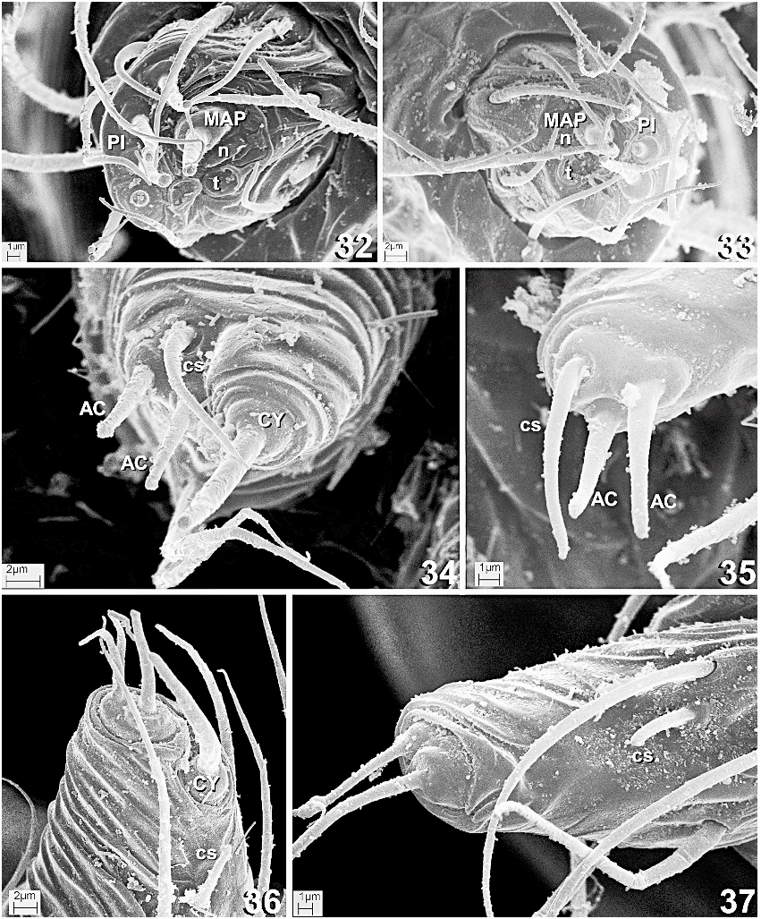
( figs. 29, 31 View Figs ). ALS ( fig. 33 View Figs ) with one MAP spigot, accompanied by a nubbin and a tartipore, separated by weak (almost nonexistent) furrow from PI field. PI field, on external side of ALS, contains three PI spigots with reduced bases, posterior PI spigot base larger. PMS ( fig. 35 View Figs ) with two AC spigots, one chemosensory seta (can be confused with a spigot) located anteriorly, its base deepens around shaft. PLS ( fig. 37 View Figs ) with two spigots of slightly different morphology, clumped in same field. Internal one with rounded, larger base and more cylindrical shaft, external one with oval base and tapering shaft. Short thick chemosensory seta (can be confused with a small spigot), located more basally on internal side of distal PLS segment. Palp ( figs. 38, 39, 42–54): Enormous, compressed ( figs. 2, 6, 8 View Figs ). Tibia rounded retrolaterally, without apophyses, pressed toward the bulb retrolaterally ( figs. 8 View Figs , 46, 47, 52 View Figs ). One tibial trichobothrium located dorsal and distally, close to cymbial base ( figs. 46, 47). Cymbium long, narrow, thicker at base, then equally narrow, dorsal ( figs. 44– 49, 53, 54 View Figs ). Tarsal organ dorsal, distal, capsulated, flat, opening teardrop-shaped ( fig. 54 View Figs ). Basal retrolateral margin of cymbium with triangular paracymbium ( figs. 39, 46, 47, 53 View Figs ). Embolus filiform, long ( figs. 42–44, 47). Embolar base irregular, retrolateral, ventrally located, membranous, without expansions ( figs. 38, 42, 51 View Figs ). Embolus running clockwise (in left palp) on retrolateral side of bulb, passing to and ending on prolateral side, distally, within conductor groove ( figs. 38, 39, 50 View Figs ). Huge membranous conductor occupying most of retrolateral and distal half of prolateral bulb, with groove where embolus fits ( figs. 42–46). Small cuticular protuberances interspersed on distal area of conductor ( fig. 50 View Figs ). Conductor with prolateral pointed apophysis where groove ends ( figs. 39, 45–47, 52 View Figs ). One dorsal tegular apophysis, close to cymbium, pointed ( figs. 39, 45, 49). Spermatic duct seems to undergo two transverse loops before reaching embolar base ( fig. 38). Diameter of spermatic duct gradually increases before entering base of embolus for fraction of loop length, returning to smaller diameter before entering embolus (arrow in fig. 38).
FEMALE (range of nine measured paralectotypes): Total length 0.90 (0.85–0.96). Carapace length 0.36 (0.35–0.38), width 0.35 (0.33–0.37), height 0.16 (0.14–0.19). Clypeus height 0.11 (0.09–0.12), ca. 4.25 (3–5) AME diameters. One seta located on clypeus along
TABLE 1 Length of Right Leg for Four Male Paralectotypes ( MNHN-AR 1059 ) of Cepheia longiseta ( Simon 1881) Measurements are in millimeters, ranges in parentheses.
TABLE 2 Length of Right Leg for Nine Female Paralectotypes ( MNHN-AR 1059 ) of Cepheia longiseta ( Simon 1881) Measurements are in millimeters, ranges in parentheses.
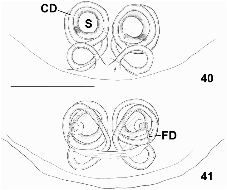
midline ( fig. 7 View Figs ). Sternum length 0.23 (0.23– 0.27), width 0.24 (0.24–0.28), length/width 0.95 (0.87–1.02). Palp without claw ( figs. 1 View Figs , 12). Abdomen oval, length 0.60 (0.57–0.66), width 0.52 (0.47–0.56), height 0.51 (0.45–0.55). Legs: Leg formula 451523. Leg measurements: see table 2. Spinnerets ( fig. 30 View Figs , see also Lopardo et al., 2007): Colulus large, fleshy, triangular, about half length and width of ALS, with four setae ( figs. 28, 30 View Figs ). Spinnerets as in male, except: three PI spigots (instead of two) on ALS ( fig. 32 View Figs ); one external (ectal) CY spigot on PMS ( fig. 34 View Figs ); one internal (mesal) CY on PLS ( fig. 36 View Figs ). Epigynum ( figs. 40, 41 View Figs , 56–59, 61): Slightly sclerotized, translucent, with medial depression bearing copulatory openings ( figs. 56, 57). Copulatory ducts initially coiled posteriorly in one loop ( fig. 40 View Figs ), then directing anterior and dorsally, then wrapped around spermathecae in four loops ( figs. 40, 41 View Figs , 59). Spermathecae cylindrical ( figs. 59, 61). Fertilization ducts slightly coiled, arising at dorsal edge of spermathecae ( figs. 41 View Figs , 61).
NATURAL HISTORY: See generic natural history.
DISTRIBUTION: See generic distribution.
OTHER MATERIAL EXAMINED: No locality data, no collector, 13 ( MNHN-AR 1063 ) 4; FRANCE: Banyuls , no date, no collector, 13 1 sub 3 ( MNHN-AR 1070 ); ITALY: South Tirol , Bolzano Province , Bolzano / Guntschna, 470 m, 27.vi.1988, Noflatscher, 2♀ 13 (NMW- 14994) .


| PM |
Pratt Museum |
No known copyright restrictions apply. See Agosti, D., Egloff, W., 2009. Taxonomic information exchange and copyright: the Plazi approach. BMC Research Notes 2009, 2:53 for further explanation.
|
Kingdom |
|
|
Phylum |
|
|
Class |
|
|
Order |
|
|
Family |
|
|
Genus |
Theonoe longiseta Simon 1881: 132–133
| LOPARDO, LARA & HORMIGA, GUSTAVO 2007 |
Cepheia longiseta
| Lopardo, L. & G. Hormiga & A. Melic 2007: 9 |
| Marusik, Y. M. & P. T. Lehtinen 2003: 151 |
| Heimer, S. & W. Nentwig 1991: 306 |
| Thaler, K. & M. - T. Noflatscher 1990: 173 |
| Wunderlich, J. 1980: 267 |
| Brignoli, P. M. 1970: 1410 |
| Levi, H. W. & L. R. Levi 1962: 18 |
| Denis, J. 1933: 564 |
| Denis, J. 1933: 93 |
| Simon, E. 1926: 313 |
| Simon, E. 1894: 589 |
Theonoe longiseta
| Bertkau, P. 1890: 10 |
Theonoe longiseta Simon 1881: 132–133
| Simon, E. 1881: 133 |