
Thouarella hicksoni Thomson, 1911
|
publication ID |
https://doi.org/ 10.11646/zootaxa.3602.1.1 |
|
publication LSID |
lsid:zoobank.org:pub:10304FBF-3969-4EFA-83F1-BB8A5E2B37F3 |
|
persistent identifier |
https://treatment.plazi.org/id/EE36E867-FFBA-FFA9-FF0A-A885FE8408BC |
|
treatment provided by |
Felipe |
|
scientific name |
Thouarella hicksoni Thomson, 1911 |
| status |
|
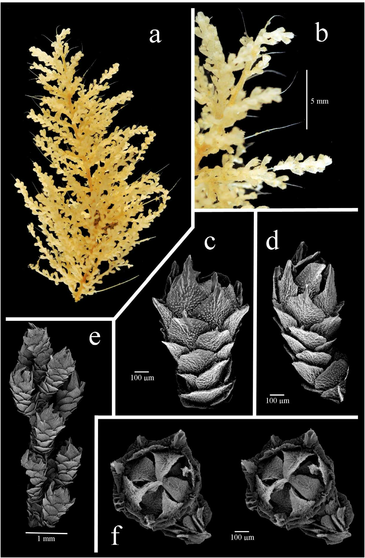
12. Thouarella hicksoni Thomson, 1911 View in CoL
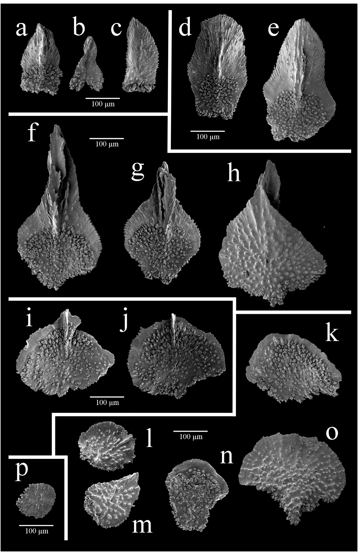
Figs 26 View FIGURE 26 , 27 View FIGURE 27
Thouarella hicksoni Thomson, 1911: 886–887 View in CoL , pl. 44, figs 3, 3a, pl. 45, fig. 1; Kükenthal 1919: 439–40; 1924: 301–2; Stiansy 1940: 32, text fig. G, pl. 4, fig. 21; Williams 1992: 277–280, fig. 1H, 66–68; Cairns & Bayer 2009: 27 (listed)
Thouarella Hicksoni Tixier-Durivault 1954: 625 View in CoL
Material examined: Holotype, NHM 1962.7 .20.36, off Cape St. Francis , South Africa, 135 m, rocky substratum, 19 Feb 1902, 8.5 cm and 4.5 cm fragments seen.
Other material: USNM 53911, south of Port Elizabeth, South Africa, 34˚15’S, 25˚05’E, 11 m, SCD 8–J, collected by J. Day, 1958, 2 colonies; USNM 53912 + USNM 53813 (same location), SCD 3–D, South Africa, 34˚30’S, 24˚40’E, collected by J. Day, 102 m, 18 Apr 1958.
Description
The holotype is bottlebrush in shape, tapering at the tip ( Fig. 26a View FIGURE 26 ). The branchlets emanate in 4 directions approximately perpendicular to the main stem, and are tightly placed at 1 mm intervals ( Fig. 26b,e View FIGURE 26 ). Secondary and tertiary ramification is common and occurs close to the branchlet base. The axis is yellow, iridescent, and has fine longitudinal striations.
The polyps rarely occur on the stem but are usually isolated on branchlets and can be placed in a spiral in 3 directions, but this is inconsistent. The polyps are modestly flared ( Fig. 26c,d View FIGURE 26 ), angled at 30 ° to being appressed against the branchlet, and are 1–1.25 mm high, with a H:W of 1.79–1.95, with a clustered arrangement ( Fig. 26e View FIGURE 26 ) of 16–22 per cm, less towards the branchlet base. Each polyp has 7 longitudinal rows of body-wall scales reducing to 4 rows at the base. There are 4–5 scales in the abaxial rows ( Fig. 26c View FIGURE 26 ), 3–4 in the outer and inner lateral rows, and 2–3 scales in the adaxial rows.
There are 8 operculars, measuring 300–360 µm high (average 330 µm), 170–210 µm wide (average 190 µm), with an average H:W of 1.7. Two to three of the largest operculars are lanceolate with a pointed distal edge ( Fig. 27e View FIGURE 27 ) whilst the remainder are tongue-shaped ( Fig. 27d View FIGURE 27 ). The operculars have a longitudinally concave outer surface, which is smooth, with tubercles sparsely arranged across the proximal area. The inner surface of the operculars have longitudinal median striations or a small simple keel, and the proximal third is tuberculate.
Beneath the 8 operculars are 2–4 (perhaps more in other colonies) accessory operculars ( Fig. 27a–c View FIGURE 27 ) that are 200–210 µm high (average 200 µm), 110 µm wide, a fraction the size of regular operculars, with a H:W of 1.75–1.9 (average 1.8). They have a relatively smooth outer surface with tubercles visible at the proximal edge and small median longitudinal striations or a small keel ( Fig. 27a View FIGURE 27 ) on the inner surface, as is present on the larger operculars.
The marginals form 2 alternate circles of 4 inner and 4 outer scales, although the pattern is not strict. They have a wide circular to oval body and are pointed distally ( Fig. 27f–h View FIGURE 27 ) with a channelled keel on the inner surface (lateral keel projections can sometimes be visible from an abaxial view). Their dimensions are 350–480 µm high (average 400 µm), 220–320 µm wide (average 280 µm), with an average H:W of 1.4 (range of 1.1–2). The adaxial marginals are often shorter with a reduced or absent distal point (probably because these sclerites are compressed against the polyp and thus reduced in size). The inner surface of all marginals is tuberculate with smooth lateral areas adjacent to the keel. The outer surface is covered with granules which are placed more densely in the proximal central area, gradually diminishing in number towards the distal edge.
The submarginals are elliptical with those of the abaxial side often having a moderately pointed distal edge ( Fig. 27i,j View FIGURE 27 ). They are 250–270 µm high (average 260 µm), 260–280 µm wide (average 270 µm), with an average H:W of 0.9. The inner surface has small, simple single or double (and rarely treble) keels. The central area is tuberculate with a smooth band along the distal edge lateral to the keels. The outer surface is identical to that of the body-wall scales, as described below.
Body-wall scales are semi-circular ( Fig. 27k View FIGURE 27 ), fan-shaped ( Fig. 27o View FIGURE 27 ), circular ( Fig. 27l View FIGURE 27 ) or irregular in shape ( Fig. 27n,m View FIGURE 27 ), often with an irregularly lobate proximal edge. Body-wall scales are 190–270 µm high (average 220 µm), 140–310 µm wide (average 240 µm), average H:W of 0.9 (range of 0.7–1.4). The outer surface is covered with granules proximally, with some tubercles at the proximal edge; the inner surface is tuberculate with a smooth band along the distal edge.
The coenenchymal scales are circular ( Fig. 27p View FIGURE 27 ), 110–150 µm high (average 130 µm), 100–180 µm wide (average 140 µm), with an average H:W of 1, with a smooth outer surface and tuberculate inner surface.
Distribution
This species is only known from the Port Elizabeth region, South Africa, at depths of 11–135 m.
Remarks
Contrary to Thomson’s description (1911), the branchlets at the colony base tend to be shorter; likely as a result of damage when collected.
A small, circular, pebble-encrusted casing (perhaps an egg case) was found within the branchlets of the holotype.
Comparisons
Thouarella hicksoni and T. pendulina are very similar, sharing a similar number of abaxial body-wall scales, polyp length, and a clustered polyp arrangement (see Table 3). However, T. hicksoni has fewer polyps per cm and some operculars that have a tongue-shaped distal edge ( Table 3).
The polyps of T. striata , T. variabilis , and T. andeep have a similar number of scales in the abaxial row as the polyps of T. hicksoni , however, these three species have large, distally flared polyps that are arranged less densely on the branchlets than is found on specimens of T. hicksoni .
The polyps of T. hicksoni have a similar number of scales in the abaxial rows, and similar marginal shapes as found in the polyps of T. brucei , however, the former species generally has smaller (noting that some additional specimens of T. brucei have smaller polyps than the holotype), more pointed polyps, and more clustered, finer, flexible branchlets.
No known copyright restrictions apply. See Agosti, D., Egloff, W., 2009. Taxonomic information exchange and copyright: the Plazi approach. BMC Research Notes 2009, 2:53 for further explanation.
|
Kingdom |
|
|
Phylum |
|
|
Class |
|
|
Order |
|
|
Family |
|
|
Genus |
Thouarella hicksoni Thomson, 1911
| TAYLOR, M. L., CAIRNS, S. D., AGNEW, D. J. & ROGERS, A. D. 2013 |
Thouarella Hicksoni Tixier-Durivault 1954: 625
| Tixier-Durivault, A. 1954: 625 |
Thouarella hicksoni Thomson, 1911: 886–887
| Cairns, S. D. & Bayer, F. M. 2009: 27 |
| Williams, G. C. 1992: 277 |
| Kukenthal, W. 1924: 301 |
| Kukenthal, W. 1919: 439 |
| Thomson, J. A. 1911: 887 |