
Trimma hollemani, Winterbottom, Richard, 2016
|
publication ID |
https://doi.org/ 10.11646/zootaxa.4144.1.1 |
|
publication LSID |
lsid:zoobank.org:pub:0BDD56E3-A657-46B2-B5E5-3CF7F8E8D3A6 |
|
DOI |
https://doi.org/10.5281/zenodo.6087311 |
|
persistent identifier |
https://treatment.plazi.org/id/B0AE4C16-8333-471E-9CAD-2CB0ACCBEEF7 |
|
taxon LSID |
lsid:zoobank.org:act:B0AE4C16-8333-471E-9CAD-2CB0ACCBEEF7 |
|
treatment provided by |
Plazi |
|
scientific name |
Trimma hollemani |
| status |
sp. nov. |
Trimma hollemani View in CoL n.sp.
Holleman’s Pygmygoby Figs. 11 View FIGURE 11 , 28–32 View FIGURE 28 View FIGURE 29 View FIGURE 30 View FIGURE 31 View FIGURE 32
Trimma tevegae View in CoL : (non Cohen & Davis, 1969:321)— Allen & Erdmann, 2012:948 (partim, Raja Ampat View in CoL ); Winterbotton et al., 2014:88 (Group 4 only); Winterbottom & Hoese, 2015:87 View Cited Treatment (identification uncertain, Queensland only)
Material examined. All type specimens collected from the Raja Ampat Regency , West Papua Province of Indonesia . The description is based primarily on up to 16 specimens (15.4–27.6 mm SL). Additional material is from Halmahera , Indonesia and Rabaul, New Britain (identifications confirmed by COI), the Great Barrier Reef of Australia, northern Sulawesi, Malaysia, and the Philippines (no genetic confirmation).
Holotype. ROM 84878, 25.5 mm SL female, Keruo Id off Penemu Id, Fam Ids, 00°35'15.4"S, 130°17'41.1"E, various hard and soft corals, tunicates, and sponges, 18.3–27.4 m, clove oil, 25 Jan., 2010, R. Winterbottom & L. Katz.
Paratypes. MZB.23013 (was ROM 87543), (21.4), Keruo Id , steep slope with overhangs, sand and gorgonians, 0°35'17.48" S, 130°18'34.80" E, 70 m, clove oil, 27 Aug., 2010, M.V. Erdmann GoogleMaps . ROM 84886, (17.1), Keruo Id off Penemu Id, Fam Ids, 00°35'15.4" S, 130°17'41.1" E, 70 m, clove oil, 25 Jan., 2010, M. V. Erdmann (plus T07709) GoogleMaps . ROM 85207, 2 (19.5–21.4), Wofoh Id , west coast near south end, 00°15'21.9" S, 130°17'32.0" E, many small corals, Tubastrea , sea whips, some black coral, sea fans, tunicates, sponges, some hydroids, 18.3−22.9 m, 29 Jan., 2010, R. Winterbottom & W. Kaka (plus T07751) GoogleMaps . ROM 85256, (17.4), Fam Id , Keruo Wall, 00°35'15.4" S, 130°17'41.1" E, 70 m, clove oil, 24 May, 2010, M. V. Erdmann GoogleMaps . ROM 85341, (27.6), E side of " Barracuda Rock " 200 m N of Wayil Id (02°11'43.4"S, 130°25'37.9"E), huge cave with lots of sea fans & sea whips near entrance at top part, large variety of soft & hard corals just outside, 19.8−27.4 m, rotenone, 1 Feb., 2010, R. Winterbottom & CI team GoogleMaps . ROM 87533, 2 (16.4–21.9), Keruo Id , cave in steep slope with sand and gorgonians, 0°35'17.48" S, 130°18'34.80" E, 66 m, clove oil, 26 Aug., 2010, M.V. Erdmann (plus five tissue specimens, T07772–T07776) GoogleMaps . ROM 87938, 2 (15.4–16.7), ' Spoon' sea-mount off Misool, 14 km SE of Femin Id, 02°19'53.08" S, 130°20'10.1"E, 70 m, clove oil, 2 Mar., 2011, M. V. Erdmann (plus T10814, T10815) GoogleMaps . ROM T07715, Keruo Id , off Penemu Id, Fam Ids, 00°35'15.4" S, 130°17'41.1" E, steep 80° slope with numerous small caves and overhangs, 18.3–27.4 m, clove oil, 25 Jan., 2010, R. Winterbottom & L. Katz GoogleMaps . ROM 100155, 2(20.8–20.9), collected with the holotype (plus T07715). ROM 1913 View Materials CS (was ROM 84902), 3(15.5–20.6), Penemu Id , Fam Ids, 00°35'29.5" S, 130°17'10.7" E, 75° slope, some rubble; small caves/crevices in slope, some sand, 8–25 m, clove oil, 25 Jan., 2010, R. Winterbottom & L. Katz. Specimens cleared and stained with Alizarin Red S and Alcian Blue. GoogleMaps
Additional material. Indonesia, Halmahera : ROM T12746 (tissue only), (18.6), Jailolo Patch Reef, 01°00.2928'N, 127°27.857'E, 25m, 8 May 2012, M. V. Erdmann GoogleMaps . Papua New Guinea: New Britain, Rabaul : ROM 92124, (16.3), N side of Little Pigeon Island, S of Simpson Bay, hard and soft corals, sponges, hydroids; steep slope and vertical drop-off wall below wreck of the ' Atun', small caves and overhangs, 04°16.145'S, 152°19.457'E, 31.4 m, clove oil, 23 Nov., 2011, R. Winterbottom & W. Holleman GoogleMaps . ROM 92129, 2 (21.–22.0), N side of Little Pigeon Id, S of Simpson Bay , hard and soft corals, sponges, hydroids; steep slope and vertical drop-off wall below wreck of the ' Atun', small caves and overhangs, 04°16.145'S, 152°19.457'E, 25.6 m, clove oil, 23 Nov., 2011, R. Winterbottom & W. Holleman (plus T13176 View Materials ) GoogleMaps . ROM 92137, (8.3), N side of Little Pigeon Id, S of Simpson Bay , hard and soft corals, sponges, hydroids; steep slope and vertical drop-off wall below wreck of the ' Atun', small caves and overhangs, 04°16.145'S, 152°19.457'E, 15m, clove oil, 23 Nov., 2011, R. Winterbottom & W. Holleman GoogleMaps . ROM 92169, (17.8), SE side of Little Pigeon Id off Simpson Bay, hard and soft corals, lots of Goniopora and Tubersteria, sponges, hydroid 'trees'; vertical wall with small and large caves and crevices, 0415.993S, 15219.684E, 11.9 m, clove oil, 24 Nov., 2011, R. Winterbottom & W. Holleman.
Additional material, identification not confirmed by genetic analysis. Australia: See material listed under T. tevegae in Winterbottom & Hoese (2015:87, Queensland specimens only) . Indonesia: ROM 94883, (23.8), northern Sulawesi, Bunaken Id , cave with silt, 01°36.755'N, 124°44.333'E, 70m, clove oil, 0 2 Sept., 2013, M. Erdmann GoogleMaps . Malaysia: ROM 101081 View Materials , (25.2) , Sabah, Pulau Sipidan , 04°07'00"N, 118°37'00"E, 8 m, 8 Sept., 1980, M. Swan GoogleMaps . Philippines: Cebu, ROM 48569, (20.4), Mactan Id , 10°15'N, 124°00'E, 14–21 m, 29 Apr., 1980, D.F. Hoese GoogleMaps . ROM 49222, 11 (11.8–20.3), Mactan Id near point about 0.5 km S of Hudson Beach (about 2 km south of Tambuli Beach Resort), vertical drop off with numerous hard corals and some sand and rubble, numerous small caves, 10°15'00"N, 124°00'00"E, 12–18 m, rotenone, 8 Aug., 1985, R. Winterbottom, R. & E.O. Murdy GoogleMaps . ROM 53094, 10 (19.3–24.7), Sumilon Id , west side of island, 0.75km NE of sand spit at SW tip, 09°26´12 ½N, 123°23´06 ½E, 15.2–26.0 m, rotenone, 20 May, 1987, D. Johnson et al . ROM 53095, 11 (14.2–26.7), Sumilon Id , west side of island, 09°26´11½N, 123°23´06 ½E, 9.1–22.9 m, rotenone, 21 May, 1987, D. Johnson et al . ROM 53122, (21.7), Sumilon Id , northwest side of island about 0.25km N of sand spit, 09°26'10"N, 123°23'06"E, 3.0– 10.7 m, rotenone, 20 May, 1987, D. Johnson et al. Negros Oriental GoogleMaps , ROM 53087, (24.6), Siquijor Id, Tonga Point at western tip off church on beach, 09°12'16"N, 123°27'15"E, 12.2–21.3 m, rotenone, 7 May, 1987, R. Winterbottom et al GoogleMaps . ROM 53088, 2 (23.5–26.0), Siquijor Id, Tonga Point, west side about 1 km from tip of point, 09°12'42"N, 123°26'56"E, 12.2–18.3 m, rotenone, 8 May, 1987, R. Winterbottom et al GoogleMaps . ROM 53089, (23.9), Siquijor Id, Tonga Point, drop off, 09°12'16"N, 123°27'16"E, 13.7–18.3 m, rotenone, 8 May, 1987, R. Winterbottom et al GoogleMaps . ROM 53091, 2 (21.9– 22.6), Siquijor Id, Tonga Point, 09°12'16"N, 123°27'16"E, 15.2–21.3 m, rotenone, 12 May,1987, R. Winterbottom et al GoogleMaps . ROM 101082, 4(10.3–24.0), Siquijor Id, Tonga Point, 09°12'17"N, 123°27'14"E, 9.1–21.3 m, rotenone, 22 May, 1987, R.Winterbottom et al. GoogleMaps
Diagnosis. A species of Trimma with a bony interorbital 82–100% pupil diameter, 11 usually ctenoid scales in predorsal midline, a second dorsal spine that may reach to the base of the third dorsal fin ray (in mature males) or anterior to this point, normally five free neuromasts (sensory papillae) in row r on the top of the snout, usually 14– 15 pectoral-fin rays with 1–4 branched rays in about half the individuals, and an unbranched fifth pelvic-fin ray that is 47–64% the length of the fourth ray. Freshly collected specimens have a red to yellow background colour with a light stripe along the mid-lateral body that continues forward over the top of the pupil, ending posteriorly at a pale bar across the peduncle in front of the dark caudal blotch, a light stripe below the eye bordered dorsally by a narrow red stripe (both light stripes blue in life). In preserved specimens, the central dark stripe on the snout (blue in life) on snout is usually made up of amorphous brown chromatophores, often with a few scattered rounded very dark chromatophores, there is a dark blotch made up of rounded brown chromatophores on chin just behind the symphysis, and there are at least some dark chromatophores on the lower lip.
Description. The description is based on up to 16 specimens, 15.4–27.6 mm SL (mean 19.7) from various islands in the Raja Ampat Regency , West Papua Province, Indonesia. Dorsal fin VI + I 8 (once I 7, n = 16), second spine somewhat elongated, reaching to between base of spine and third ray of second dorsal fin (mean to between bases of first and second rays, see Table 1 View TABLE 1 ), all fin rays branched except for posterior element of last (first dorsal ray unbranched in two specimens), last ray 33– 51 –69% (mean 48.4%, n = 11) of distance between its base and first dorsal procurrent caudal fin ray; anal fin I 8 (once I 7, n = 16), first ray branched (unbranched in 1), last ray 32– 42 – 81% (mean 45.5, n = 10) of distance between its base and first ventral procurrent caudal fin ray; pectoral fin 14 –15 (mean 14.3, n = 16), rays usually unbranched (between 1– 3 –4 branched rays in 7 specimens, usually on only one side of body, specimens with such rays usually> 20 mm SL), reaching posteriorly to vertical line above urogenital papilla to base of anal fin spine; pelvic fin I 5, fifth ray usually unbranched (branched in 2 specimens) and 47– 64 % (mean 54.2%, n = 13) length of fourth ray, which reaches posteriorly to between anus and base of third ray of anal fin, pelvic rays 1–4 with a single sequential branch point; basal membrane varies between 7% length of fifth ray and attached to sides of body near midline; no fraenum. Lateral scales 23– 24 (mean 23.3, n = 12); anterior transverse scales 8– 9 –10 (mean 8.5, n = 10); posterior transverse scales 7– 8 –9 (mean 7.8, n = 10); predorsal midline scales 9– 10 –12 (mean 10.3, n = 12) usually all ctenoid in specimens> 18 mm SL, scales reaching anteriorly to above anterior to middle of pupil; cheek with up to three rows of cycloid scales, uppermost row of 0– 2 scales (if present, mean 1.3, n = 6), row below it of 7– 8 scales (mean 8.0, n = 8), ventralmost row of 0– 3 –4 scales (if present, mean 2.7, n = 8); opercle with four (once three) horizontal rows of scales, often with 1–2 small dorsal supernumerary cycloid scales, larger, more central scales may be ctenoid, others cycloid, dorsalmost row with 2 –4 scales (mean 3.0, n = 3), second row 2– 3 (mean 2.7, n = 3), third row 2– 3 (mean 2.3, n = 3), ventralmost row if present of 2 (n = 2); pectoral base with 2– 3 vertical rows of cycloid scales, with 0– 2 in anterior row, 4 in next row, and 4 –5 in outer row (n = 3); 7– 8 rows of cycloid scales across midline of breast (mean 7.7, n = 7); area between pelvic spine and ventral margin of pectoral fin base and midline of belly just behind pelvic fin base with cycloid scales, those adjacent to bases of dorsal and anal fins ctenoid. Upper jaw with outer row of closely spaced, curved conical teeth, decreasing slightly in size and curvature posteriorly, reaching almost to posteroventral tip of premaxilla; about 2 irregular rows of small, conical, slightly curved inner teeth at symphysis reducing to single row at bend of premaxilla, continuing posteriorly almost to proximal tip of premaxilla, innermost row may be slightly enlarged and directed posteriorly near symphysis or directed medially more laterally. Lower jaw with outer row of about 4–7 enlarged, slightly curved, spaced canines ending at bend in dentary, 2 irregular inner rows of conical, slightly curved teeth at symphysis grading into single row to bend of dentary, innermost row somewhat enlarged, teeth in single row beyond dentary bend mixed between smaller and larger teeth, largest teeth in this series on coronoid process (description of teeth based on cleared and stained material). Tongue truncately rounded. Gill opening extending anteroventrally to below mid-pupil; gill rakers 3– 4 –5 + 14 –16 = 17– 18 –19 (mean 3.5 + 14.4 = 17.9, n = 14). Anterior nares a short tube reaching anteriorly to above anterior margin of upper lip, posterior opening pore-like with raised rim, separated from bony front of orbit by 2.5–3 times its diameter, nasal sac raised and on anterior one-third of snout. Bony interorbital 82– 100 % (mean 94.8%, n = 10) pupil diameter; shallowly concave with slight median fleshy ridge forming broadly rounded W in cross section; epaxialis extending anteriorly to point above posterior of pupil. Caudal peduncle depth as percentage caudal peduncle length 34.8– 40 – 49.0 (mean 40.0, n = 10); head length as percentage SL 29.5– 30.3 –33.3 (mean 31.3, n = 11); as percentage head length, horizontal eye diameter 30.8– 34.6 –35.8 (33.6, n = 11); snout length 20.9– 22.8 –26.7 (mean 23.3, n = 11), cheek depth 9.5– 12.8 –19.0 (mean 15.0, n = 11). Cephalic sensory papillae as in Fig. 28 View FIGURE 28 , number of papillae in each row given in Table 2 View TABLE 2 . Papillae in row p at position 3 (from anteriormost papilla) present as short transverse row of two papillae in 8 of 16 (including holotype) specimens. In two of these specimens, transverse row present only one side of body. At position 5 of row p, extra papilla present in 1 specimen. In papillae below eye, lines 2, 5 (including holotype) and 6 (of Winterbottom, 2011, fig, 2B) may have one extra papilla. Abdominal/caudal vertebral transition Type A, with haemal arches of first two caudal vertebrae expanded (based on cleared and stained material).
Colour pattern. Live ( Fig. 29 View FIGURE 29 ). Specimen from Sulawesi ( Fig. 29 View FIGURE 29 A) with body reddish-brown with olive-green overtone, grading to orange on head, prominent blue stripe in midline of snout continuing posteriorly to first dorsal fin and intermittently to end of second dorsal fin, lateral blue stripe from anterior margin of eye over top of pupil, widening somewhat posteriorly and ending with a saddle over dorsal part of peduncle just anterior to caudal spot; thin red stripe on cheek just below eye from maxilla to vertical limb of preopercle, cheek below stripe pale. Second dorsal fin with basal and distal purple stripes separated by broad orange-yellow stripe. Posterior edge of caudal spot margined with magenta, caudal fin with purple and light orange wash. Second Sulawesi specimen similar ( Fig. 29 View FIGURE 29 B), but with more reddish background, body sharply demarcated along line from tip of lower lip to end of anal fin with reddish above and white below, blue saddle over dorsal part of peduncle with ventral counterpart that is discontinuous with lateral stripe, first dorsal fins and anal fin lavender with central, broad, diffuse yellow stripe, caudal lavender with diffuse yellowish streaks. A third Sulawesi specimen ( Fig. 29 View FIGURE 29 C) more brownish than red except for head, which is yellow-green to orange, bluish bar over peduncle continuous, unpaired fins apparently hyaline. Halmahera specimen ( Fig. 29 View FIGURE 29 D) orange-red with whitish lateral and dorsal stripes margined with red, full whitish bar over peduncle anterior to caudal spot, white part of venter separated from main body colour by diffuse red stripe, and fin membranes apparently hyaline.
Freshly collected. (Descriptions based on photos of 12 freshly collected individuals, including 3 tissue specimens from Raja Ampat and Rabaul ). Colour somewhat variable. Background brownish-orange in 24.8 mm SL male ( Raja Ampat , Fig. 30 View FIGURE 30 A), with half-pupil width lighter stripe from upper eye margined dorsally and ventrally with brick red and curving along scale row above midlateral line to end in front of large (<eye diameter) black blotch on dark red background covering posterior peduncle and anterior bases of caudal fin rays. Dorsal part of body above stripe with dark chromatophores, somewhat concentrated along scale margins. Abdomen with numerous amorphous and very dark subdermal chromatophores reaching posteriorly almost to peduncle spot. Top of snout with central dark blue stripe, snout with numerous small dark chromatophores as far as ventral margin of eye. Area below this and whole of cheek yellow, a somewhat diagonal red stripe from lower part of maxilla passing posteroventrally just below eye to bend of preopercle. Breast and belly whitish with slight reddish tint. Dorsal fin elements reddish, membranes without much pigment except for faint basal band of black chromatophores followed by even fainter yellow stripe in second dorsal fin. Anal fin with some small scattered xanthophores and dark chromatophores, pelvic and pectoral fins hyaline, pectoral rays reddish. Iris mottled yellow with many dark chromatophores, especially anteroventrally; light lateral stripe continues over pupil anteriorly across iris as dark blue to black, narrower stripe. A 17.0 mm SL female tissue specimen ( Raja Ampat , Fig. 30 View FIGURE 30 B) basically similar, but more yellow-brown in background colour. Light lateral stripe bordered by thin brick-red line both dorsally and ventrally, cheek below red stripe under eye pale. No dark basal stripe in second dorsal fin visible (may be due to orientation of specimen), slight reddish tinge to posterior part of second dorsal fin. Body below lateral stripe yellowish with numerous amorphous large darker chromatophores. Diffuse, pupil-diameter whitish bar across peduncle anterior to caudal spot, better defined dorsally than ventrally. Male (16.7 mm SL, Raja Ampat , Fig. 30 View FIGURE 30 C) with thin reddish stripe along dorsum from interorbital region to end of second dorsal fin, a yellowish stripe about 1 scale wide below this, followed by the median light stripe margined with red; area of body below this yellow with many scattered, amorphous, darker chromatophores, yellow grading to rose above and behind anal fin. Snout dusky yellow, cheek below red stripe with short pale stripe about one-third pupil diameter in height, red stripe continuing anteriorly onto lower jaw; eye, upper cheek, opercle, and pectoral fin base yellow, central region of lower belly offwhite with faint pink tinge. Second dorsal fin with very faint yellow stripe in middle of fin. Diffuse pale bar across peduncle anterior to caudal spot, which is more dark red than black. A 16.3 mm SL female from Rabaul ( Fig. 30 View FIGURE 30 D) with heavily pigmented body (except for lateral stripe and saddle over peduncle), red-brown above and yelloworange below stripe, upper cheek, opercle and pectoral fin base orange-yellow, area immediately below red cheek stripe pale, lower cheek and lower jaw with diffuse red and yellow blush, throat, breast and lower abdomen offwhite. Orange red stripe in second dorsal and posterior half of anal fins. Stripe over pupil margined dorsally with red. Juvenile (8.4 mm SL, Fig. 30 View FIGURE 30 E, from Rabaul) with translucent dorsum margined dorsally by 4 elongate narrow saddles of red and black preceded by small red spot on nape just lateral to midline, first saddle at bases of spines 2– 5 of first two fin and extending a short distance into fin membranes, second over bases of spine and first ray of second dorsal fin, third at bases of last three rays of second dorsal fin, and fourth about half way along peduncle. Braincase with large black blotches, vertebrae and neural canal diffusely dark, lower trunk behind abdominal cavity suffused with light red, with some black blotches posterior to anal fin, bar over peduncle translucent and almost as wide as large, intensely black caudal spot. Opercle red (probably due to gill filaments), snout red with black stripe through nares, tip of upper jaw red, head below eye and most of abdomen translucent or off-white. Iris densely speckled with dark chromatophores with flecks of gold. Posterodorsal part of head dark red, snout and lips yellow-brown, large amorphous black chromatophores on dorsal braincase, and over opercular region, abdomen and vertebral column (where densely scattered). Caudal blotch intensely black, no trace of lateral light stripe.
Preserved. Similar to above, but all colour faded. Background pale straw, body heavily pigmented with amorphous dark brown chromatophores (which may be somewhat concentrated at scale margins, especially below second dorsal fin) mixed with small round melanophores ( Fig. 31 View FIGURE 31 A). Fewer chromatophores in scale row above midlateral line, forming a pale diffuse stripe, and on lower belly and adjacent to anal fin base. Abdomen and area above anal fin base with subcutaneous, amorphous grey-brown chromatophores. Dorsal surface of snout ( Fig. 31 View FIGURE 31 B) with diffuse central dark stripe beginning just behind upper lip, tapering posteriorly to end at first predorsal scale, usually (67%, n = 18) made up of amorphous brown chromatophores mixed with a few rounded and darker pigment cells, stripe more or less constricted to central ridge between eyes; upper lip with numerous similar brown chromatophores, with a few such cells present at middle of lower lip, a dark diffuse blotch on chin just behind symphysis of lower jaw. In 33% of specimens (n = 6), posterior half of snout stripe made up mainly of small round black melanophores which look like Rapidograph stipple ( Fig. 31 View FIGURE 31 C). Caudal spot of subcutaneous light and dark brown chromatophores with similar pigmentation on surface. Small juvenile (8.4 mm SL, Rabaul) with rounded black melanophores on top of head and internally over brain, a few dark chromatophores on nasal capsule and posterior to anal fin, one or two black melanophores at base of origin of first dorsal fin and on dorsal surface at mid-peduncle, and caudal spot well developed with internal and external large, irregular, very dark brown chromatophores with smaller ones between bases of procurrent caudal fin rays, remainder of head and body without pigmentation.
Etymology. The name is in honour of Wouter Holleman, friend, diving partner, collector extraordinaire, and processor of fishes both small and large on my expeditions for too many decades to detail. He is also a world expert on the systematics of tripterygiid and clinid fishes.
Distribution. Definitely known only from Raja Ampat, Halmahera and Rabaul. The presence of this species at the Philippines, Sabah, Sulawesi and on the Great Barrier Reef needs confirmation, preferably by genetic analysis.
Comparisons. See under Trimma tevegae for differences between various similar-looking species.
Discussion. The length of the second spine of the first dorsal fin is very similar between the sexes. Based on specimens from Raja Ampat the spine in adults (<17 mm SL) is slightly shorter in females (adpressed tip reaching to between the base of the first and second rays of the second dorsal fin, n = 5) than in males (bases of the second to third ray in males, n = 9).
Although T. hollemani is genetically distinct from T. burridgeae based on COI (see below), it has proved difficult to find morphological characters that separate the two. The specimens from localities other than Raja Ampat have, for the most part, the central dark snout stripe made up of small stipple-like melanophores rather than amorphous large brown chromatophores as in two-thirds of the specimens from the type locality. The broad diffuse internal dark stripe over the abdominal cavity which narrows and continues posteriorly on and just below the vertebral column in freshly collected and in preserved T. burridgeae is less obvious or absent in T. hollemani , but this character is rather subjective. The only other character that seems to provide separation (albeit with some overlap) is the length of the second dorsal spine of the first dorsal fin. At all localities where there is more than a single specimen, the average posterior extent of this spine is to between the bases of the first and second rays of the second dorsal fin except for specimens from the Philippines, where the average distance is to between the second and third rays ( Table 1 View TABLE 1 ). In contrast, the average for T. burridgeae is to just behind the last dorsal fin ray. It is on this basis that the Philippine specimens are tentatively identified as being T hollemani . Clearly, this identification needs to be tested against the COI barcode, but tissues are currently unavailable.
Genetics. An analysis of the partial mitochondrial 5' cytochrome c oxidase I gene (DNA barcode) sequences of 105 specimens of the T. tevegae subgroup was conducted ( Fig. 32 View FIGURE 32 , Table 4 View TABLE 4 ). The resulting Neighbour-Joining network suggests that there are five discreet haplogroups corresponding to the species described here represented in the material ( Fig. 32 View FIGURE 32 ). The detailed geographic breakdown is: T. burridgeae , 8 specimens, Palau only, Main Islands (3) and South-West Islands (type locality, 5); T. caudomaculatum , 18 specimens, Japan (type locality, 11), Timor-Leste (2), Port Moresby (2) and Rabaul (3); T. corerefum , 41 specimens, Palau (type locality, 38) and Sulawesi (3); T. hollemani , 12 specimens, Raja Ampat (type locality, 10), Halmahera (1) and Rabaul (1); T. tevegae , 26 specimens, Rabaul (type locality, 14), Raja Ampat (5), Halmahera (4) and Palawan (3).
The COI gene of the 26 specimens of T. tevegae from four localities analysed (see above for localities) showed a variation of about 0.4% between individuals, but there does not appear to be any population structuring by locality in this species.
For T. caudomaculatum , 18 specimens from Japan, Timor Leste, Port Moresby and Rabaul have now been sequenced for the COI gene. There is a minimum difference of 1.6% between the Timor Leste (n = 2) and the Japanese samples (n =11), and a minimum distance of 1.9% between the Timor Leste specimens and the five specimens from Rabaul (n = 3) plus Port Moresby (n = 2). The difference between the latter group and Japanese specimens is 1.6%. While this is suggestive of structuring in the three populations, much more material from intervening areas is needed before the issue can be resolved. Trimma caudomaculatum had a minimum of 7.9% difference from T. tevegae in this gene.
Eight specimens of T. burridgeae were sequenced for the COI gene, including specimens from both the Main and the South West Islands of Palau. The species differs from all other members of the T. tevegae subgroup as defined in Winterbottom et al. (2014) by a minimum of 8.9% of the COI base pairs (which is the difference from T. hollemani ), and by 9.1% from T. caudomaculatum . It has a within species variance of 0.5% (op.cit., table 1, Group 5).
The COI gene of 41 specimens of T. corerefum from three lots were sequenced. Of these, 38 specimens in two lots were from Palau, and three specimens were from northern Sulawesi, Indonesia. Within-group variance was 0.6%, with the Sulawesi samples nested within those from Palau. The species differs from all other members of the T. tevegae subgroup as defined in Winterbottom et al. (2014) by a minimum of 20.8% of the COI base pairs.
For T. hollemani , 10 specimens from Raja Ampat and 1 specimen each from Sulawesi, Halmahera and Rabaul were sequenced for the COI gene. The species differs from all other members of the T. tevegae subgroup by a minimum of 8.9% of the COI base pairs (from T. burridgeae ), and has a within species variance of 0.6%. Note that the Halmahera and Rabaul specimens nest with three specimens from Raja Ampat at the top of the dendrogram ( Fig. 32 View FIGURE 32 ), and are thus phenetically more similar in their COI to these specimens than any of them are to the rest of the Raja Ampat material.
The results of the COI analysis thus support and strengthen those reached on the basis of traditional morphology, suggesting that the species described in this paper are valid entities. Further coverage of geographic range is necessary to explore some of the within-species variation uncovered (especially for T. caudomaculatum ), and to ascertain whether the specimens here identified as T. hollemani from the Philippines and from Queensland, Australia are indeed that species, whether they are T. burridgeae , or whether they represent other as yet undescribed species.

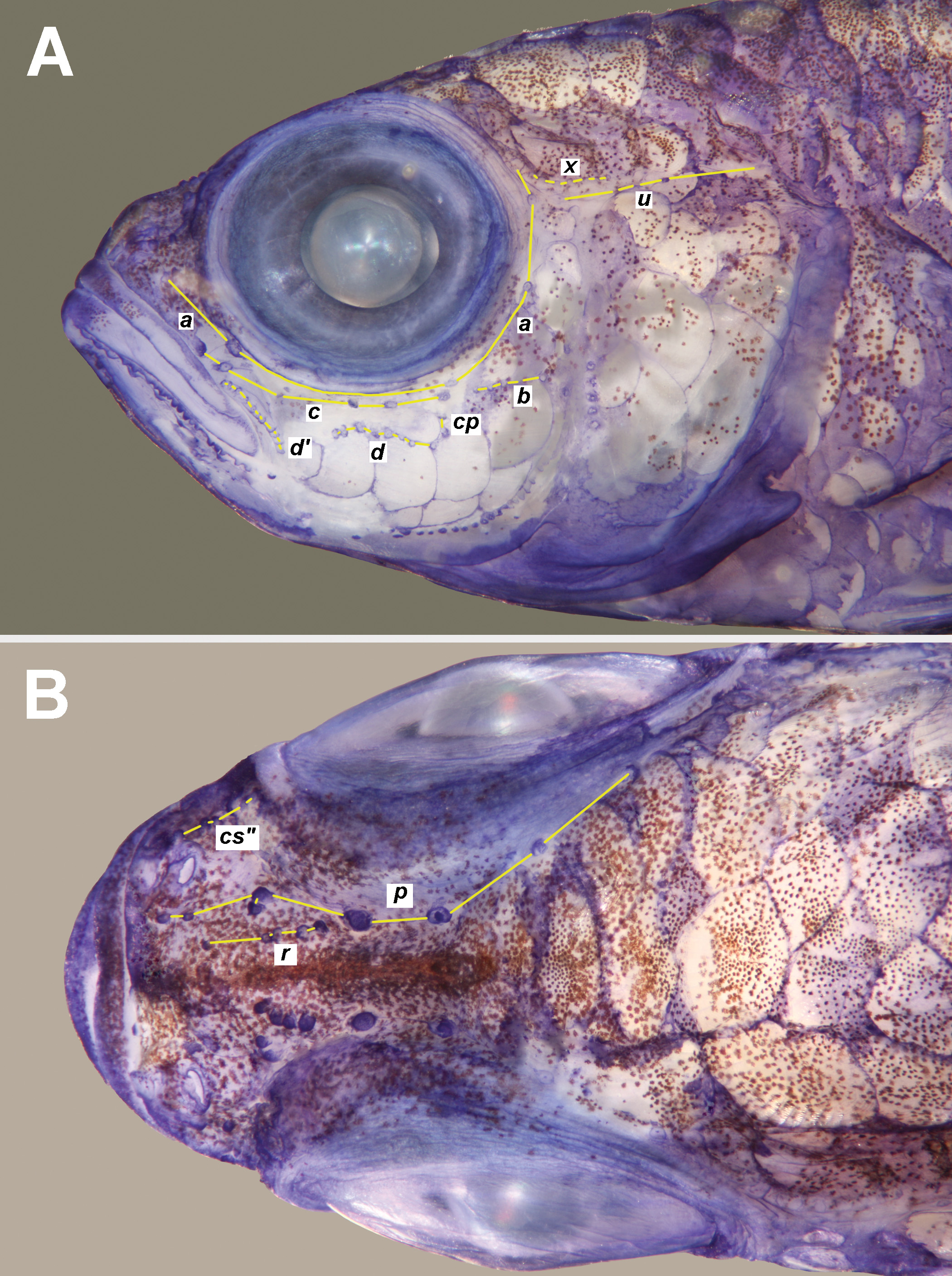


TABLE 4. Results from a barcode (COI) analysis of 105 specimens of Trimma tevegae subgroup, giving localities represented, number of specimens per species, the maximum variation within each group, followed by the minimum distances between groups (as percentages). Locality abbreviations are: Ha = Halmahera; P = Palawan; PM = Port Moresby; RA = Raja Ampat; Ra = Rabaul; Su = Sulawesi; TL = Timor Leste.
| Species | Locality | n | Var. | hollemani | burridgeae | caudomaculatum tevegae |
|---|---|---|---|---|---|---|
| T. corerefum | Palau/Su | 41 | 0.6 | 21.1 | 21.5 | 22.4 20.8 |
| T. hollemani | Ha/RA/Ra | 12 | 0. 6 | - | 8.9 | 9.8 10.2 |
| T. burridgeae | Palau | 8 | 0.5 | - | 9.1 9.5 | |
| T. caudomaculatum | PM/RA/Ra/TL | 18 | 1.7 | - 7.9 | ||
| T. tevegae | Ha/P/RA/Ra | 26 | 0.4 | - |
No known copyright restrictions apply. See Agosti, D., Egloff, W., 2009. Taxonomic information exchange and copyright: the Plazi approach. BMC Research Notes 2009, 2:53 for further explanation.
|
Kingdom |
|
|
Phylum |
|
|
Class |
|
|
Order |
|
|
Family |
|
|
Genus |
Trimma hollemani
| Winterbottom, Richard 2016 |