
Cletocamptus cecsurirensis, Gómez & Scheihing & Labarca, 2007
|
publication ID |
https://doi.org/ 10.1080/00222930601141476 |
|
persistent identifier |
https://treatment.plazi.org/id/03CA87FD-FFF5-0350-88BB-65A3FCA46A7F |
|
treatment provided by |
Felipe |
|
scientific name |
Cletocamptus cecsurirensis |
| status |
sp. nov. |
Cletocamptus cecsurirensis sp. nov.
( Figures 1–11 View Figure 1 View Figure 2 View Figure 3 View Figure 4 View Figure 5 View Figure 6 View Figure 7 View Figure 8 View Figure 9 View Figure 10 View Figure 11 )
Type material
One female holotype (EMUCOP-1004-01) and one male allotype (EMUCOP-1004-02) preserved in alcohol. Thirteen female (EMUCOP-1004-14 to EMUCOP-1004-26) and 10
male (EMUCOP-1004-04 to EMUCOP-1004-13) dissected paratypes, and 10 female and nine male paratypes preserved in alcohol (EMUCOP-1004-03). Collected on 19 October 2004 and 19 October 2005; small freshwater stream with sandy bottom (18 ° 47924.70S, 69 ° 05917.60W), two shallow ponds (18 ° 519430S, 69 ° 07959.30W and 18 ° 47934.60S, 69 ° 05927.70W) with muddy bottom; 4180 m a.s.l.; coll. Rodrigo Scheihing.
Type locality
Salar de Surire, Chilean high Andean Plateau (18 ° 47924.70S, 69 ° 05917.60W). Etymology
The specific epithet refers to the Centro de Estudios Científicos where two of us ( R.S. and P.L.) work, and to the type locality where the species was found .
Female
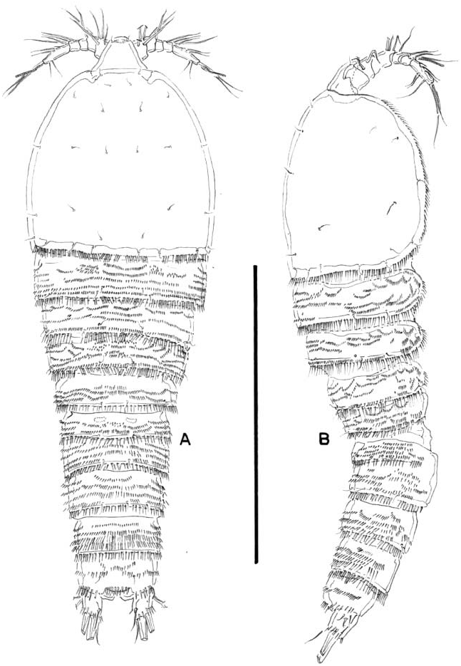
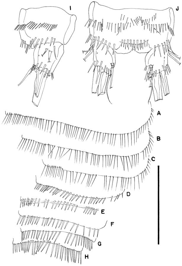
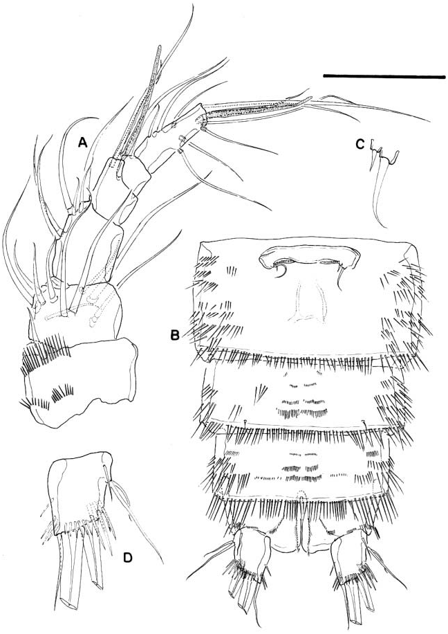
Habitus ( Figure 1A, B View Figure 1 ) tapering posteriorly; total body length measured from tip of rostrum to posterior margin of caudal rami ranging from 680 to 820 Mm (mean 737 mm, n 518; holotype 740 mm). Rostrum defined at base, triangular, with pair of setules subapically and ornamented with small spinules distally on ventral surface. Cephalic shield ( Figure 1A, B View Figure 1 ) with small, fine spinules along margin dorsally and laterally. Dorsal and lateral surface of free thoracic somites (P2–P4-bearing somites) with transverse rows of minute spinules, with longitudinal row of small spinules close to posterior margin and with long spinules along posterior margin ( Figure 2A–C View Figure 2 ). Dorsal and lateral surface of first urosomite (P5-bearing somite) with transverse rows of minute spinules, with row of small spinules close to posterior margin and with long spinules along posterior margin ( Figure 2D View Figure 2 ). Genital double-somite with subcuticular rib dorsally and laterally indicating former division between second and third urosomites ( Figure 1A, B View Figure 1 ), but completely fused ventrally ( Figure 3B View Figure 3 ); dorsal and lateral surface of second and third urosomite (first and second genital somites) with transverse rows of minute spinules, with row of long spinules along posterior margin ( Figure 2E, F View Figure 2 ), and with relatively longer spinules laterally, ventrally with spinules as figured ( Figure 3B View Figure 3 ). Fourth and fifth urosomites as in previous somite dorsally, ventrally with spinular pattern ( Figure 3B View Figure 3 ).
Dorsal surface of anal somite ( Figures 1A, B View Figure 1 , 2J View Figure 2 ) with transverse rows of spinules and with dorsolateral strong spinules close to joint with caudal rami ( Figure 3I, J View Figure 3 ); rounded anal operculum furnished with two rows of strong spinules. Caudal rami ( Figures 1A, B View Figure 1 , 2I, J View Figure 2 , 3D View Figure 3 ) nearly as long as wide; dorsal and ventral surface smooth, except for spinules close to posterior margin; with seven elements.
Antennule ( Figure 3A View Figure 3 ) six-segmented; surface of segments smooth except for two rows of spinules on first segment. Armature formula, 1-(1), 2-(10), 3-(6), 4-(1+[1+ae]), 5-(1), 6- (9+[1+ae]).
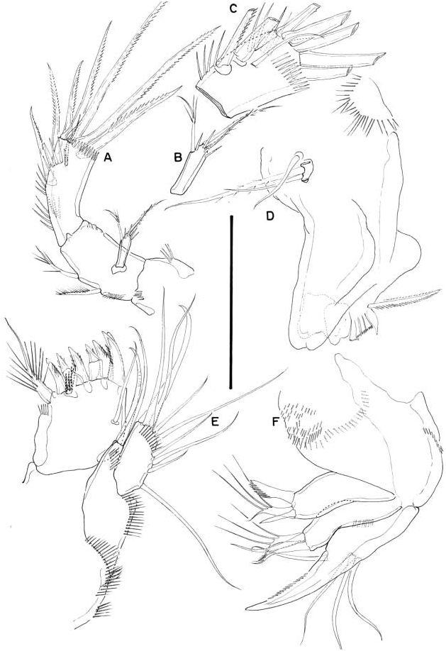
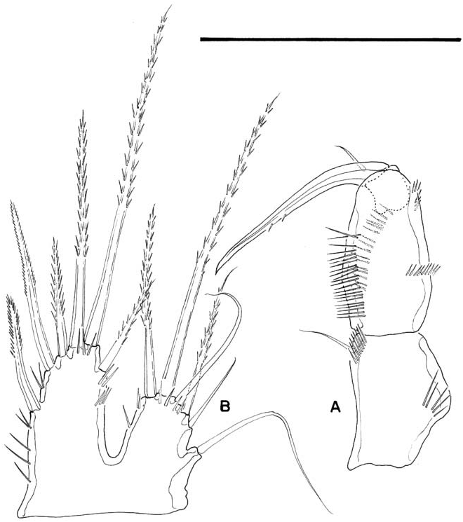
Antenna ( Figure 4A View Figure 4 ) with small coxa. Allobasis armed with two abexopodal setae. Free endopodal segment ornamented with inner strong spinules proximally and subdistally; with two lateral inner spines and a slender seta and five distal elements ( Figure 4C View Figure 4 ). Exopod one-segmented; about five times longer than wide; with few spinules, and with one lateral and two apical setae ( Figure 4B View Figure 4 ).
Mandible ( Figure 4D View Figure 4 ) robust; chewing edge with two bicuspidate teeth, four multicuspidate teeth, one pyriform element and one lateral seta. Palp one-segmented, with two setae unequal in length and one small seta arising nearby.
Maxillule ( Figure 4E View Figure 4 ) robust; arthrite of praecoxa with few spinules, with one surface seta, seven distal spines and one lateral strong seta, the latter spinulose. Coxa with some spinules and with two slender setae. Basis with some median spinules. Homology of the setae on basis, exopod and endopod difficult to assess. Basis seemingly with three apical and two lateral setae, endopod and exopod seemingly represented by three and one seta, respectively.
Maxilla ( Figure 4F View Figure 4 ): syncoxa with minute spinules along inner margin; with two endites, each bearing three setae as figured. Allobasis drawn into strong claw bearing one accompanying seta. Endopod represented by three setae.
Maxilliped ( Figure 5A View Figure 5 ) subchelate. Syncoxa with rows of spinules and with a small seta on inner distal corner. Basis without armature; with one anterior and one posterior longitudinal row of spinules along inner margin; with small spinules medially and subapically. Endopod drawn into long and slender claw with one accompanying small seta.
P1 ( Figure 6A View Figure 6 ): praecoxa with spinules close to joint with coxa. The latter with transverse spinule rows on anterior face, and with spinule row near outer distal corner on posterior face. Basis with inner and outer spines; with median spinule row, and with stronger spinules at base of exopod, between rami and at base of inner basal spine. Exopod three-segmented. Endopod two-segmented, reaching the middle of EXP 3.
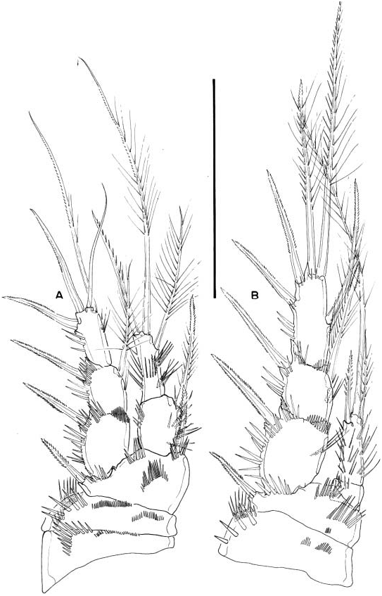
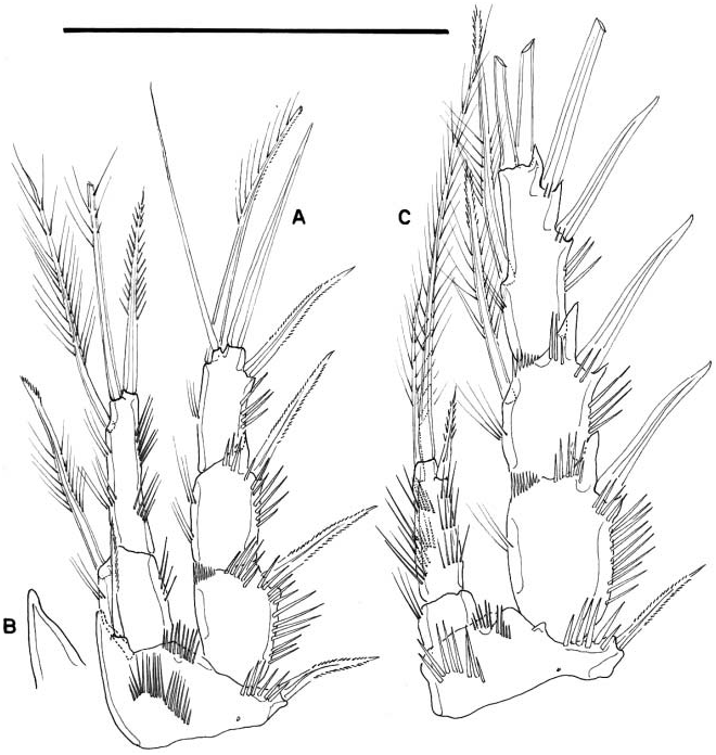
P2 ( Figure 6B View Figure 6 ): coxa as in P1. Basis as in P1 except for inner spine; outer element spinelike. Exopod three-segmented and ornamented as figured; EXP 2 and 3 with inner seta. Endopod two-segmented, barely reaching beyond tip of EXP 1; ENP 1 small, slightly wider than long and with outer spinules; ENP 2 with long spinules as shown, and with one outer spine, one apical and one inner seta.
P3 ( Figure 7A View Figure 7 ): coxa as in P2. Basis as in P2 except for seta-like outer element. Exopod and endopod as in P2.
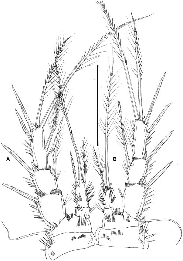
P4 ( Figure 7B View Figure 7 ): coxa and basis as in P3. Exopod three-segmented; EXP 2 with, EXP 3 without inner seta. Endopod two-segmented, barely reaching the tip of P4 EXP 1; ENP 1 small, slightly wider than long; ENP 2 with inner and outer slender spinules and armed with two apical setae (innermost shorter).
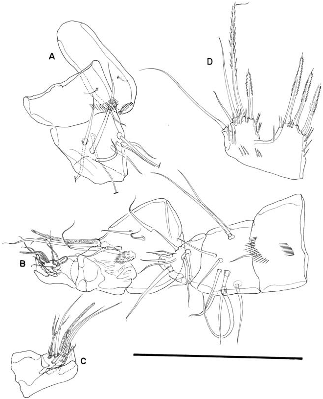
P5 ( Figure 5B View Figure 5 ): exopod and baseoendopod fused. Baseoendopodal lobe about twice as long as exopod, with sets of spinules along inner and outer margin, with spinules at base of apical seta; with one outer, one apical, and four inner setae; relative length of setae as figured. Exopod with spinules as figured, with five setae, plus outer seta of basis.
Armature formula of female P1–P5 as follows:
P6 ( Figure 3C View Figure 3 ) represented by median plate in anterior half of second urosomite (first genital somite); each vestigial leg represented by one outer long and one slender inner seta. Copulatory pore in the middle of genital double-somite.
Male
Body (not shown) as in female except for genital double-somite ( Figure 8A, B View Figure 8 ). Rostrum ( Figure 9A View Figure 9 ) sexually dimorphic, elongate, with two lateral setules and set with small spinules apically on ventral surface. Second to fifth urosomites ornamented with spinules as figured. Anal somite and caudal rami ( Figure 8A, B View Figure 8 ) as in female.
Antennule ( Figure 9A–C View Figure 9 ) six-segmented, subchirocer; last segment as in Figure 9C View Figure 9 and with three teeth. Armature formula difficult to define, but probably as follows 1-(1), 2-(9), 3-(8), 4-(6+[1+ae]), 5-(0), 6-(7+[1+ae]).
Antenna, mandible, maxillule, maxilla, and maxilliped (not shown) as in female.
P1 ( Figure 10A View Figure 10 ) as in female except for inner projection of basis in the male ( Figure 10B View Figure 10 ).
P2 ( Figure 10C View Figure 10 ) as in female except for dimorphic inner spine of ENP2, and stronger and bare outer dimorphic spines of EXP 1–3.
P3 exopod ( Figure 11A, B View Figure 11 ) as in female except for stronger and bare outer dimorphic spines; endopod dimorphic, three-segmented, second segment with apophysis reaching far beyond ENP 3.
P4 ( Figure 11C View Figure 11 ) as in female except for stronger outer dimorphic spines.
P5 ( Figure 9D View Figure 9 ): both legs distinct; baseoendopod and exopod fused; exopod with four setae plus basal seta; baseoendopod with three setae.
P6 ( Figure 8B View Figure 8 ) represented by a plate, without armature.
Variability
Females (13 females analysed). The second and third innermost setae of the P5 baseoendopod of about the same length in four specimens; the anal operculum of one female possesses one row of spinules; the left P4 endopod of one female possesses three instead of two setae on the last segment; the left P3 endopod of one female possesses four instead of three setae on the last segment.
Males (10 males analysed). One male was observed without spinular ornamentation on the anal operculum; the ventral spinular rows of urosomites are longer in two males; the anal operculum of one male is furnished with only one row of spinules; the inferior spinules on the anal operculum of one male are very small; one male was found with a two-segmented left exopod of P2 (the second and third segments partially fused); the last segment of the left exopod of P2 of one male is shorter; the left P4 ENP 3 View Materials of one male possesses three instead of two setae and the right P3 EXP 3 of the same animal possesses two instead of one inner setae; the outermost spine of the P5 baseoendopod of one male is shorter; the P5 baseoendopod of one male possesses four instead of three setae .


| R |
Departamento de Geologia, Universidad de Chile |
No known copyright restrictions apply. See Agosti, D., Egloff, W., 2009. Taxonomic information exchange and copyright: the Plazi approach. BMC Research Notes 2009, 2:53 for further explanation.
|
Kingdom |
|
|
Phylum |
|
|
Class |
|
|
Order |
|
|
Family |
|
|
Genus |