
Sinopoda cochlearia, Zhang, Bao-Shi, Zhang, Zhi-Sheng & Zhang, Feng, 2015
|
publication ID |
https://doi.org/10.11646/zootaxa.3974.1.4 |
|
publication LSID |
lsid:zoobank.org:pub:C2B68D50-6CC0-4726-9141-73BB45606036 |
|
DOI |
https://doi.org/10.5281/zenodo.6100730 |
|
persistent identifier |
https://treatment.plazi.org/id/03869D4F-FFAF-CB09-90DC-FC40B030307C |
|
treatment provided by |
Plazi |
|
scientific name |
Sinopoda cochlearia |
| status |
sp. nov. |
Sinopoda cochlearia View in CoL sp. nov.
( Figs 1–16 View FIGURES 1 – 2 View FIGURES 3 – 5 View FIGURES 6 – 9 View FIGURES 10 – 16 )
Type material: Holotype: ♂, CHINA: Chongqing Municipality: Jinfo Mountain Natural Reserve, Wolongtan, 29°04′N, 107°13′E, 757 m, native forest, by hand, 24 July 2011, L.Y. Wang leg. ( MHBU, SP-CQ-11-0704).
Paratypes: CHINA: Chongqing Municipality: 1♀, same data as holotype ( MHBU, SP-CQ-11-0705); 1♂, Jinyun Mountain Natural Reserve, 29°49′N, 106°22′E, 510 m, native forest, by hand, 8 July 2010, Z.S. Zhang leg., ( SWUC, SP-CQ-10-0701); 1♂, Simian Mountain Forest Park, 28°40′N, 106°25′E, 730 m, 18 July 2011, M.X. Liu leg. ( SWUC, SP-CQ-11-0702).
Etymology. The specific name is derived from the Latin word ‘cochlear’, meaning ‘cucullar’, referring to the shape of the cucullar conductor; adjective.
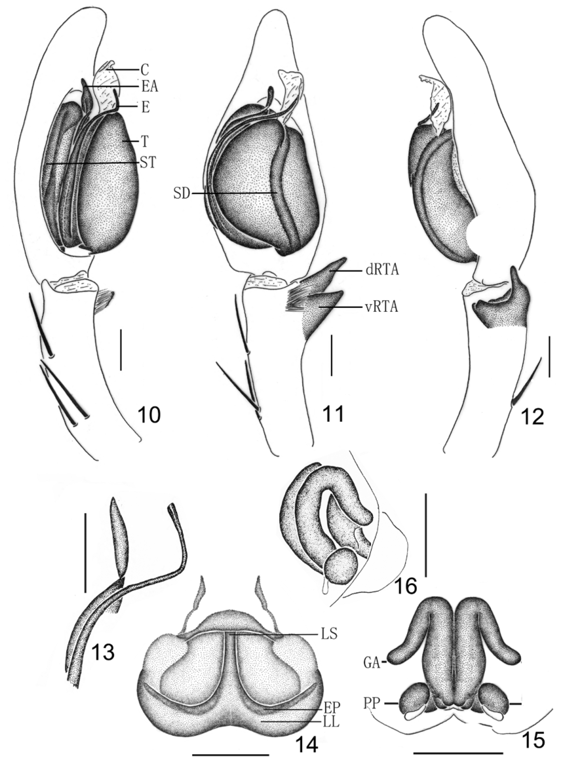
Diagnosis. Based on the reduced vRTA and the presence of a distinct brush of stiff setae at the base of the RTA, males of S. cochlearia sp. nov. belong to the Sinopoda okinawana -group as defined by Jäger and Ono (2002), but can be separated from S. albofasciata Jäger and Ono, 2002 , S. derivata Jäger and Ono, 2002 , S. nuda Liu et al., 2008 , S. tanikawai Jäger and Ono, 2000 and S. wangi Song and Zhu, 1999 by the developed EA; separated from S. fasciculata Jäger et al., 2002 , S. koreana (Paik, 1968) and S. okinawana Jäger and Ono, 2000 by the long EA (about half the length of embolus); and separated from S. hamata (Fox, 1937) by the slightly bent SD in ventral view, short dRTA and the narrow embolic base ( Figs 3–5 View FIGURES 3 – 5 , 10–13 View FIGURES 10 – 16 ). The female of S. cochlearia sp. nov. resembles those of S. suang Jäger, 2012 , S. tham Jäger, 2012 and S. triangula Liu et al., 2008 by: epigynal pockets with a narrow lobal septum, posterior margin of lateral lobes forming a semi-circle, posterior margin of epigyne slightly bilobate as well as internal duct system with long parallel part. It can be distinguished from S. suang and S. tham by: lobal septum long, glandular appendages long, extending posteriorly in posterior half of internal duct system, and distinctly diverging posteriorly; and distinguished from S. triangula by small posterior part of spermathecae ( Figs 8–9 View FIGURES 6 – 9 , 14–16 View FIGURES 10 – 16 ).
Description. Male: Total length (n=3) 18.91–19.10. Holotype: total length 19.10; prosoma 9.20 long, 8.40 wide; opisthosoma 9.90 long, 5.80 wide. Dorsal prosoma reddish-brown, lateral and posterior margins dark brown, with yellow submarginal transversal band posteriorly. Fovea and radial furrows distinctly dark brown. Eye diameters and interdistances: AME 0.42, ALE 0.60, PME 0.44, PLE 0.62; AME–AME 0.29, AME–ALE 0.08, PME–PME 0.36, PME–PLE 0.60. MOA 1.43 long, anterior width 1.07, posterior width 1.30. Clypeus height 0.37. Chelicerae deep reddish-brown with dark pattern, promargin with 3 teeth and retromargin with 4 teeth, and with 43 denticles in elongated patch close to promarginal teeth. Labium and gnathocoxae reddish-brown, both with distal parts brighter. Sternum reddish-brown, margin dark. Legs reddish-brown except femora brown and with dark spots. Ventral metatarsus III with sparse double row of bristles in proximal half, IV with dense double row of bristles along entire length. Leg measurements: I 54.70 (13.10, 4.80, 14.80, 15.90, 6.10), II 59.90 (15.50, 5.90, 15.80, 17.60, 5.10), III 40.80 (10.10, 4.10, 10.60, 12.10, 3.90), IV 46.90 (12.30, 4.80, 11.40, 13.80, 4.60). Leg formula: 2143. Leg spination: palp 131, 101, 2121; femur I–III 333, IV 331; patella I–III 101, IV 100; tibia I–II 2226, III 2326, IV 2226; metatarsus I–II 1014, III 2026, IV 3036. Dorsal opisthosoma dark brown, heart patch and muscle impressions yellow; venter dark brown, with two longitudinal yellow lines in front of spinnerets ( Figs 1–2 View FIGURES 1 – 2 ).
Palp ( Figs 3–5 View FIGURES 3 – 5 , 10–13 View FIGURES 10 – 16 ). Conductor cucullar, and with a short handle in ventral view; embolus long and slender, arising in 6- to 7-o'clock-position from the swollen tegulum (left palp in ventral view), embolus distinctly curved, basal part of embolus only partly visible in ventral view; tip of embolic apophysis bent at obtuse angle; sperm duct slightly curved in ventral view; dRTA slender and pointed, vRTA short and stout in retrolateral view; cymbium longer than tibia.
Female: Paratype: total length (n=1) 24.17; prosoma 9.69 long, 9.07 wide; opisthosoma 14.48 long, 9.99 wide. Eye diameters and interdistances: AME 0.42, ALE 0.60, PME 0.44, PLE 0.62; AME–AME 0.29, AME–ALE 0.08, PME–PME 0.36, PME–PLE 0.60. MOA 1.43 long, anterior width 1.07, posterior width 1.30. Clypeus height 0.37. Leg measurements: I 36.51 (10.20, 3.21, 11.37, 9.18, 2.55), II 39.87 (11.73, 3.25, 12.25, 9.79, 2.85), III 32.31 (9.58, 3.02, 9.72, 7.65, 2.34), IV 35.37 (10.10, 2.93, 9.91, 9.58, 2.85). Leg formula: 2143. Leg spination: palp 131, 101, 2121, 1012; femur I–III 323, IV 331; patella I–II 101, III 0 0 1, IV 000; tibia I–IV 2026; metatarsus I–II 2028, III 2016, IV 2036. Dorsal prosoma darker than that in male, dorsal opisthosoma brighter than that in male. Anterior part of dorsal opisthosoma with two wide and short yellow patches. Otherwise, shape, color and markings of body as in male ( Figs 6–7 View FIGURES 6 – 9 ).
Epigyne ( Figs 8–9 View FIGURES 6 – 9 , 14–16 View FIGURES 10 – 16 ). Epigynal field wider than long, with two long and narrow anterior bands; lateral lobes fused, posteriorly with only slight median indentation; epigynal pockets running from latero-posterior to medio-anterior, where copulatory openings are situated; lobal septum narrow and long; internal ducts running parallel along the median line, glandular appendages slightly narrower than posterior part of internal ducts; glandular appendages extending posteriorly in posterior half of internal duct system; posterior part of spermathecae bulging laterally; fertilisation ducts arising posterio-laterally from ovate posterior part of spermathecae. Distribution. China (Chongqing).
Remarks. The okinawana -group of the genus Sinopoda was established by Jäger and Ono (2002). This group can be diagnosed by: reduced embolic apophysis and vRTA, and distinct brush of stiff hairs at the base of the RTA.
The okinawana View in CoL -group is distributed in Japan, Korea and China. The embolic apophysis and the vRTA of westernmost species are well developed. The females of the okinawana View in CoL -group can hardly be diagnosed without conspecific males, but there is a recognizable trend in some female genitalia: dorsal side of the vulva lengthened and bent dorsal ( Jäger & Ono 2002; Jäger 2006). The males of S. cochlearia View in CoL sp. nov. belong to the S. okinawana View in CoL - group according to the characters of RTA and the presence of a distinct brush of stiff setae at the base of the RTA ( Fig. 3 View FIGURES 3 – 5 ). The females of S. cochlearia View in CoL sp. nov. have a dorsally bent vulva ( Fig. 16 View FIGURES 10 – 16 ).


No known copyright restrictions apply. See Agosti, D., Egloff, W., 2009. Taxonomic information exchange and copyright: the Plazi approach. BMC Research Notes 2009, 2:53 for further explanation.
|
Kingdom |
|
|
Phylum |
|
|
Class |
|
|
Order |
|
|
Family |
|
|
Genus |