
Prionospio gayheadia, Delgado-Blas & Peraza, 2024
|
publication ID |
https://doi.org/ 10.11646/zootaxa.5432.1.5 |
|
publication LSID |
lsid:zoobank.org:pub:ACE338D6-8F97-4462-AC8C-F2E9A3ED05A3 |
|
DOI |
https://doi.org/10.5281/zenodo.10909271 |
|
persistent identifier |
https://treatment.plazi.org/id/038B87AE-FFB2-FFDC-FF11-A2E0F9304719 |
|
treatment provided by |
Plazi |
|
scientific name |
Prionospio gayheadia |
| status |
sp. nov. |
Prionospio gayheadia sp. nov.
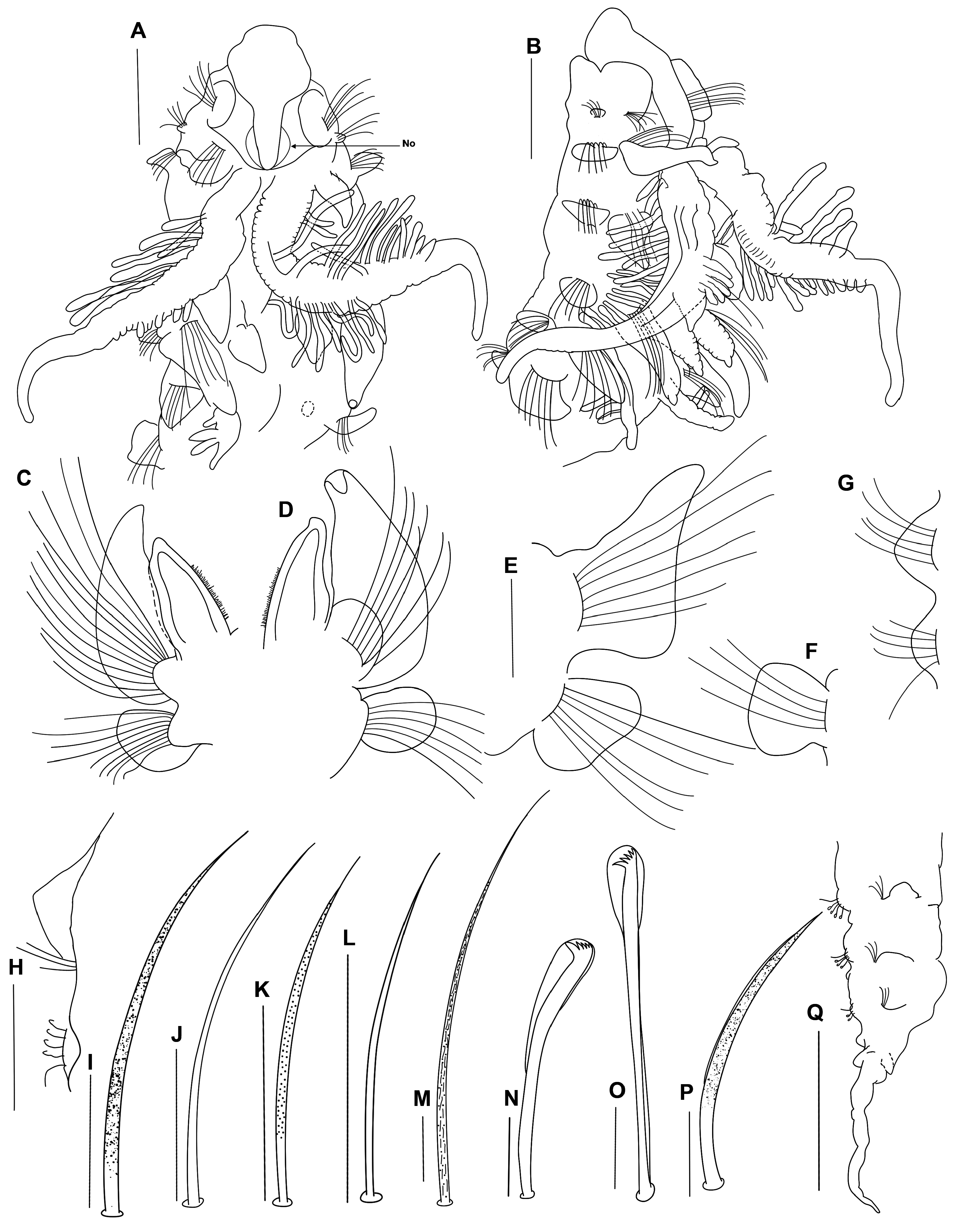
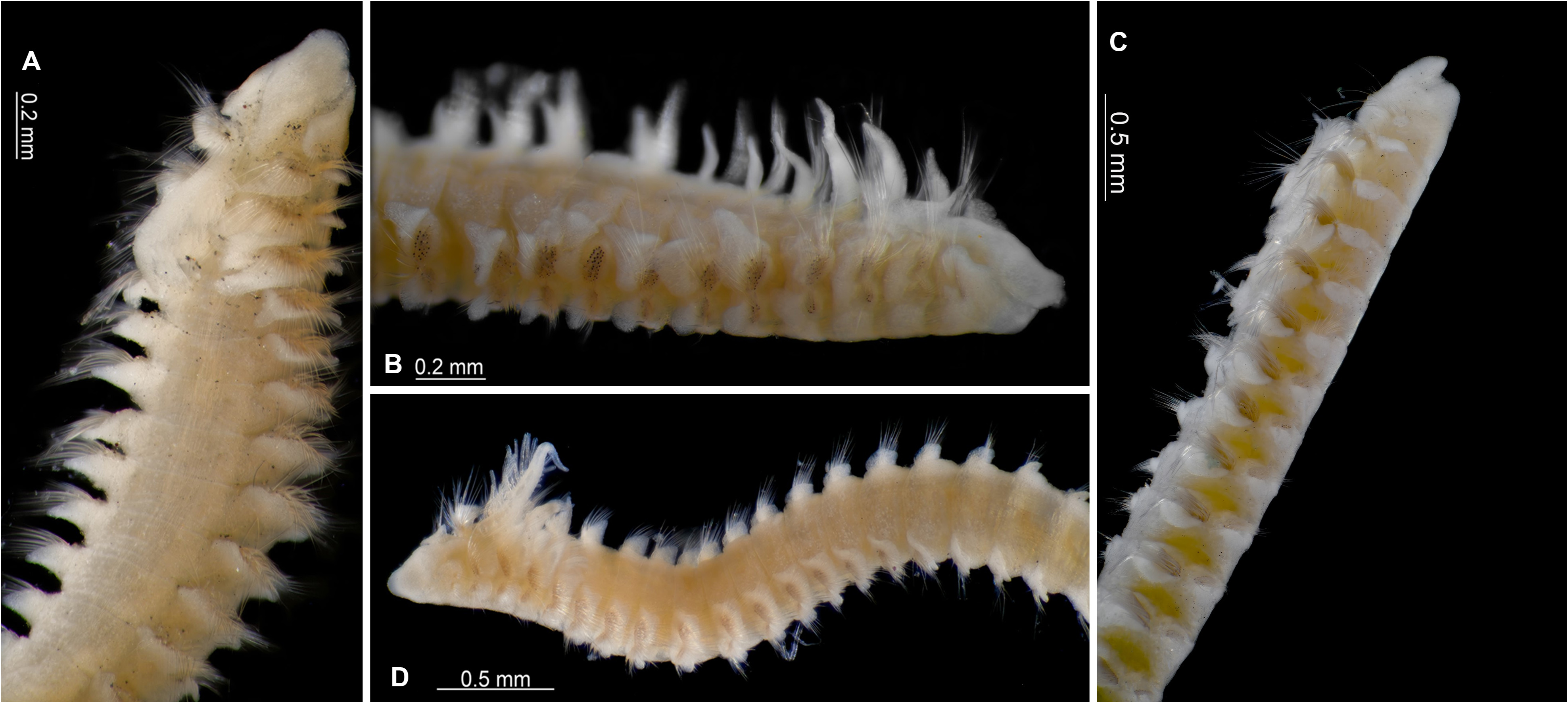
Figures 2A–Q View FIGURE 2 ; 3A–D View FIGURE 3
Prionospio (Prionospio) dubia Maciolek, 1985: 336-339 View in CoL , figs. 2, 3 (in part, Western North Atlantic Ocean specimens).
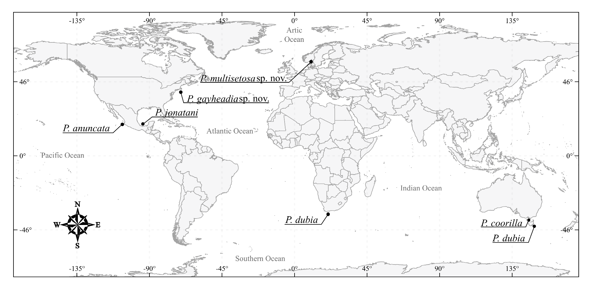
Material examined: WESTERN NORTH ATLANTIC OCEAN. Off New England, south of Gay Head, Martha’s Vineyard , “Gay Head-Bermuda Transect”, coll. H. L. Sanders, 28 August 1962, Atlantis cruise 283, WH0I, Sta. Slope 3, 39°58.9’N, 70°40.3’W, 300 m, anchor dredge, holotype ( LACM-AHF-Poly 14388 ) GoogleMaps ; WH0I, Sta Slope. 3, 39°58.9’N, 70°40.3’W, 300 m, coll. H. L. Sanders, 28 August 1962, 222 paratypes ( LACM-AHF- Poly 6272 ) GoogleMaps . 28 August 1962, WH0I, Sta. Slope 2, 40º01.81’N, 70º42’W, 200 m, 2 specimens ( LACM-AHF Poly 6273 ) GoogleMaps .
Description. Holotype complete: 34.5 mm long for 96 chaetigers, 0.4 mm wide. Complete paratypes: 18–23 mm long for 76–94 chaetigers, 0.3–0.5 mm wide. Incomplete paratypes: 2–12 mm long for 15–63 chaetigers, 0.3–0.4 mm wide. Smaller specimens: 7.5–13 mm long for 53–61 chaetigers, 0.25–0.3 mm wide. Color in alcohol, brown. The numbers in parentheses refers to the variation in the largest and smallest paratypes. Some specimens with oocytes in chaetigers 17–78 (holotype, 22–76). Prostomium flask-shaped, anteriorly narrow, truncate, widening in the mid-region, posteriorly tapered, with a long, narrow caruncle extending to the anterior edge of chaetiger 2 ( Figs 2A, B View FIGURE 2 , 3A, B View FIGURE 3 ), and with large nuchal organs on either side ( Fig. 2A View FIGURE 2 ). Eyes absent. Palps lost. Peristomium short, collar-like, surrounding prostomium, fused dorsally with large, rounded notopodial lamellae of chaetiger 1 ( Figs 2A, B View FIGURE 2 , 3A–C View FIGURE 3 ). Neuropodial postchaetal lamellae on chaetiger 1 very short, rounded ( Figs 2A, B View FIGURE 2 , 3B–D View FIGURE 3 ), much smaller than 1/5 of the size of the notopodial lamellae.
Four pairs of branchiae present on chaetigers 2–5, first pair longest and thickest, up to six times longer than the fourth pair ( Fig. 2B View FIGURE 2 ); and extending up to chaetiger 6, fourth pair longer than notopodial lamellae ( Fig. 2A, B View FIGURE 2 ). Both first and fourth pairs with very long, thick, dense digitiform pinnules, pair 1 with pinnules present from the base of the branchiae and extending up to 2/3 along the posterior face ( Figs 2A, B View FIGURE 2 , 3D View FIGURE 3 ), and pair 4 with fewer pinnules, arranged along the outer lateral margin; branchiae with long, naked, smooth distal tips ( Figs 2A, B View FIGURE 2 , 3D View FIGURE 3 ). Axes of branchial pairs 1 and 4 pinnate, elongate; pair 1 with articulations, pair 4 smoother (less articulated). Branchial pairs 2 and 3 apinnate, subtriangular, wide, thick, heavily ciliated laterally, with sharply pointed short tips; and shorter than fourth pair. ( Figs 2C, D View FIGURE 2 , 3D View FIGURE 3 ); subequal in length, smaller than the notopodial lamellae ( Figs 2C, D View FIGURE 2 , 3B View FIGURE 3 ).
Notopodial postchaetal lamellae of chaetigers 2–5 triangular, slender with sharply pointed long tips, and with wide, rounded bases ( Figs 2C, D View FIGURE 2 , 3A View FIGURE 3 ); lamellae of chaetiger 2 with dorsal edges touching between them (when branchiae lost or removed); lamellae longest on chaetigers 3–4, noticeably shorter on chaetigers 5 and 6 ( Fig. 3A View FIGURE 3 ), thereafter gradually decreasing in size; notopodial lamellae on chaetigers 6–10 subtriangular with short, blunt tips ( Fig. 3B View FIGURE 3 ); notopodial lamellae on chaetigers 11–13 square ( Fig. 2F View FIGURE 2 ); notopodial lamellae on chaetigers 14–20 rounded, wider and smaller than those on previous chaetigers ( Figs 2G View FIGURE 2 , 3D View FIGURE 3 ), extending slightly onto dorsum from chaetigers 21–32 (holotype 26) ( Fig. 2H View FIGURE 2 ), but never connected along the dorsal ridge or crest; subsequent notopodial lamellae rounded, small on far posterior chaetigers and with the chaetal row located in the ventral region of the noto-lamella ( Fig. 2H View FIGURE 2 ). Notopodial prechaetal lamellae very low, rudimentary throughout. No dorsolateral skin folds.
Neuropodial postchaetal lamellae of chaetiger 2 oval, each with a rounded ventral lobe and slightly elongated dorsal edge ( Fig. 2E View FIGURE 2 ); lamellae of chaetigers 3–4 oval ( Figs 2C, D View FIGURE 2 , 3B–C View FIGURE 3 ); subsequent neuropodial lamellae rounded ( Figs 2G View FIGURE 2 , 3B, C View FIGURE 3 ), smallest on far posterior chaetigers ( Fig. 2H View FIGURE 2 ). Neuropodial prechaetal lamellae very low ( Figs 1 View FIGURE 1 , 2B View FIGURE 2 ), rudimentary throughout. Inter-parapodial pouches lacking.
Chaetiger 1 with short, alimbate and moderately granulated capillary chaetae, arranged in one row in both tiers ( Fig. 3B–D View FIGURE 3 ); notopodial capillaries unilimbate with very long tips of chaetigers 2–12/14 (holotype, 2–12) arranged in three rows ( Fig. 3B View FIGURE 3 ); chaetae moderately granulated on posterior rows ( Fig. 2I View FIGURE 2 ), and smooth on anterior row ( Fig. 2J View FIGURE 2 ); both notopodial chaetae and neuropodial chaetae longest on the most posterior row, but with the latter being shorter than the former ( Fig. 2K, L View FIGURE 2 ); neuropodial chaetae arranged in two rows ( Fig. 3B View FIGURE 3 ). Chaetae on middle chaetigers with notopodial and neuropodial capillaries striated, lightly granulated and narrowly sheathed ( Fig. 2M View FIGURE 2 ). Middle notopodial capillaries very long (up to 70 um), so long (up to 70 um) that they can be coiled, and up to five times longer than the neuropodial capillaries ( Fig. 2M View FIGURE 2 ). Posterior chaetiger with slender alimbate, smooth capillaries. Sabre chaetae from chaetigers 15–18 (holotype, 16) (smaller specimens, 15–17), numbering one or two per neuropodium, each chaeta heavily granulated, with a very narrow sheath ( Fig. 2P View FIGURE 2 ). Neuropodial hooded hooks ( Fig. 2N View FIGURE 2 ) from chaetigers 19–20 (holotype, 22) (smaller specimens, 17–18), up to 11 per fascicle, accompanied by slender capillaries. Notopodial hooded hooks ( Fig. 2O View FIGURE 2 ) from chaetigers 70–76 (holotype, 70) (smaller specimens, 56–59), up to three per fascicle, accompanied by four or five capillaries; all hooks with four pairs of teeth above the main tooth, secondary hood absent ( Fig. 2N, O View FIGURE 2 ).
Pygidium with a very long, thick mid-dorsal cirrus and two short lateral lobes ( Fig. 2Q View FIGURE 2 ).
Methyl green staining pattern. Dorsal surfaces of the prostomium, lateral peristomium and the first four notopodial and neuropodial lamellae intensely stained; staining discrete in the following segments; dorsal and ventral surfaces intensely stained from around chaetiger 10 up to the middle chaetigers; rest of the body unstained.
Remarks. Prionospio gayheadia sp. nov. is very similar to P. dubia Day, 1961 , P. jonatani Delgado-Blas, 2015 and P. multisetosa sp. nov. P. gayheadia sp. nov. is similar to P. dubia Day, 1961 , due to the absence of a dorsal crest or ridges, anterior notopodial prechaetal lamellae very low, and the absence of secondary hoods on the hooded hooks. However, Prionospio gayheadia sp. nov. can be distinguished from the original description of P. dubia Day, 1961 and the redescriptions of P. dubia given by Wilson (1990), in that the former has a flask-shaped prostomium and anteriorly truncate, whereas in P. dubia the prostomium is triangular, anteriorly rounded; P. gayheadia has no eyes in any of the specimens examined independent of their size; the first pair of branchiae shorter and with very long, naked, smooth distal tips; the fourth pair of branchiae with a few pinnules on the outer lateral margin; the apinnate branchial pairs 2 and 3 are shorter than the notopodial lamellae; the notopodial lamellae extend slightly onto the dorsum; and smooth chaetae on the anterior rows of the anterior notopodial and neuropodial chaetigers. In addition, the point at which the sabre chaetae, and the notopodial and neuropodial hooded hooks start from differs: in P. gayheadia sp. nov. the sabre chaetae are present from chaetigers 15–18 (smaller specimens, 15–17) vs. 17–20 ( Wilson 1990) and 18–20 (South African specimens, Sigvaldadóttir & Mackie, 1993); the neuropodial hooded hooks are present from chaetigers 19–22 (smaller specimens, 17–18) vs. 18–19 ( Wilson 1990) and 18–20 (South African specimens; Sigvaldadóttir & Mackie, 1993); and the notopodial hooded hooks are present from chaetigers 70–76 (smaller specimens, 56–59) vs. 46–50 ( Wilson 1990) and 48 (South African specimens; Sigvaldadóttir & Mackie, 1993); and finally, P. gayheadia sp. nov. has four pairs of accessory teeth on the hooded hooks vs. three to four pairs ( Wilson 1990).
In addition, Prionospio gayheadia sp. nov. shows some morphological similarities with P. jonatani Delgado-Blas, 2015 , in that neither species have any eyes, first pair of branchiae of equal size, the branchiae pinnate with very long, naked distal tips, the fourth pinnate branchial pair is longer than the apinnate pairs, 2 and 3, the number of rows of the chaetae in the anterior region is the same, and the hooded hooks have the same number of accessory teeth. However, P. gayheadia sp. nov. can be distinguished from the original description of P. jonatani , in that the former species is larger in size; the prostomium is anteriorly truncate and flask-shaped, whereas in P. jonatani the prostomium is anteriorly rounded and skittle-shaped; furthermore, in P. gayheadia sp. nov. the notopodial prechaetal lamellae are not basally fused with the notopodial postchaetal lamellae in the branchial region; in P. gayheadia sp. nov. the notopodial lamellae extend slightly onto the dorsum on chaetigers 21–32, whereas in P. jonatani the notopodial lamellae do not extend onto the dorsum. In addition, P. gayheadia sp. nov. the neuropodial postchaetal lamellae are oval on chaetigers 2–5, whereas in P. jonatani the lamellae are square. The shape of anterior chaetae and sabre chaetae also differ between the two species: in P. gayheadia sp. nov. the chaetae are smooth on the anterior row of the anterior notopodial and neuropodial chaetigers, and the sabre chaetae are heavily granulated and have very narrow sheaths, whereas in P. jonatani the sabre chaetae are moderately granulated and alimbate.
Prionospio gayheadia sp. nov. also shows some morphological similarities with P. multisetosa sp. nov. in that eyes are absent in both species; the fourth pinnate branchial pair is longer than the second and third apinnate pairs; the notopodial lamellae extend slightly onto the dorsum of the middle chaetigers; and the neuropodial postchaetal lamellae of chaetigers 4 and 5 are oval and rounded, respectively. However, P. gayheadia sp. nov. differs from P. multisetosa sp. nov. in that the former species is smaller in size, the prostomium is flask-shaped and anteriorly truncate, whereas in P. multisetosa sp. nov. the prostomium is skittle-shaped and anteriorly rounded. Furthermore, in P. gayheadia sp. nov. the first branchial pair is longer, with the distal tips are long, naked and smooth. In P. gayheadia sp. nov. the apinnate branchial pairs, 2 and 3, are shorter than the notopodial lamellae. In P. gayheadia sp. nov. the postbranchial notopodial prechaetal lamellae are very low, whereas in P. multisetosa sp. nov. the notopodial prechaetal lamellae are well-developed on chaetigers 13–33. In P. gayheadia sp. nov. the first neuropodial lamellae are very short, rounded and much smaller than 1/5 of the size of the notopodial lamellae, and the neuropodial lamellae of chaetiger 3 are oval, whereas in P. multisetosa sp. nov. the first neuropodial lamellae are large, tongue-shaped, and only slightly smaller than the notopodial lamellae, and the neuropodial lamellae of chaetiger 3 are subtriangular with blunt dorsal edges, to oval. In addition, P. gayheadia sp. nov. has notopodial chaetae from chaetiger 2 onwards, arranged in three rows, with moderately granulated chaetae on the posterior rows, and smooth chaetae on the anterior row, whereas in P. multisetosa sp. nov. although the notopodial chaetae also start on chaetiger 2, they are arranged in four rows, and all the chaetae are heavily granulated. The shape of the sabre chaetae, the chaetigers from which they start, and the number of teeth on the hooded hooks also differ between the two species: in P. gayheadia sp. nov. the sabre chaetae have very narrow sheaths and are present from chaetigers 15–18, the hooded hooks have four pairs of accessory teeth, and the notopodial hooded hooks are present from chaetigers 56–76; whereas in P. multisetosa sp. nov. the sabre chaetae are alimbate and are present from chaetigers 19–24, the hooded hooks have three pairs of accessory teeth, and the notopodial hooded hooks are present from chaetigers 46–55.
The differences between this new species from other, very similar, species are given in the key provided and Table 1 View TABLE 1 .
Etymology. The species name refers to the beginning of the transect between Gay Head, Massachusetts, and the islands of Bermuda, where the benthic samples were collected.
Type locality. New England, off the coast of Massachusetts, USA, 200–300 m.
No known copyright restrictions apply. See Agosti, D., Egloff, W., 2009. Taxonomic information exchange and copyright: the Plazi approach. BMC Research Notes 2009, 2:53 for further explanation.
|
Kingdom |
|
|
Phylum |
|
|
Class |
|
|
Order |
|
|
Family |
|
|
Genus |
Prionospio gayheadia
| Delgado-Blas, Víctor Hugo & Peraza, Russell Giovanni Uc 2024 |
Prionospio (Prionospio) dubia
| Maciolek, N. J. 1985: 339 |