
Rhinogobius maculagenys, Wu & Deng & Wang & Liu, 2018
|
publication ID |
https://doi.org/ 10.11646/zootaxa.4476.1.11 |
|
publication LSID |
lsid:zoobank.org:pub:B918E9B6-B810-4319-87C1-20B09E811C83 |
|
DOI |
https://doi.org/10.5281/zenodo.5998523 |
|
persistent identifier |
https://treatment.plazi.org/id/038C879D-FF90-F333-FF1E-FB7994B6DC8B |
|
treatment provided by |
Plazi |
|
scientific name |
Rhinogobius maculagenys |
| status |
sp. nov. |
Rhinogobius maculagenys sp. nov. Wu, Deng, Wang, & Liu
( Tables 1–2; Fig.1–4 View FIGURE 1 View FIGURE 2 View FIGURE 3 View FIGURE 4 ).
Holotype. HUNNULS2016-12-2801, male, 49.50 mm SL; Zhong Water , the upper reaches of Xiangjiang River, Lanshan Country, Hunan Province, China, 25°21′36.98″N 112°11′6.55″E; collected by Qianqian Wu on 28 December 2016. GoogleMaps
Paratypes. Fifteen specimens (seven males and eight females, 41.68–52.82 mm SL), same locality as holotype: HUNNULS2016-12-2802, male, 48.12 mm SL; HUNNULS2016-12-2803, male, 41.68 mm SL; HUNNULS2016-12-2804, male, 51.10 mm SL; HUNNULS2016-12-2805, male, 46.57 mm SL; HUNNULS2016- 12-2806, male, 52.82 mm SL; HUNNULS2017-12-0602, male, 46.67 mm SL; HUNNULS 2017-12-0613, male, 39.85 mm SL; HUNNULS2016-12-2807, female, 44.87 mm SL; HUNNULS2016-12-2808, female, 46.22 mm SL; HUNNULS2016-12-2809, female, 47.31 mm SL; HUNNULS2016-12-2810, female, 47.87 mm SL; HUNNULS2016-12-2811, female, 44.89 mm SL; HUNNULS2016-12-2812, female, 46.77 mm SL; HUNNULS2017-12-0621, female, 49.79 mm SL; HUNNULS2017-12-0612, female, 44.50 mm SL.
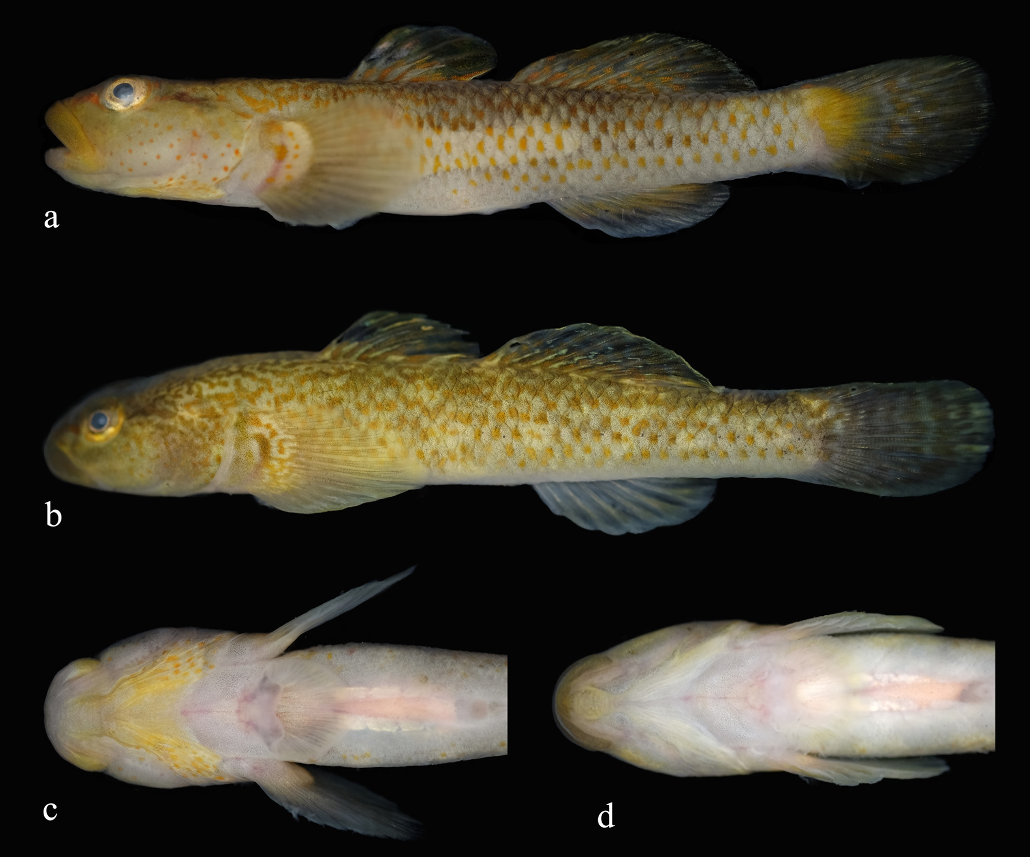
Diagnosis. Rhinogobius maculagenys is distinguished from all congeners by a combination of the following features: second dorsal-fin rays I/7–9; anal-fin rays I/6–8; pectoral-fin rays 16; longitudinal scale series 32–34; transverse scale series 9–13; predorsal scale series 0; vertebral count 11+16=27; pore ω 1 missing; head and body yellowish brown; cheek and opercle yellowish brown with over 30 small orange spots, branchiostegal membrane yellow with over 10 small orange spots in males and white and spotless in females; first dorsal fin trapezoidal in males and nearly semicircular in females, with large bright blue blotch in front of second spine; spines 4 and 5 longest, rear tip extending to base of second branched ray of second dorsal fin in males when adpressed, but just reaching or not reaching anterior margin of second dorsal fin in females; caudal fin with 5–6 vertical rows of brown spots; flank with several longitudinal rows of blackish-brown spots; belly pale white.
Description. First dorsal-fin rays VI* (16); second dorsal-fin rays I/7 (1), I/8 (10) or I/9 * (5); anal-fin rays I/6 (2), I/7 * (13), I/8 (1); pectoral-fin rays 16* (16); pelvic-fin rays I/5 * (16); branched caudal fin-rays 8+7* (5); longitudinal scales 32 (2), 33*(13), or 34 (1); transverse scales 9* (3), 10 (8), 11(1), 12 (3), or 13 (1); scales between origin of dorsal and pectoral fin 6 (2), 7* (12), or 8 (2); predorsal scales 0* (16); P-V 3/II IIII 0/9* (5); vertebral counts 11+16=27* (5); gill rakers 9+20 (1).
Morphometric data presented in Table 1. Body slender, sub-cylindrical anteriorly and compressed posteriorly. Head moderately large, eye high and large, lips thick. Upper lip more prominent than lower lip. Mouth oblique, corner of mouth extending to vertical of anterior margin of orbit in males, almost reaching the vertical of anterior margin of orbit in females. Both jaws with 3–4 rows of conical and inwardly curved teeth. Anterior nares short tubes, posterior nares with round openings. Gill opening extends ventrally reaching middle vertical line of operculum.
…….continued on the next page ……continued on the next page
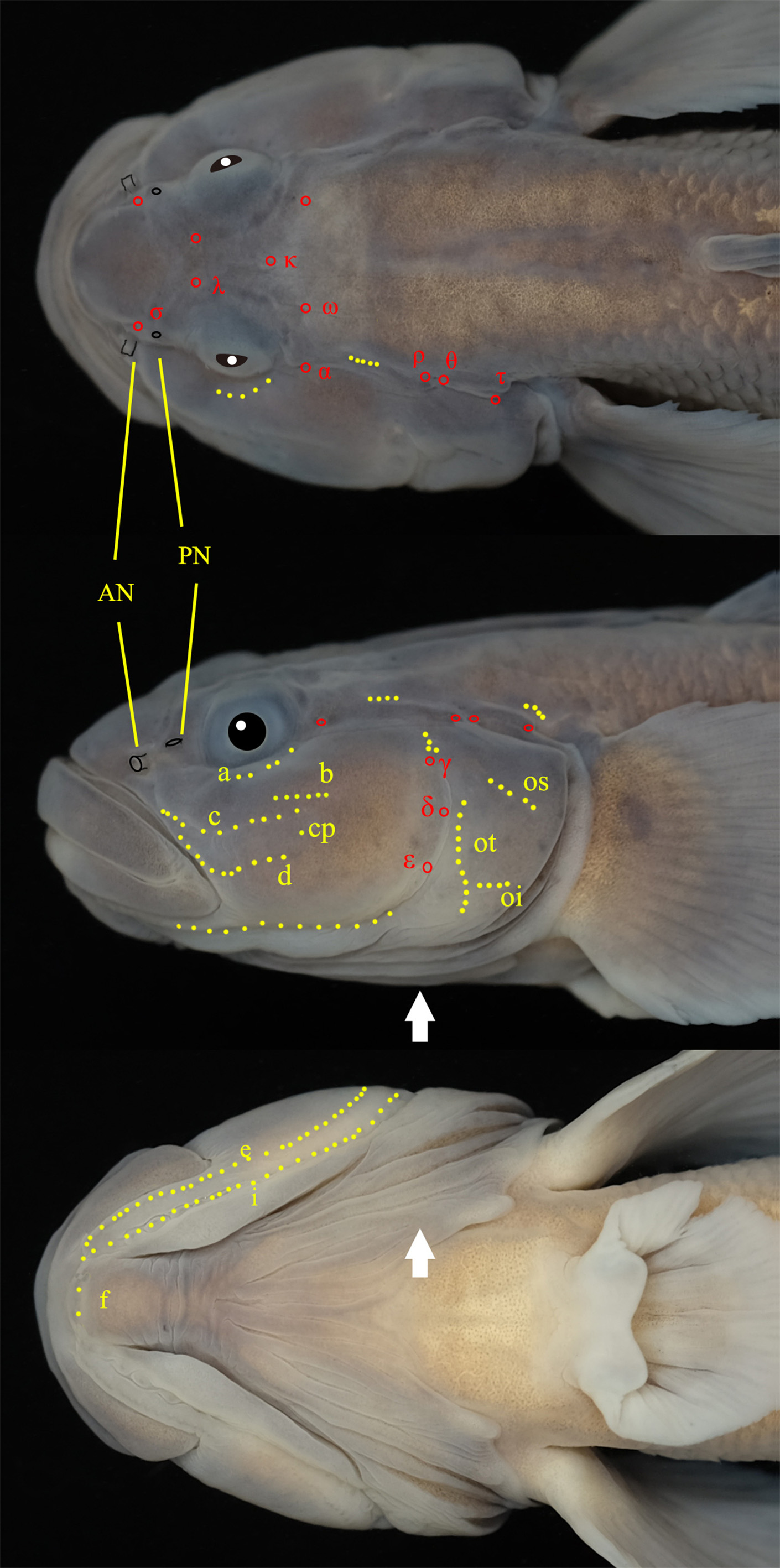
Abbreviations: D1, first dorsal fin; D2, second dorsal fin; *, segmented ray base of D2; **, of D2. Completed, cephalic canal pores consisting of σ, λ, κ, ω, ω1, α, ρ, θ, τ, γ, δ and ε; most deficiency, cephalic canal pores consisting of σ, κ and α; completed deficiency, without cephalic canal pores.
Fins. First dorsal fin trapezoidal in males and nearly semicircular in females; spines 4 and 5 longest, rear tip extending to base of second branched ray of second dorsal fin in males when adpressed, just reaching or not reaching anterior margin of second dorsal fin in females. Anal fin origin inserted below third and fourth branched soft ray of second dorsal fin. Pectoral fin broad, rear tip not extending to vertical of anus when adpressed in both sexes. Pelvic fin disc rounded, spinous ray with pointed membranous lobe. Caudal fin elliptical, posterior edge rounded with 5–6 vertical rows of brown spots.
Scales. Body covered with moderately large ctenoid scales. Opercle, preopercle, prepelvic area, and pectoralfin base always scaleless.
Head canals. Nasal extension of anterior oculoscapular canal with terminal pores σ located in vertical between anterior and posterior nares. Anterior interorbital sections of oculoscapular canal separated with paired pore λ. A single pore Κ in posterior region. Pore ω present near posterior edge of eye. Pore ω 1 absent. Lateral section of anterior oculoscapular canal with pore α and terminal pore ρ. Posterior oculoscapular canal with two terminal pores θ and τ. Gap between anterior and posterior oculoscapular canals smaller than the length of posterior oculoscapular canal. Preopercular canals present with three pores: γ, δ, and ε.
Sensory papillae. Row a oblique and uniserial, composed of loosely-arranged papillae, extending anteriorly to orbit. Row b longitudinal, extending anteriorly to a vertical through posterior margin of eye; its length slightly shorter than orbit diameter. Row c and d composed of densely-arranged papillae. Row c extending posteriorly to a vertical through posterior margin of eye. Row d extending posteriorly to vertical of posterior margin of pupil, approximately equivalent to orbit diameter. Row cp comprised of single papilla. Row f comprisedof paired papillae. Opercular papilla with ot, oi, and os; anterior end of row oi well-separated from a vertical row ot.
Coloration of fresh specimens. Head and body yellowish brown (some specimens head and body dark yellowish brown) in both sexes. Body scale pockets with brown margins, darker on dorsal half. Flank always with several longitudinal rows of blackish-brown round spots, some females with fewer rows. Belly pale white. Dorsum of snout with pair of thin, reddish-orange lines united at tip. Cheek and opercle yellowish brown with over 30 small orange spots in both sexes; branchiostegal membrane yellow with over 10 small orange spots in males, membranes white, and spotless in females. First dorsal fin with dark brown spinous rays and transparent fin membrane. First dorsal fin with large bright blue blotch in front of second spine in both sexes. Second dorsal-fin membrane orange to translucent with 5 or 6 horizontal rows of dark brown spots. Anal-fin ray 1/3 near the base yellow, distal 2/3 region black, outer margin white; fin membrane black in males, pale yellow to whitish in females. Caudal-fin base with large black brown blotch, fin membrane between rays pale yellow with 5–6 vertical rows of brown spots in both sexes. Pectoral fin grayish, basal portion with semicircular milky-yellow background and single horizontal, brown bar and row of blackish-brown spots or arc lines; distal 2/3 region grayish black in males and females. Pelvic-fin membrane grayish.
Distribution and habitat. The species is only known from Zhong Water, in the upper reaches of the Xiangjiang River on Lanshan County, Hunan Province. This species may be endemic within this basin.
Etymology. The specific name, maculagenys , from the Latin macula meaning spot and genys meaning cheek, in reference to the diagnostic feature of round orange spots on cheek. To be treated as a noun in apposition.
Comparison. Rhinogobius maculagenys , with 27 vertebrae, belongs to a group of 37 nominal species of Rhinogobius having 27 or more vertebrae. Forty-six congeners having high or unknown vertebral counts are listed in Table 2 with selective characters. Rhinogobius maculagenys can be easily distinguished from all but 6 species (viz. R. changtinensis , R. cliffordpopei , R. genanematus , R. honghensis , R. liui , and R. multimaculatus ) by having the following combination of characters: 0 predorsal scales; a head pore pattern lacking only pore ω1; first dorsalfin spines 4 and 5 longest in males, reaching to the base of the second branched ray of the second dorsal fin when adpressed; and 5 or 6 distinct vertical rows of brown spots on the caudal fin.
Rhingobius maculagenys can be distinguished from R. changtinensis , R. genanematus , and R. cliffordpopei by cheek color patterning (cheeks with spots in R. macculagenys vs. cheeks with stripes in R. changtinensis and R. genanematus , and cheeks without stripes or spots in R. cliffordpopei ). Rhingobius maculagenys can be distinguished from R. honghensis by the following features: cheeks with over 30 small orange spots in both sexes (vs. cheeks with 65–70 brown spots in males and no spots in female R. honghensis ), longitudinal scale series 32–34 (vs. 28–29), and lower number of vertebrae (27 vs. 28). Rhingobius maculagenys can be distinguished from R. liui by the following features: body with several longitudinal rows of blackish-brown spots (vs. 8–11 lateral blackbrown blotches), lower number of vertebrae (27 vs. 29), pectoral-fin rays (16 vs. 19), anal-fin rays (I/7 vs. I/8), and longitudinal scale series (32–34 vs. 36–39). Rhingobius maculagenys can be distinguished from R. multimaculatus by the following features: cheeks with over 30 spots (vs. with many tiny black spots), flank with several longitudinal rows of blackish-brown spots (vs. spots present regularly on base of the scales) longitudinal scale series (32–34 vs. 34–37), pectoral-fin base with a light grayish yellow semicircular mark (vs. with many tiny black spots), and lower number of vertebrae (27 vs. 29).
Comments. The color of the head and body is dark yellowish brown on some fresh specimens, while the holotype is substantially lighter. Colors of the species appear to adapt to the environment. For example, individuals that shelter under aquatic plants or stones for some time appear darker. The exact mechanism of this environmental adaptability is unclear and is of interest in future study.


No known copyright restrictions apply. See Agosti, D., Egloff, W., 2009. Taxonomic information exchange and copyright: the Plazi approach. BMC Research Notes 2009, 2:53 for further explanation.
|
Kingdom |
|
|
Phylum |
|
|
Class |
|
|
Order |
|
|
Family |
|
|
Genus |