
Scyphoproctus
|
publication ID |
https://doi.org/ 10.5281/zenodo.215310 |
|
publication LSID |
lsid:zoobank.org:pub:07C06068-9160-4AB4-AAF4-0451679D9F13 |
|
DOI |
https://doi.org/10.5281/zenodo.6175635 |
|
persistent identifier |
https://treatment.plazi.org/id/03913362-FFA1-FFB9-99BE-FA9499A14AE4 |
|
treatment provided by |
Plazi |
|
scientific name |
Scyphoproctus |
| status |
|
Figures 33 View FIGURE 33 D–F, 36 A–B, 37 A–E
Material examined. Oahu Island: Maunalua Bay, among branches of the invasive green alga Avrainvillea amadelpha , intertidal depths, 21°16ʹ49.4ʺ N, 157°43ʹ48.5ʺ W, coll. W. Magalhães & B. Dugan, Mar. 2010: Sta. A1R3 (1), Sta. A9R2 (8, BPBM R3631); Sta. A7R1 (2, BPBM R3632); Sta. S2R1 (2, BPBM R3633); Sta. A1R2 (7).
Description. Complete specimens 6.5–12 mm long, 0.1–0.2 mm wide for 50–61 chaetigers. Body elongate, slender, widest on mid-thoracic chaetigers. Color in alcohol pale yellow. Largest specimen incomplete 20 mm long, 1.5 mm wide for 27 chaetigers.
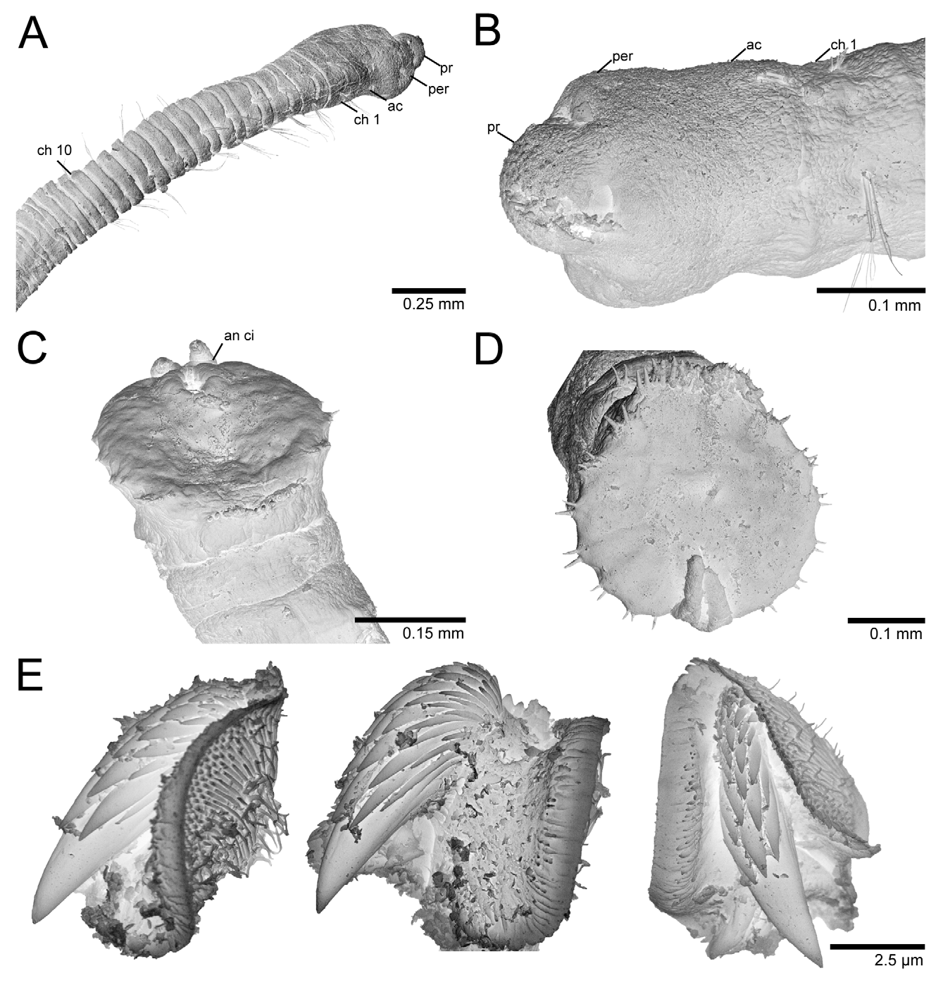
Prostomium rounded anteriorly, wider than long basally ( Figs 36 View FIGURE 36 A, 37A, B); nuchal organs not observed; postero-lateral eyespots forming a pair of oval, densely pigmented areas. Proboscis finely papillated. Peristomium achaetous, twice as long as anterior thoracic segments, sometimes partially retracted ( Fig. 37 View FIGURE 37 A, B).
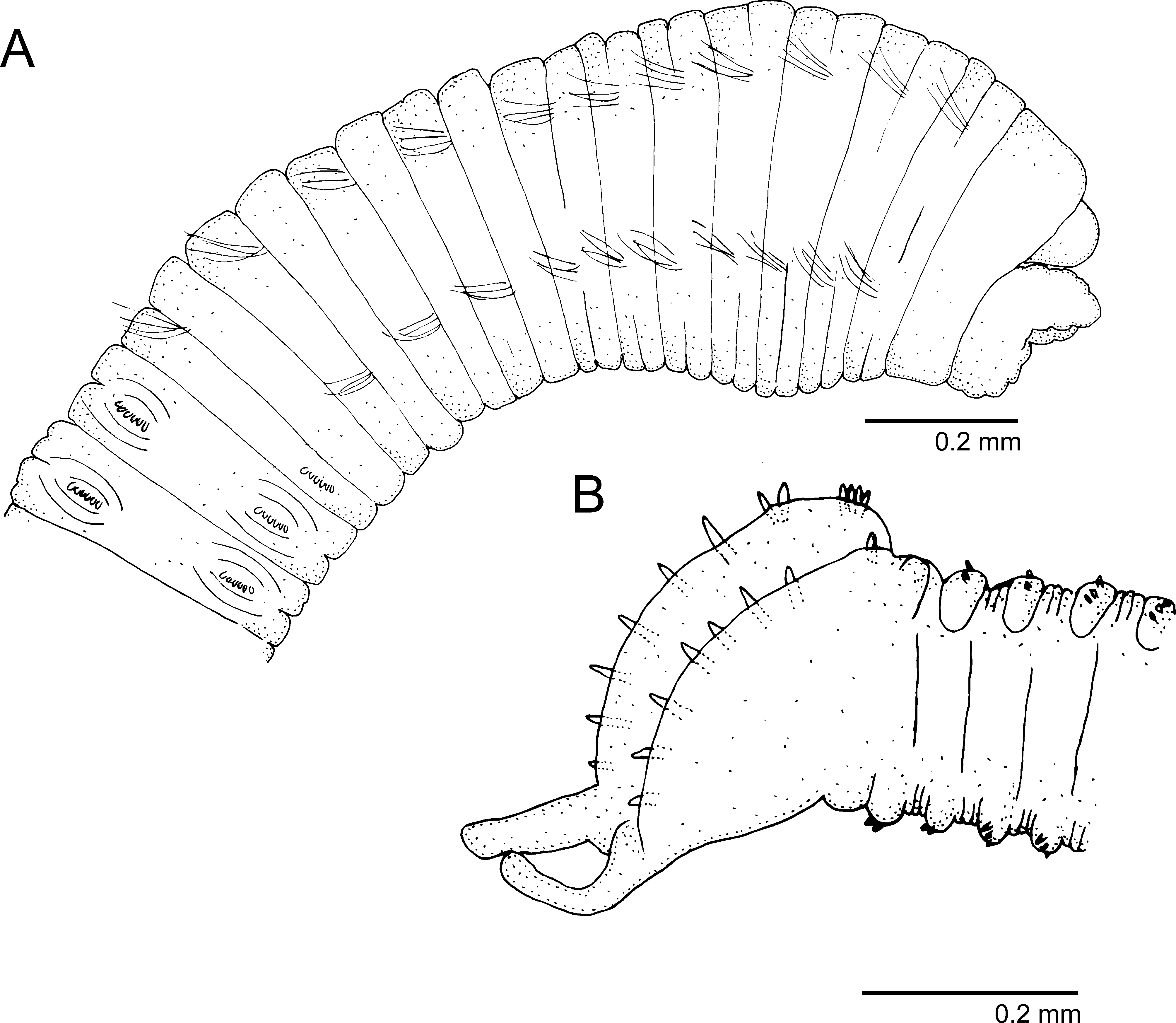
Thorax with 11–12 segments made up of one achaetous segment (not including peristomium) and 10–11 chaetigers ( Figs 36 View FIGURE 36 A, 37A). Thorax smooth, segments biannulate with deep inter-segmental grooves and bilimbate capillaries except last thoracic chaetiger which has notochaetae and neuropodial hooks. First chaetiger biramous. Parapodial lobes well separated; notopodia inserted dorso-laterally and neuropodia laterally. Lateral organs present throughout, between noto- and neuropodia, but closer to notopodia. Genital pores not observed.
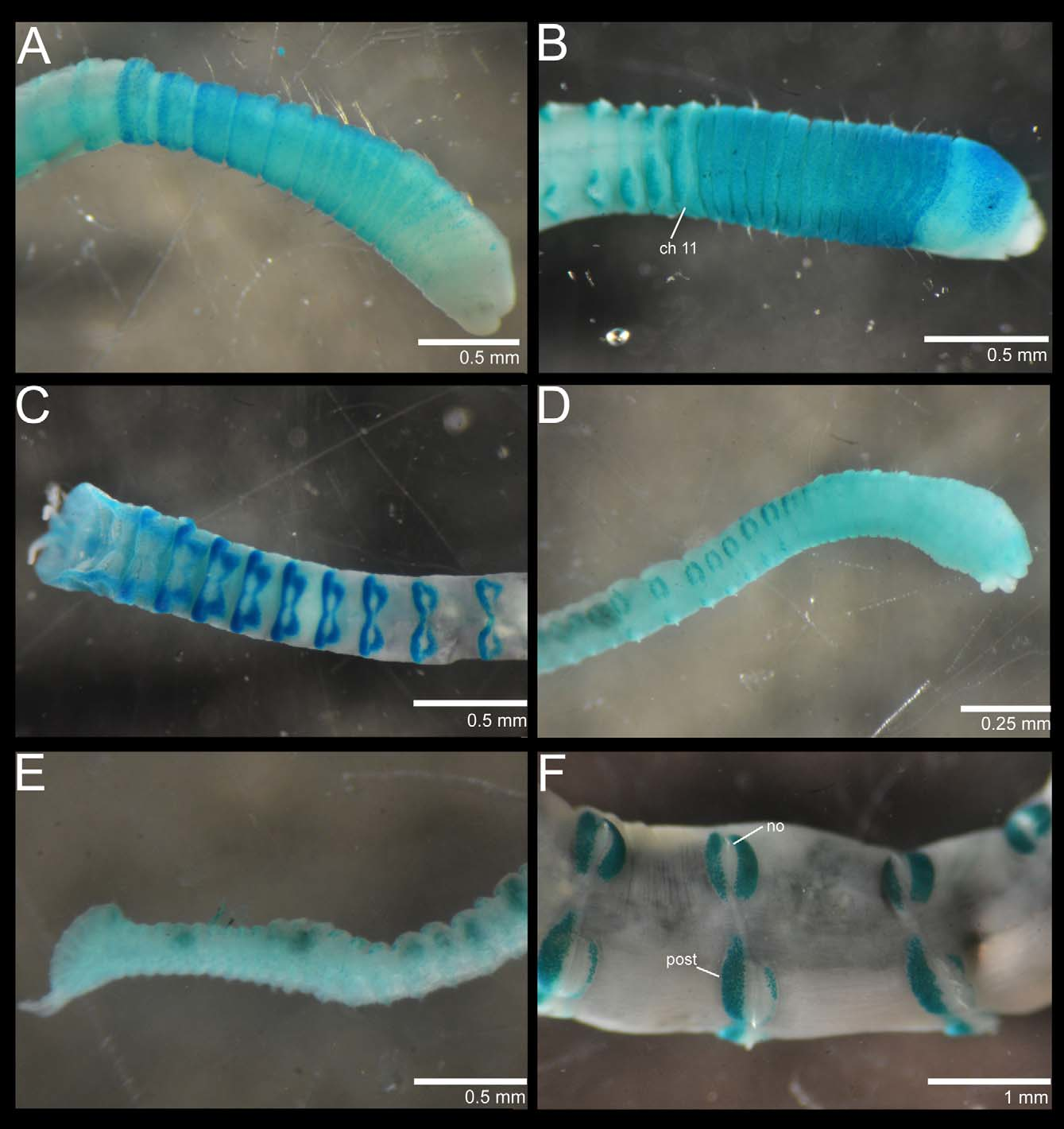
Transition between thorax and abdomen marked by change in shape of segments and chaetae; abdominal segments multiannulated with hooded hooks throughout ( Fig. 37 View FIGURE 37 A). Abdominal noto- and neuropodia with wellseparated glandular tori pads. Number of hooks on noto- and neuropodia similar; anterior tori with 5–6 (specimens 0.1 mm wide) or 20–21 hooded hooks (specimen 1.5 mm wide) reducing to 3–4 hooks in far posterior end in specimens 0.1 mm wide. Largest specimen with notochaetae on first abdominal segment. Abdominal hooks with short hoods, not extending beyond main fang nor covering hooks superiorly ( Fig. 37 View FIGURE 37 E). Hooks with multiple teeth, in frontal view with several rows; 3–4 teeth in basal row, 4–5 in middle row and 3–4 in superior rows ( Fig. 37 View FIGURE 37 E). Pre-pygidial segments with hooded hooks in both rami ( Fig. 36 View FIGURE 36 B). One pre-plaque segment with up to eight acicular hooks per notopodial lobe ( Fig. 37 View FIGURE 37 C, D). Anal plaque with up to seven sets of spines (e.g. 4:3:2:2:2:1:1; 2:1:1:1:1:1:1; 3:2:2:2:1:1; 4:3:1:1:1). Branchiae absent. Plaque funnel-shaped, short anus not extending to edge of plaque ( Fig. 37 View FIGURE 37 C, D). Two short cirri with a short median membrane ( Figs 36 View FIGURE 36 B, 37C, D).
Methyl green staining pattern. Anterior end and thoracic region stain uniformly light green ( Fig. 33 View FIGURE 33 D). Posterior half of peristomium and achaetous segment with scattered dark speckles that become more visible on the edges of all thoracic segments ( Fig. 33 View FIGURE 33 D). Abdominal tori with distinct staining; notopodial pre- and postchaetal tori staining intensely with dark green speckles but only the neuropodial postchaetal tori stained ( Fig. 33 View FIGURE 33 D, F). Anal plaque without a distinct staining pattern ( Fig. 33 View FIGURE 33 E).
Distribution. Collected from shallow reef flats of south coast of Oahu, Hawaii.
Remarks. This species is most similar to the group of species with pre-pygidial segments with hooded hooks only in both rami, including Scyphoproctus djiboutiensis , S. gravieri Okuda, 1940 and S. sp. 4 sensu Green, 2002. It differs most notably by the last thoracic chaetiger having notochaetae and neurohooks, notopodial tori completely stained, only the postchaetal neuropodial tori stained, and in the number of acicular spines on the anal plaque. Specifically, Scyphoproctus sp. has up to seven sets of spines (although this value is for specimens 0.1 mm wide), S. djiboutiensis has 11 groups of spines (0.5 mm wide, see Green 2002), S. sp. 4 sensu Green (2002) has eight (0.6 mm wide), and S. gravieri has ten (0.9 mm wide, see Green 2002). The range size of Scyphoproctus sp. is not known and the largest specimen (i.e. 1.5 mm wide) is incomplete, lacking pygidial structures. This specimen has the same distinct MGSP as the smaller ones, as well as the distinctly broad prostomium and the presence of notochaetae and neuropodial hooks in the last thoracic chaetiger, and was collected from the same locality. It would be unwise to name this species, as the complete range of several pygidial features is unknown for the large material, although it does not fit in any described species of Scyphoproctus .
| BPBM |
Bishop Museum |
No known copyright restrictions apply. See Agosti, D., Egloff, W., 2009. Taxonomic information exchange and copyright: the Plazi approach. BMC Research Notes 2009, 2:53 for further explanation.