
Mithraculus forceps A. Milne-Edwards, 1875
|
publication ID |
https://doi.org/ 10.1080/00222933.2021.1927229 |
|
persistent identifier |
https://treatment.plazi.org/id/0392F016-395C-FF82-FEF6-FCF3903C9E31 |
|
treatment provided by |
Plazi |
|
scientific name |
Mithraculus forceps A. Milne-Edwards, 1875 |
| status |
|
Gonopods of Mithraculus forceps A. Milne-Edwards, 1875 View in CoL
( Figures 3–7 View Figure 3 View Figure 4 View Figure 5 View Figure 6 View Figure 7 )
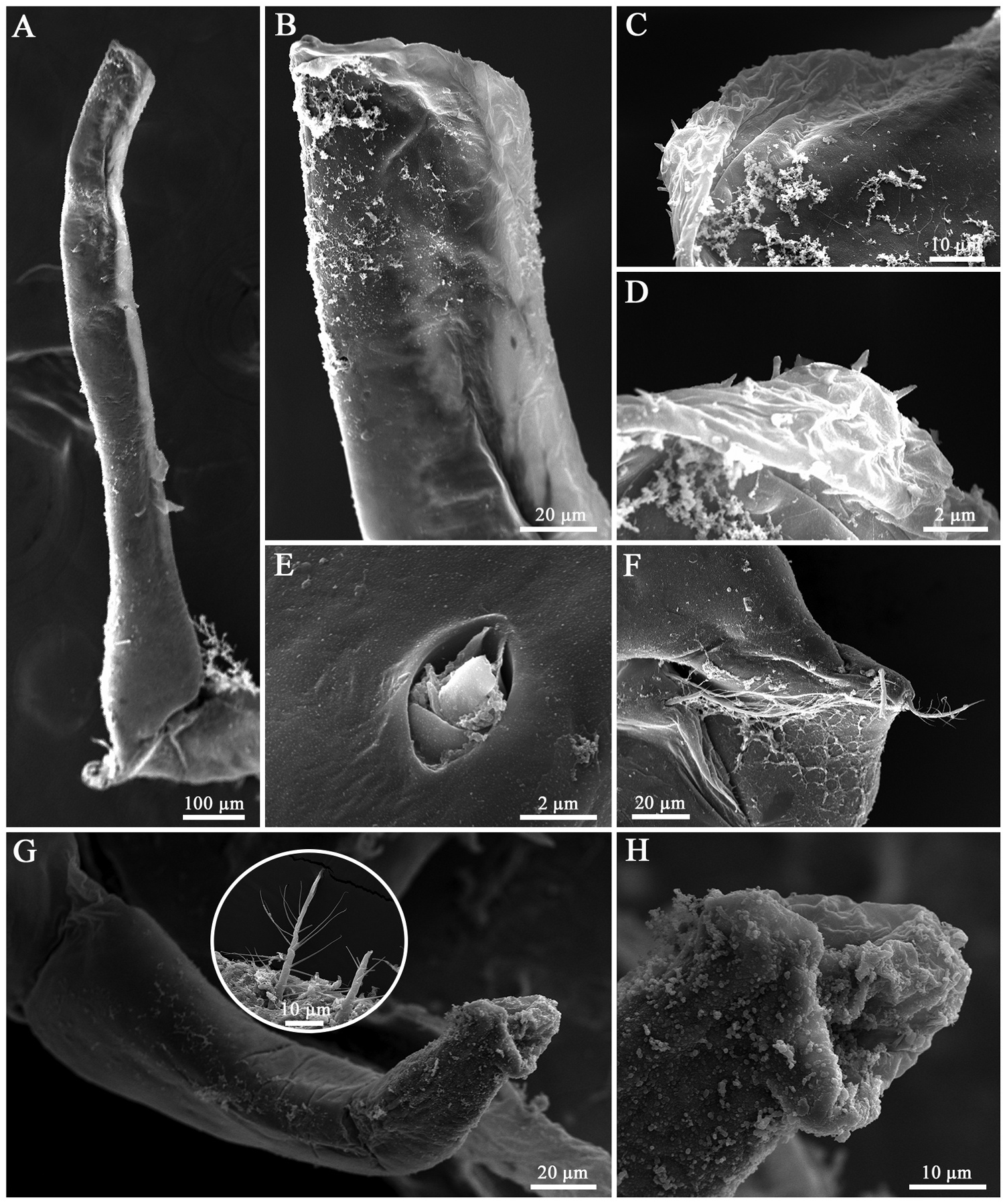
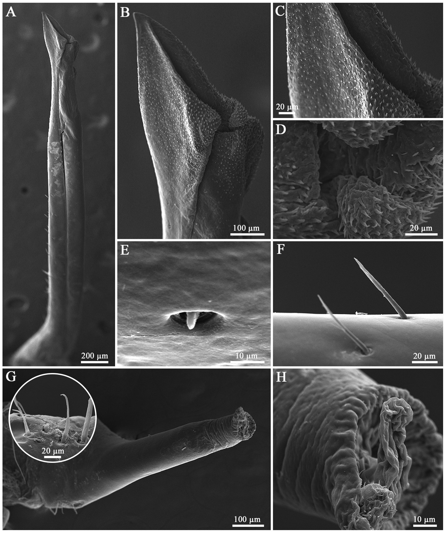
In the size class 1, the first gonopod (G1) is slightly flattened dorsoventrally, with an apical curvature and initial formation of the mesial suture ( Figure 3 View Figure 3 (a,b)). The apical surface is blunt, with rounded margins and a closed tip ( Figure 3 View Figure 3 (b,c)). The apical plate has few and small spines arranged in a row around the tip ( Figure 3 View Figure 3 (c,d)). Sub triangular sensillum is distributed parallel to the lateral margin of the apex ( Figure 3 View Figure 3 (e)). The G1 base shows a few plumose setae ( Figure 3 View Figure 3 (f)). The second gonopod (G2) is short, cylindrical, and with the enlarged basal region tapering in the last distal third ( Figure 3 View Figure 3 (g)). Sparse plumodenticulate setae are present at G2 base ( Figure 3 View Figure 3 (g) detail). The apical region is a wrinkled area and a blunt median process centrally located ( Figure 3 View Figure 3 (h)).
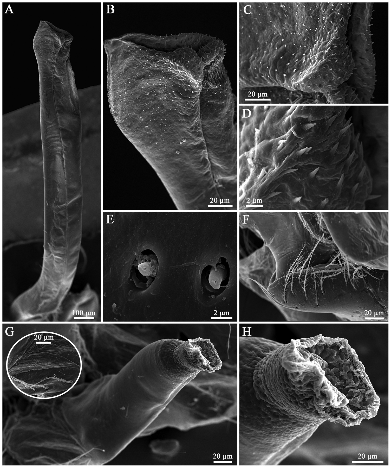
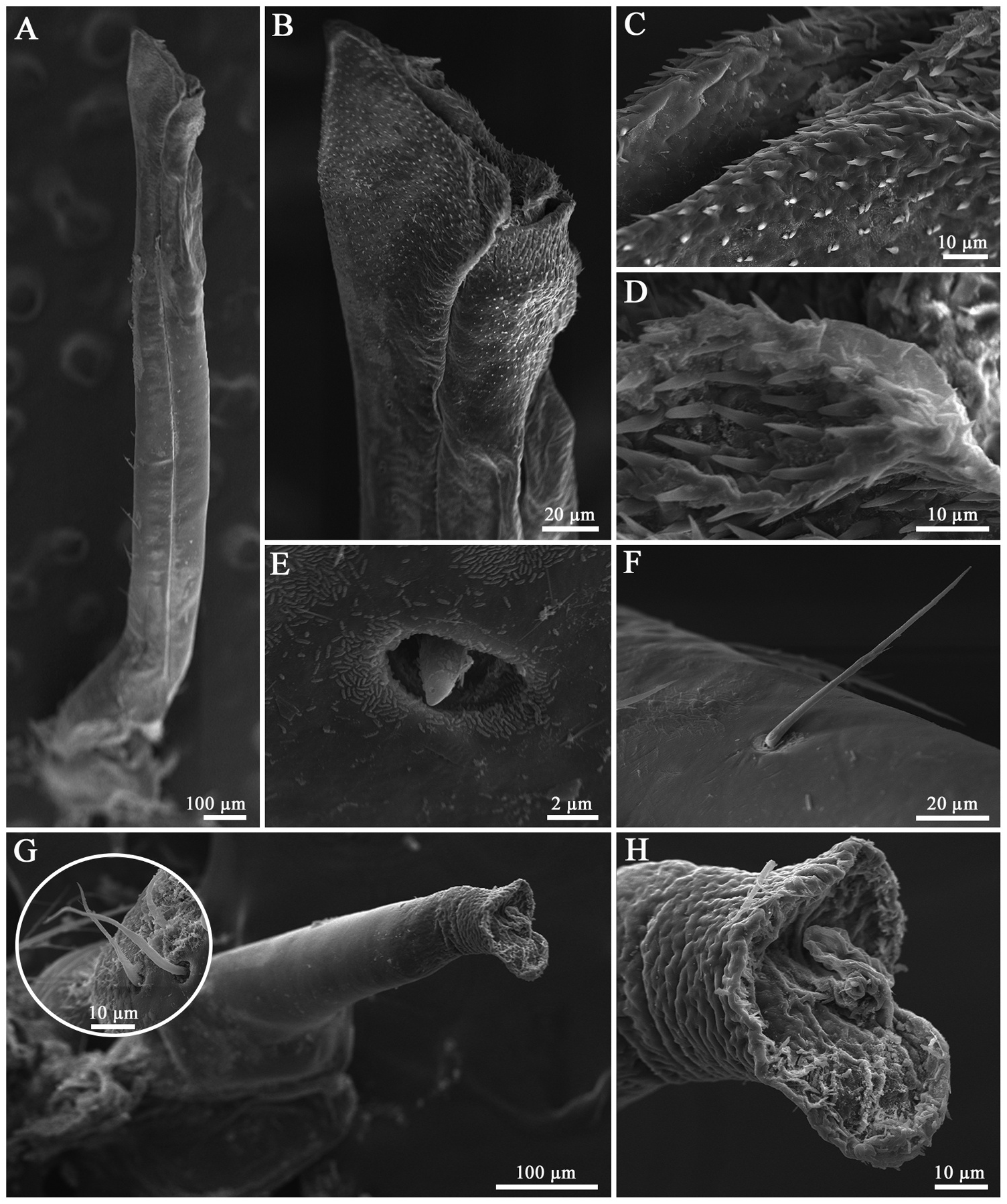
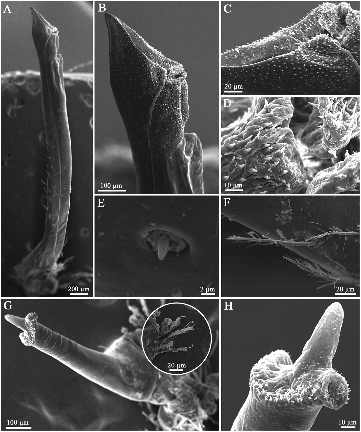
In successive stages during ontogenetic development, G1 increases in length, and one can observe the dorsoventral flatness,and the marked basal curvature ( Figures 4–7 View Figure 4 View Figure 5 View Figure 6 View Figure 7 ). From the fifth size class ( Figure 4 View Figure 4 ) it is possible to note the mesial suture extending longitudinally from the base to the apex, to form the aperture of the ejaculatory canal ( Figure 4 View Figure 4 (a,b)). The setae gradually increase in quantity and size: In the smaller individuals they are visible only in the basal region ( Figure 3 View Figure 3 (a,f)), in the larger individuals the setae extend through the G1 shaft until the second distal third ( Figures 6 View Figure 6 (a); 7(a)).In the base of the G1, plumodenticulate setae can be observed ( Figures 3 View Figure 3 (f), 4(f), 6(f)), while on the G1 shaft the lateral margin there are only simple setae arranged in rows inclined to the G1 distal region ( Figures 5 View Figure 5 (f); 7(f)). The most significant ontogenetic changes are concentrated in the apical plate of the G1. In the apical plate we notice the transition from a blunt structure with rounded margins ( Figure 3 View Figure 3 (a-c)) to the development of two distinct lobes (lateral and mesial) ( Figure 4 View Figure 4 (a,b); 5(a,b); 6(a,b)), with the lateral lobe tapering along the development ( Figure 7 View Figure 7 (a,b)). The aperture of the ejaculatory canal gradually increases in size, becoming diagonal to the apical plate ( Figure 4 View Figure 4 (a-c); 5(a-c); 6 (a-c); 7(a-c)). Also, the number of spines bordering the ejaculatory canal aperture increases considerably ( Figure 4 View Figure 4 (b-d); 5(b-d); 6(b-d); 7(b-d)), with some inclined to the canal interior in the larger specimens ( Figures 5 View Figure 5 (c); 6(c);7(c)).The sensillum increases in number and reduces in size along the ontogeny ( Figures 3 View Figure 3 (e); 4(e); 5(e); 6(e); 7(e)), being subtriangular in smaller individuals ( Figure 3 View Figure 3 (e)) and smaller with an acute apex in larger individuals ( Figures 6 View Figure 6 (e); 7(e)).
The second gonopod (G2) does not show abrupt changes during the ontogenetic development. A gradually formation of a girdle in the apical region, with the median process becoming more prominent and tapered ( Figure 3 View Figure 3 (g,h); 4(g,h); 5(g,h); 6(g,h); 7(g,h)) is verified. The number of setae at the base of the G2 has a slight increase ( Figure 3 View Figure 3 (g) detail; 4(g) detail; 5(g) detail; 6(g) detail; 7(g) detail), with spines appearing in the apical region, specifically around the apical girdle, and increasing in quantity during development ( Figures 4 View Figure 4 (h); 5(h); 6(h); 7(h)).
The specimens are considered physiologically mature from size class 5, diagnosed by the presence of white testicles and ductus deferens with spermatophores/sperm inside. Until size class 2, the reproductive system is not found in the juvenile individuals, and from size class 3 are found only as translucent ductus and not clearly differentiated, therefore no spermatophores are present.
No known copyright restrictions apply. See Agosti, D., Egloff, W., 2009. Taxonomic information exchange and copyright: the Plazi approach. BMC Research Notes 2009, 2:53 for further explanation.
|
Kingdom |
|
|
Phylum |
|
|
Class |
|
|
Order |
|
|
Family |
|
|
Genus |