
Perinereis bairdii ( Webster, 1884 )
|
publication ID |
https://doi.org/10.5852/ejt.2021.787.1619 |
|
publication LSID |
lsid:zoobank.org:pub:6E595BC0-37AB-460E-B0EB-435576CDD207 |
|
DOI |
https://doi.org/10.5281/zenodo.5841097 |
|
persistent identifier |
https://treatment.plazi.org/id/03948791-CB24-2936-FDA1-FC319B45D694 |
|
treatment provided by |
Felipe |
|
scientific name |
Perinereis bairdii ( Webster, 1884 ) |
| status |
|
Perinereis bairdii ( Webster, 1884) View in CoL reinstated
Figs 3–6 View Fig View Fig View Fig View Fig , 13 View Fig
Nereis bairdii Webster, 1884: 312–313 View in CoL , pl. 8 figs 22a–24, 25–26, 27–28.
Nereis (Perinereis) melanocephala McIntosh, 1885: 216–219 View in CoL , pl. 34 figs 14–17, pl. 16a figs 8–9.
Perinereis bairdii View in CoL – Monro 1933b: 41 (synonym and new combination only).
Type material
Lectotype (hereby designated) BERMUDA • 1 spec.; Bermuda; 1876; G.B. Goode leg.; USNM 4786 About USNM .
Paralectotypes (hereby designated) BERMUDA • 4 specs; Bermuda; 1876; G.B. Goode leg.; USNM 1660576 About USNM .
Additional material
BERMUDA • 1 spec.; Bermuda , SW of Whalebone Bay; 17 Nov. 1979; M.L. Jones leg.; USNM 1480190 About USNM • 3 ♂♂; Bermuda , Ferry Reach; 11 Oct. 1982; Manning and Hart leg.; with night light; USNM 1480197 About USNM • 2 ♂♂; Bermuda , Ferry Reach; 9 Oct. 1982; Manning and Hart leg.; with night light; USNM 1480191 About USNM .
Description
Atoke
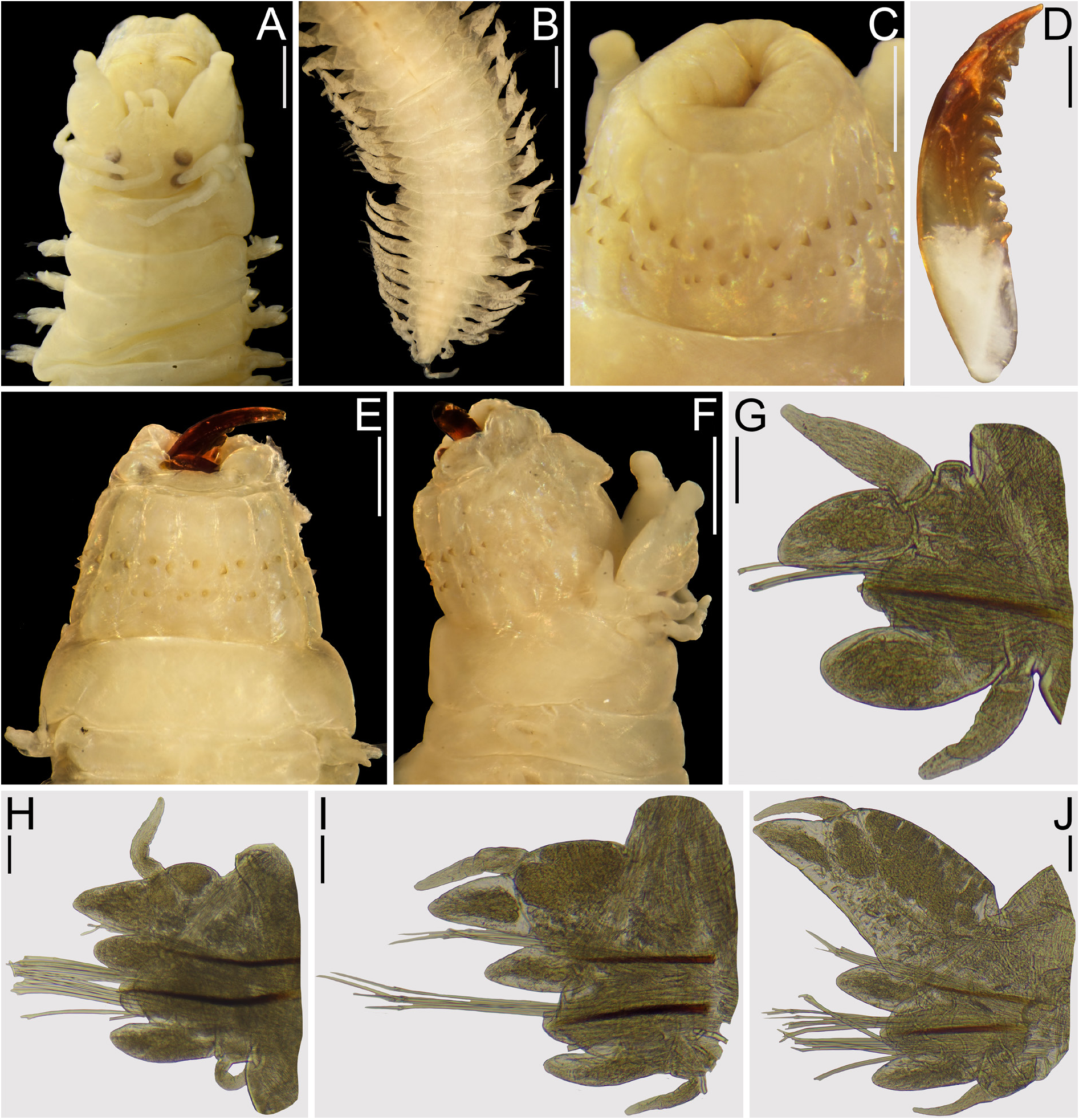
BODY AND MEASUREMENTS. Specimen designated as lectotype (USNM 4786) complete, 26 mm long, 1.7 mm wide at chaetiger 10 excluding parapodia, 64 chaetigers ( Fig. 3A–B View Fig ). Specimens designated as paralectotypes (USNM 1660576) ( Fig. 3D–F View Fig ) 4 complete, in good condition, largest paralectotype 48 mm long, 2 mm wide at chaetiger 10 excluding parapodia, 118 chaetigers. Additional non-type specimen (USNM 1480190) complete, in excellent conditions, 50 mm long, mm wide at chaetiger 10 excluding parapodia, 77 chaetigers ( Fig. 4A–B View Fig ). Pigmentation faded out in type and additional specimens.
PROSTOMIUM. Subpyriform, as long as wide, anterior region distally entire, as long as posterior region, dorsal groove present ( Figs 3A View Fig , 4A View Fig ); anterolateral gap between antenna and palpophore as long as diameter of antennae ( Figs 3A View Fig , 4A View Fig ).
ANTENNAE. Digitiform or subconical, not passing palps, 0.3× as long as prostomium, gap between them as long as basal wide of antennae ( Figs 3A View Fig , 4A View Fig ).
PALPS. Palpophores subconical, swollen, 1.2 × as long as wide, shorter than prostomium, subdistal transverse groove present ( Figs 3A, F View Fig , 4A View Fig ). Palpostyles rounded or digitiform ( Figs 3A, F View Fig , 4A View Fig ).
EYES. Rounded, anterior and posterior pairs subequal, in trapezoidal arrangement, sometimes posterior pair partly covered by anterior margin of tentacular belt ( Figs 3A View Fig , 4A View Fig ).
TENTACULAR BELT. 1.5–2.0× as long as chaetiger 1, covering posterior pair of eyes, anterior dorsal margin straight and sometimes omega-shaped ( Figs 3A, E–F View Fig , 4A View Fig ).
TENTACULAR CIRRI. Moniliform, not joint, basal segment largest, remaining ones decrease in size progressively toward distal end longest cirri reaching end of chaetigers 2–3 ( Figs 3A, F View Fig , 4A View Fig ).
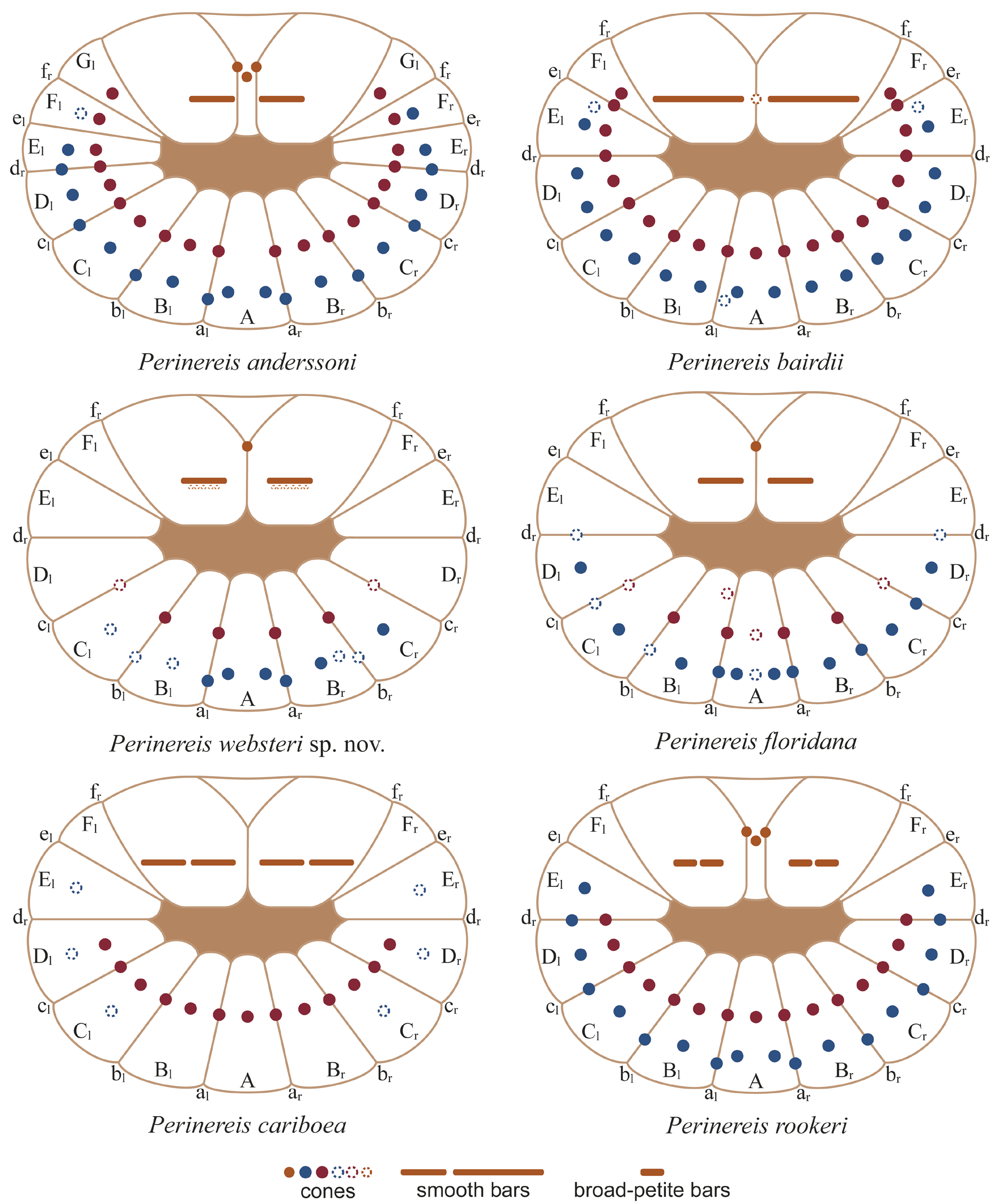
PHARYNX. Everted in both types and additional material; jaws brown, cutting edge with 8–11 teeth ( Figs 3D View Fig , 5E View Fig ), proximal ones sometimes ensheathed ( Fig. 4E View Fig ). Maxillary ring: I = 7 cones in triangle (5–18); II = 15–16 cones in arc (11–28); III = 6 in ellipse (6–22); IV = 21–24 cones in triangle (14–52) ( Figs 3C, E–F View Fig ). Oral ring: V = 1 cone (0–1); VI = 1–1 smooth bar (1–1); VII–VIII = 39 cones (33–39) in two bands: anterior band with 10 paragnaths in furrow row with 1 cone on the regions a–e, and 11 paragnaths in ridge row with 1 cone on the regions A–F; posterior band with 18 paragnaths in a single ridge row with two cones on the regions A–E ( Figs 3C, E–F View Fig , 13 View Fig ). Furrow pattern of areas VI–V–VI, λ-shaped ( Figs 3A View Fig , 13 View Fig ).
DORSAL CIRRI. Digitiform in first chaetigers, subconical with blunt tip thereafter, attached basally to dorsal ligule in anteriormost chaetigers, medially in middle chaetigers, and subdistally in posterior chaetigers ( Figs 3G–J View Fig , 4F–K View Fig ); 1× length of distal lobe of dorsal ligule in chaetigers 1–2, 1.2 × as long as in chaetigers 10–34, 1 × in chaetigers 41–59, 1.7× in chaetiger 72 ( Figs 3G–J View Fig , 4F–K View Fig ); 4× than proximal lobe of dorsal ligule in chaetigers 1–2, 1.5 × in chaetigers 10–11, 1.2× in chaetigers 21–41, 0.58 × length in chaetigers 57–59, 0.3 × in chaetiger 72 ( Figs 3G–J View Fig , 4F–K View Fig ).
DORSAL LIGULES. Subconical with blunt tip in anterior and middle chaetigers, becoming pennant-like toward posterior chaetigers, with distal lobes longer than proximal ones in first chaetigers, becoming as long as in anterior chaetigers, and shorter than in middle and posterior chaetigers ( Figs 3G–J View Fig , 4F–K View Fig ). Distal lobe of dorsal ligule lanceolate with blunt tip in anterior chaetigers, subconical thereafter; 1.6× as long as median ligule in chaetiger 10–11, 1.8× in chaetigers 21–41, 3.4 × length in chaetigers 57–59, 3.3× in chaetiger 72 ( Figs 3G–J View Fig , 4F–K View Fig ).
MEDIAN LIGULES. Digitiform in anterior chaetigers, becoming subconical with blunt tip thereafter ( Figs 3G–J View Fig , 4F–K View Fig ); 2.4× as long as neuroacicular ligule in chaetiger 10–11, 1.8× in chaetiger 21–34, 1.5× in chaetiger 41, 2.5× in chaetigers 57–59, 1.8× in chaetiger 72 ( Figs 3G–J View Fig , 4F–K View Fig ).
NEUROACICULAR LIGULES. Subconical throughout ( Figs 3G–J View Fig , 4F–K View Fig ); 0.6× length of ventral ligule in chaetigers 1–2, 1× in chaetigers 10–41, 0.8 × length in chaetiger 57–72 ( Figs 3G–J View Fig , 4F–K View Fig ).
NEUROPODIAL SUPERIOR AND INFERIOR LOBES. Present in anterior and middle chaetigers, both rounded, inferior one wider than superior one throughout ( Figs 3G–J View Fig , 4F–K View Fig ).
NEUROPODIAL POSTCHAETAL LOBES. Rounded, half as long as neuroacicular ligule throughout.
VENTRAL LIGULES. Digitiform throughout ( Figs 3G–J View Fig , 4F–K View Fig ).
VENTRAL CIRRI. Digitiform throughout, becoming narrower toward posterior chaetigers ( Figs 3G–J View Fig , 4F– K View Fig ); 0.7× length of ventral ligule in chaetigers 1–11, 0.4 × in chaetigers 21–34, 0.7× in chaetigers 41–72 ( Figs 3G–J View Fig , 4F–K View Fig ).
ACICULAE. Amber or dark brown throughout ( Figs 3G–J View Fig , 4F–K View Fig ); notoaciculae absent in first two chaetigers ( Figs 3G View Fig , 4F View Fig ).
NOTOCHAETAE. All homogomph symmetrical spinigers. Blades of spinigers with pectinate, minute teeth, teeth decreasing in size toward distal end.
NEUROCHAETAE. Homogomph symmetrical spinigers and heterogomph falcigers in supra-acicular fascicles, heterogomph spinigers and falcigers in sub-acicular fascicles. Neuropodial homogomph and heterogomph spinigers with blades as notopodial ones. Heterogomph falcigers pectinate, narrow teeth, two third of inner edge of blade dentate, distal tips stout ( Fig. 4C–D View Fig ); shafts of supra-acicular falcigers stouter than in sub-acicular ones ( Fig. 4C–D View Fig ).
PYGIDIUM. Crenulated ( Figs 3B View Fig , 4B View Fig ); anal cirri subulate, as long as last 3–4 chaetigers ( Figs 3B View Fig , 4B View Fig ).
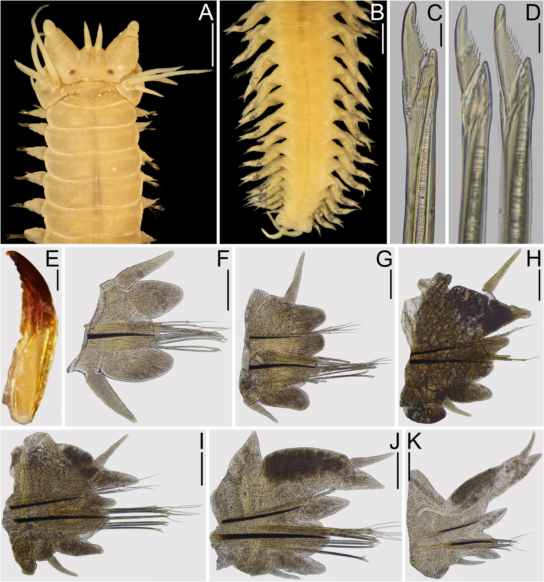
Male
BODY AND MEASUREMENTS. Three specimens (USNM 1480197) complete, in excellent conditions, specimen used for description 15 mm long, 1.2 mm wide at chaetiger 10 excluding parapodia, 78 chaetigers ( Fig. 5A View Fig ). Pigmentation present in anterior end, brown pigment in prostomium and tentacular belt; two transverse, parallel lines of brown pigment in lateral margins of chaetigers 1–13 ( Fig. 5A–C View Fig ).
PROSTOMIUM. As long as wide, subpentagonal, anterior margin directed downward, dorsal groove present ( Fig. 5A–C View Fig ).
ANTENNAE. Subconical with sharp tip, oriented downward, not extending beyond palps ( Fig. 5A–C View Fig ).
PALPS. Palpophores ovoid, swollen, 1.2 × as long as wide, shorter than prostomium, subdistal transverse groove present ( Fig. 5C View Fig ). Palpostyles rounded.
EYES. Black, rounded, subequal, in rectangular arrangement, anterior and posterior pairs overlapped, lenses visible, posterior pair not covered by tentacular belt ( Fig. 5A–C View Fig ).
TENTACULAR BELT. As long as chaetiger 1, with straight anterior margin ( Fig. 5A–C View Fig ).
TENTACULAR CIRRI. Smooth, cirrophores conspicuous, longest cirri extending backwards up to chaetiger 3 ( Fig. 5A–C View Fig ).
PHARYNX. Dissected ( Fig. 5E View Fig ); jaws brown, cutting edge with 7–8 rounded teeth ( Fig. 6A View Fig ). Maxillary ring: I = 7 cones in triangle; II = 17–16 cones in arc; III = 21 cones in ellipse; IV= 27–23 cones in triangle ( Fig. 5E View Fig ). Oral ring: V = 1 cone; VI = 1–1 smooth bar; VII–VIII = 37 cones in two bands: anterior band with 10 paragnaths in furrow row with 1 cone on the regions a–e, and 11 paragnaths in ridge row with 1 cone on the regions A–F; posterior band with 16 paragnaths in a single ridge row with 2 cones on the regions A–D and 1 cone on the regions E ( Fig. 5E View Fig ). Furrow pattern of areas VI–V–VI, λ-shaped.
BODY REGIONS. Two regions: 1) pre-natatory region includes chaetigers 1–13, subdivided into two subregions: a) dorsal cirri in chaetigers 1–7 and ventral cirri in chaetigers 1–5 pyriform or cattail-like, and 2) chaetigers 8–13 with both dorsal and ventral cirri cirriform; 2) natatory region includes chaetigers 14 to end of body, chaetiger 14 with upper lamella in dorsal cirrus and both upper and lower lamellae in ventral cirrus, in chaetiger 15 and remaining ones also appear lower lamella in dorsal cirrus, a lamella below median ligule, ventral lamella and a basal, dorsal protrusion in neuropodial ventral ligules.
PRE- NATATORY REGION. Parapodia resembling atokous ones ( Fig. 6B–E View Fig ). Dorsal cirrus pyriform in chaetigers 1–3, cattail-like in chaetiger 4–7, and filiform in remaining chaetigers ( Fig. 6B–E View Fig ); 2 × as long as distal lobe of dorsal ligule in chaetiger 2, 2.7× in chaetiger 3, 2.0 × in chaetigers 7 and 10 ( Fig. 6B–E View Fig ); 2.6 × as long as proximal lobe of dorsal ligule in chaetiger 2, 2.9× in chaetiger 3, 2.4× in chaetiger 7, 1.5× in chaetiger 10 ( Fig. 6B–E View Fig ). Dorsal ligule digitiform in first chaetigers, becoming subconical toward posterior ones; distal lobe of dorsal ligule 1 × length of median ligule in chaetiger 3, 1.5× in chaetiger 7, 1.5× in chaetiger 10 ( Fig. 6B–E View Fig ). Median ligule digitiform throughout; 2.3× as long as neuroacicular ligule in chaetiger 3, 1.7× in chaetiger 7, 1.5× in chaetiger 10 ( Fig. 6B–E View Fig ). Neuroacicular ligule subconical throughout; 1.7 × as long as ventral ligule in chaetiger 2, 0.8× length in chaetiger 3, 1.2× in chaetiger 7, 1.3 × in chaetiger 10 ( Fig. 6B–E View Fig ). Neuropodial superior and inferior lobes rounded, inferior one longer and wider than superior one throughout ( Fig. 6B–E View Fig ); neuropodial postchaetal lobe rounded, half as long as neuroacicular ligule ( Fig. 6B–E View Fig ). Ventral ligule digitiform throughout; 0.7× length of ventral cirrus in chaetiger 2, 1× in chaetiger 3, 1.3× in chaetigers 7 and 10 ( Fig. 6B–E View Fig ). Ventral cirrus pyriform in chaetigers 1–5, filiform thereafter ( Fig. 6B–E View Fig ).
NATATORY REGION. Parapodia distinct from atokous ones ( Fig. 6F–J View Fig ). Dorsal cirrus filiform, crenulations present from chaetiger 15, decreasing in size and number toward posterior chaetigers until disappear ( Fig. 6F–J View Fig ); 1.5× as long as distal lobe of dorsal ligule in chaetiger 14, 2× in chaetiger 15, 1.6 × in chaetiger 30, 1.2× in chaetiger 50, 1.2 × in chaetiger 72 ( Fig. 6F–J View Fig ). Upper lamella of dorsal cirrus present since chaetiger 14, flabellate throughout, larger in middle chaetigers, 0.7× length of dorsal cirrus in chaetiger 30 ( Fig. 6F–J View Fig ); ventral lamella after chaetiger 15, flabellate and smaller than upper lamella of dorsal cirrus throughout ( Fig. 6G–J View Fig ). Dorsal ligule subconical in chaetiger 14, digitiform thereafter, becoming narrower toward posterior chaetigers ( Fig. 6F–J View Fig ); distal lobe of dorsal ligule 2.7× as long as median ligule in chaetiger 14, 0.7× in chaetiger 15, 1× in chaetigers 30–50, 1.2× in chaetiger 72 ( Fig. 6F–J View Fig ). Notopodial prechaetal lobe rounded, lamelliform, present since chaetiger 15 ( Fig. 6G– J View Fig ); 0.5× length of dorsal ligule in chaetiger 15, 0.3 × in chaetigers 30–72 ( Fig. 6G–J View Fig ). Median ligule digitiform throughout, becoming narrower toward posterior chaetigers ( Fig. 6F–J View Fig ); 1.4× as long as neuroacicular ligules in chaetigers 15–50, 1.8× in chaetiger 72 ( Fig. 6F–J View Fig ); small basal lamella of median ligule present since chaetiger 15, flabellate throughout ( Fig. 6G–J View Fig ). Neuroacicular ligule subconical in chaetiger 14, digitiform thereafter ( Fig. 6F–J View Fig ), superior and inferior lobes absent; 1.2 × as long as ventral ligule in chaetiger 14, 2 × in chaetiger 15, 1.2× in chaetigers 30–50, 0.8× in chaetiger 72 ( Fig. 6F–J View Fig ). Neuropodial postchaetal lobe rounded in chaetiger 14, transformed into broad flabellate ventral lamella with a basal, dorsal protrusion thereafter ( Fig. 6F–J View Fig ); 2× as long as neuroacicular ligule in chaetiger 15, 2.5–3× in chaetigers 30–50, 2× in chaetiger 75 ( Fig. 6G–J View Fig ). Ventral ligule subconical in chaetiger 14, and digitiform with a small basal, dorsal lobe thereafter ( Fig. 6F–J View Fig ); 0.7× length of ventral cirrus in chaetigers 14–15, 1× in chaetigers 30–50, 1.2× in chaetiger 72 ( Fig. 6F–J View Fig ). Ventral cirrus filiform throughout ( Fig. 6F–J View Fig ); upper lamella divided in two subequal, digitiform lobes ( Fig. 6F–J View Fig ); lower lamella flabellate, 2–3× as wide as upper ones throughout ( Fig. 6F–J View Fig ).
ACICULAE. Basally amber and distally dark brown, amber region enlarges toward posterior chaetigers ( Fig. 6B–J View Fig ). Notoaciculae absent in first two chaetigers ( Fig. 6B View Fig ), proximal end rectangular in nonnatatory region and becoming flabellate in natatory one.
NOTO- AND NEUROCHAETAE. Resembling atokous ones in non-natatory region, replaced with paddle-like, heterogomph chaetae with short bosses in natatory region ( Fig. 6B–J View Fig ).
PYGIDIUM. Crenulated, with a rosette of papillae formed by two or three rows of papillae ( Fig. 5D View Fig ); anal cirrus as long as last 4–5 chaetigers ( Fig. 5D View Fig ).
Remarks
Webster (1884) did not indicate where the type series was deposited. USNM records state that a group of specimens called ‘Annelids collected in Bermuda in 1876-7’ was received from the Wesleyan University as a gift, entered in the Worm catalog in February 1890 with the accession number 22885, and assigned catalog number USNM 4786 for the 9 syntypes of ‘ Nereis bairdii n. sp. ’ (K.Ahlfeld, USNM, pers. com.). The original description of Webster (1884) is detailed, including the illustrations of the pharynx and parapodia from several regions of the body. Remarkably, Webster described two recently revised pharyngeal features: the description of the shape of areas VI as “which have straight inner margins” ( Webster 1884), i.e., the pattern of areas V– VI–V, and the description of the disposition of the paragnaths in areas VII–VIII into discrete regions, i.e., furrow and ridge regions of areas VII–VIII (Conde-Vela 2018; Villalobos-Guerrero 2019).
As outlined above, two different morphological patterns were observed in the syntypes of N. bairdii . For the discussion in the following paragraphs, specimens with short tentacular cirri, several paragnaths in areas VII–VIII, and large dorsal ligules in posterior chaetigers are referred to as ‘sp. 1’; and specimens with long tentacular cirri, a smaller number of paragnaths in areas VII–VIII, and short dorsal ligules in posterior chaetigers are referred to as ‘sp. 2’.
There are some indications that Webster described Nereis bairdii using several specimens because features from the two morphological patterns are mixed in the description. Webster (1884: fig. 22) detailed an anterior end with long posterodorsal tentacular cirri, which was also included in the description: “Tentacular cirri… the posterior superior longest, reaching back to the eighth segment…”, which matches with sp. 2. Webster (1884) described the arrangement of paragnaths but the number of paragnaths in most areas was not detailed, but they can be traced from his figures 22a and 23. The area I was depicted with 7 paragnaths, which matches with sp. 1, although the number of paragnaths in areas II–IV lies between the range of variation of sp.1 and sp. 2. Webster (1884) described the presence of 0–3 paragnaths in area V, but all syntypes examined have 0–1 paragnaths in such area. Webster (1884) described areas VII–VIII as follows: “vii and viii in two series, the anterior composed of a few denticles, the posterior more numerous and smaller”, which could refer to both sp. 1 and sp. 2, but the figure ( Webster 1884: fig. 23) clearly shows an anterior band with a furrow row, which is absent in sp. 2. Therefore, the pharynx depicted and described ( Webster 1884) belongs to a specimen from sp. 1.
Webster (1884) described the parapodia of his new species in a paragraph and depicted them in figures 24, 25, and 26. At the end of this paragraph, Webster mentioned that “other specimens, certainly belonging to this species, have the feet more delicate, the dorsal and ventral cirri a trifle longer. (figs 24a, 26a.)”. Webster described the dorsal ligules as follows: “The superior lingula is enlarged (fig. 25), the dorsal cirrus moves nearer the apex of its lingula, and on the extreme posterior feet becomes a little more delicate (fig. 26)”; figure 26 shows a posterior parapodium with dorsal ligule several times longer than median ligule and ventral ligule slightly longer than neuroacicular ligule. Conversely, posterior parapodium depicted from ‘other specimens’ ( Webster 1884: fig. 26a) shows dorsal ligule faintly longer than median ligule and ventral ligule shorter than neuroacicular ligule. Therefore, the former parapodial description is from sp. 1, whereas the illustrations of the ‘other specimens’ match with sp. 2. The chaetae described are a notopodial homogomph spiniger and a neuropodial heterogomph falciger, but both drawings are very schematic and match either sp. 1 or sp. 2. Finally, Webster (1884) described anal cirri as long as last ten chaetigers, matching with sp. 2.
All these pieces of information point to the fact that Webster used more than one specimen from the syntypes when describing Nereis bairdii : descriptions of pharynx and parapodia refer to sp. 1, whereas the descriptions of the anterior end, additional illustrations of the parapodia, and the anal cirri, refer to sp. 2. Despite the mix of features in the original description, the idea that Nereis bairdii has short tentacular cirri and enlarged dorsal ligules in posterior chaetigers, i.e., corresponding with sp. 1, prevailed among the contemporary authors, explaining the synonymy of Nereis bairdii with P. anderssoni because they have a very similar morphology. For example, Augener (1927) reported Nereis ( Perinereis) bairdii for Curaçao, and proposed the synonymy of Nereis ( Perinereis) melanocephala McIntosh, 1885 with this species, a species from Bermuda with tentacular cirri reaching to chaetiger 3 and dorsal ligules enlarged in posterior chaetigers; posteriorly, Augener (1936) reported the species for Bonaire. Monro (1933b) transferred N. bairdii to Perinereis and retained the synonymy proposed by Augener (1927). Later, Hartman (1944) examined the type material of P. anderssoni and concluded that Nereis bairdii and N. melanocephala are junior synonyms and maintained this viewpoint in subsequent works ( Hartman 1948, 1951). Hartman (1951) reported P. anderssoni for the Gulf of Mexico (and including N. bairdii as a junior synonym) and highlighted that the species “is readily identified for having posterior notopodial lobes much prolonged…”, which is reinforced when describing P. floridana some paragraphs below as “…posterior parapodial lobes are short, resembling those of median segments and are thus not be confused with those of P. anderssoni (see above).”. Other records of P. anderssoni retained the synonymy of N. melanocephala but not mentioned N. bairdii (e.g., Díaz-Díaz & Liñero-Arana 2002).
With all these pieces of information, it is concluded that the name Nereis bairdii is represented with specimens of sp. 1 and that specimens of sp. 2 are not N. bairdii , so the split of the syntypes is required. To redefine Nereis bairdii and to preserve the stability of the name and its application through time ( ICZN 1999, Recomm. 74A), specimens of sp. 1 were selected lectotype and paralectotypes; the terms lectotype and paralectotypes have been stated in the material examined and in the description of P. bairdii , they have been described and illustrated and their data updated for its recognition ( ICZN 1999, Art. 74.7, Recomm. 74A–G). Specimens of sp. 2 are shown to be an undescribed species and are identified as the new species, Perinereis websteri sp. nov.
The synonymy of Nereis ( Perinereis) melanocephala with P. bairdii is retained here. Nereis ( Perinereis) melanocephala was described from Bermuda from a single specimen ( McIntosh 1885), and based on the original description, it agrees with P. bairdii in most features, the following being the most relevant ones: 1) anterior end with brown pigment, 2) tentacular cirri reaching chaetiger 3, 3) tentacular belt twice longer than chaetiger 1, 4) arrangement of paragnaths are almost identical and especially the size of smooth bars in areas VI, 5) dorsal ligules 2–3 × longer than median ligules, 6) dorsal cirri not extending beyond the tip of the distal lobe of dorsal ligules in posterior chaetigers, 7) ventral ligules longer than neuroacicular ligules in posterior chaetigers.
Conversely, there are several differences between Perinereis bairdii and P. anderssoni : 1)in P.anderssoni , the pattern of areas V–VI–V is Π- shaped, whereas in P. bairdii it is λ- shaped; 2) in P. anderssoni , the area V has 3 paragnaths in a triangle and are not horizontally aligned with smooth bars in areas VI but they are posteriorly displaced, whereas P. bairdii has 0–1 cones that are horizontally aligned with smooth bars; 3) in P. anderssoni , the smooth bars in areas VI are half as long as wide in area VI, whereas in P. bairdii they are almost as long as; 4) in P. anderssoni , the posterior band has one furrow and one ridge row, whereas in P. bairdii has a single ridge row only; 5) in P. anderssoni , there are 13 ridge regions (A–G) in areas VII–VIII, whereas in P. bairdii there are 11 ridge regions (A–F); 6) in P. anderssoni , the dorsal and ventral ligules in first two chaetigers are subconical, whereas in P. bairdii they are globose; 7) in P. anderssoni the dorsal cirri are longer than dorsal ligules in anterior chaetigers and becoming shorter toward posterior ones, whereas in P. bairdii they are shorter than dorsal ligules throughout body; 8) in P. anderssoni , the neuropodial heterogomph falcigers are stouter and the length of inner margin edentate is longer than in P. bairdii .
It seems reasonable to assume that most reports of Perinereis anderssoni from the Caribbean Sea represent P. bairdii , but it is unclear. For example, P. anderssoni from Venezuelan coasts (Liñero-Arana & Reyes- Vásquez 1979; Díaz-Díaz & Liñero-Arana 2002; Vanegas-Espinosa et al. 2007) differs from P. bairdii in the following features: 1) in Venezuelan specimens, there are 46–85 paragnaths in area III, whereas in P. bairdii they are 6–22 paragnaths; 2) in Venezuelan specimens, dorsal cirri in posterior chaetigers are 0.6× as long as proximal lobes and are 1.5× as long as distal lobes of dorsal ligules, whereas in P. bairdii , dorsal cirri are 0.3 × as long as proximal lobes and are 1.7 × as long as distal lobes of dorsal ligules; 3) in Venezuelan specimens, the dorsal ligules are at least 3.5× as long as the median ligules and the ventral ligules are 2–4× longer than the neuroacicular ligules in posterior chaetigers, whereas in P. bairdii , the dorsal ligules are 3.3× as long as the median ligules and the ventral ligules are 1.2× than the neuroacicular ligules. A record of P. anderssoni from the Mexican Caribbean ( Salazar-Vallejo & Jiménez-Cueto 1997) differs from P. bairdii in the following features: 1) in Caribbean specimens, the dorsal cirri are 1.4× as long as the distal lobes of the dorsal ligules and the median ligules are as long as the neuroacicular ligules in anterior chaetigers, whereas in P. bairdii the dorsal cirri are 1.2× longer and the median ligules are 2.4× longer; 2) in Caribbean specimens, the dorsal cirri are 0.5× as long as proximal lobes and as long as distal lobes of dorsal ligules in posterior chaetigers, whereas in P. bairdii the dorsal cirri are 0.3× as long as the proximal lobes and are 1.7× as long as the distal lobes. Further studies will clarify whether records of P. anderssoni from the Gulf of Mexico and the Mexican Caribbean, and other regions of the Caribbean Sea, are different species.
Distribution
Bermuda.

No known copyright restrictions apply. See Agosti, D., Egloff, W., 2009. Taxonomic information exchange and copyright: the Plazi approach. BMC Research Notes 2009, 2:53 for further explanation.
|
Kingdom |
|
|
Phylum |
|
|
Class |
|
|
Order |
|
|
Family |
|
|
Genus |
Perinereis bairdii ( Webster, 1884 )
| Conde-Vela, Víctor Manuel 2022 |
Perinereis bairdii
| Monro C. C. A. 1933: 41 |
Nereis (Perinereis) melanocephala
| McIntosh W. C. 1885: 219 |
Nereis bairdii
| Webster H. E. 1884: 313 |