
Pilargis papillata Rasmussen, 1973
|
publication ID |
https://doi.org/ 10.1080/00222930600594212 |
|
persistent identifier |
https://treatment.plazi.org/id/039487A1-B44B-FFDA-F6CF-FC94FBAEFB00 |
|
treatment provided by |
Carolina |
|
scientific name |
Pilargis papillata Rasmussen, 1973 |
| status |
|
Pilargis papillata Rasmussen, 1973 View in CoL
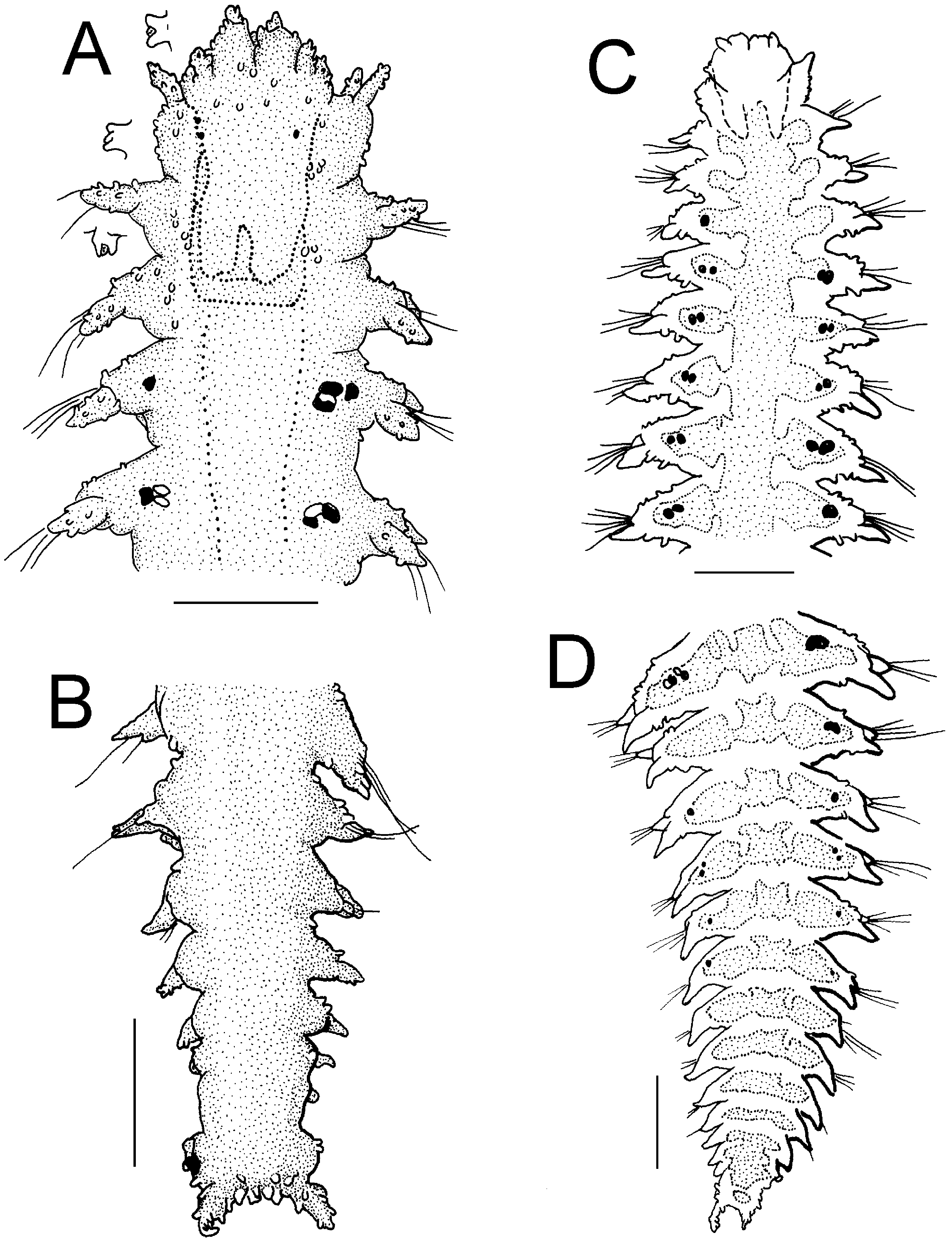
( Figure 11 View Figure 11 )
Pilargis papillata Rasmussen 1973, p 20 View in CoL –22, Figure 2 View Figure 2 .
Type material
Northeastern Atlantic Ocean : holotype (ZMUB-53527), Fensfjorden NE for Gardsenflu (60 ° 499N, 5 ° 039420E), Norway, 412 m, coll. K. Rasmussen. Complete animal; two (not three) paratypes (ZMUB-53528), Fensfjorden, NE for Gardsenflu, Norway, 580– 412 m, coll. and id. K. Rasmussen (dried out; anterior fragment 14.5 mm long, 1.0 mm wide, 73 setigers; posterior fragment with eight setigers) .
Redescription
Holotype (ZMUB-53527) complete, twisted over itself, making difficult any measurement without further damaging it. It was described as 20 mm long (it is 5.7 mm long), 1.0 mm wide (setiger 15), 80 setigers and a regenerating posterior end with eight or nine asetigers; it has 26 setigers and a regenerating portion with two inmature setigers and three preanal asetigers. It is now colorless but the posterior brain lobes are dark (occupy first setiger and slightly invade the second one). Body flat, densely covered by verrucae dorsally, verrucae abundant over tentacular and dorsal cirri ( Figure 11A View Figure 11 ). Ventrally verrucae restricted to parapodial bases, smaller, leaving a smooth midventral wide area limited by two longitudinal muscle bands. Left setigers 7–10 are folded dorsally (because of body compression in the vial).
Prostomium completely fused dorsally to peristomium; palps biarticulate, palpostyles central, directed ventrally. Lateral antennae wide, densely covered with verrucae, placed anteriorly, slightly surpass prostomial anterior margin ( Figure 11B, C View Figure 11 ). Tentacular cirri cirriform, directed anteriorly, dorsal ones slightly larger than the ventral.
First setiger with dorsal cirri globose, thin, acuminate, slightly longer than dorsal tentacular cirri, slightly shorter than dorsal cirri of second setiger. Anterior parapodia with fusiform cirri with verrrucae; dorsal cirri twice as long as ventral cirri, much thicker ( Figure 11E View Figure 11 ). Posterior parapodia with dorsal cirri foliose, larger than setal lobe, separated in cirrophore and cirrostyles ( Figure 11F View Figure 11 ). Cirrophore massive with dense cover of large verrucae, with an anterior small glandular area; cirrostyles digitate with smaller verrucae. Ventral cirri smooth placed basally to the setal lobe and shorter than it. Neurosetae mostly complete; despite adsorpted materials, they are limbates with limbus thin, smooth, distally entire (in paratype, most setae broken, some appear distally bidentate).
Posterior end with pygidium in regeneration; damaged ( Figure 11D View Figure 11 ); two setigers and three preanal asetigers. Pygidium as an inverted truncate cone, two ventrolateral anal cirri well developed, with many verrucae. Paratypes with enteric diverticula dark, clearly seen from setiger 15; few eggs can be seen from about setiger 50. Posterior fragment shows them too.
Discussion
The holotype differs in several regards to the original description, besides the difference in size. The original illustration included a ventral view, which was indicated as a dorsal view. The posterior brain lobes are dark, there are two other lateral smaller dark glands projecting towards the posterolateral corners of the tentacular segment, and the pigmented glandular area in posterior cirrophores has faded slightly. The dorsal cirri are larger in median setigers; the second dorsal cirri is not shorter than the first dorsal cirri. The neurosetae are distally entire, not bidentate, and have a thin, smooth blade.
This species belongs to the group with abundant dispersed verrucae over the back and parapodial lobes. Pilargis papillata is closely allied to P. modesta and P. rozbaczyloi n. sp., but differs from them by having the first dorsal cirri smaller than the following ones rather than larger, and fusiform verrucose dorsal cirri, contrasting with digitate verrucose cirri in P. rozbaczyloi n. sp., and smooth fusiform cirri in P. modesta .
Distribution
Restricted to the type locality in Southwestern Norway in depths of over 400 m.

No known copyright restrictions apply. See Agosti, D., Egloff, W., 2009. Taxonomic information exchange and copyright: the Plazi approach. BMC Research Notes 2009, 2:53 for further explanation.
|
Kingdom |
|
|
Phylum |
|
|
Class |
|
|
Order |
|
|
Family |
|
|
Genus |
Pilargis papillata Rasmussen, 1973
| Salazar-Vallejo, Sergio I. & Harris, Leslie H. 2006 |
Pilargis papillata
| Rasmussen 1973: 20 |