
Paraehlersia pamelae, Prado & Martín, 2024
|
publication ID |
https://doi.org/ 10.11646/zootaxa.5437.1.5 |
|
publication LSID |
lsid:zoobank.org:pub:1D2A6EBD-A64D-4A77-8455-DEE05D92909A |
|
DOI |
https://doi.org/10.5281/zenodo.10959322 |
|
persistent identifier |
https://treatment.plazi.org/id/0398879F-AF36-FFEF-FF69-428DFA1CC274 |
|
treatment provided by |
Plazi |
|
scientific name |
Paraehlersia pamelae |
| status |
sp. nov. |
Paraehlersia pamelae , n. sp.
Figs 2–5 View FIGURE 2 View FIGURE 3 View FIGURE 4 View FIGURE 5
Material examined. Alborán Sea; Holotype ( MNCNM 16.01.19285 ), 35º50’41”N, 03º13’39”W, gravel with organic detritus, 111–114 m, 25 September 2011; GoogleMaps 44 Paratypes ( MNCNM 16.01.19286 ) (7 mounted for SEM), 35º50’41”N, 03º13’39”W, gravel with organic detritus, 111–114 m, 25 September 2011; GoogleMaps 6 Paratypes ( MNCNM 16.01.19287 ) 36º00’40”N, 02º55’32”W, gravel with organic detritus, 93–101 m, 24 September 2011; GoogleMaps 1 Paratype ( MNCNM 16.01.19288 ), 35º53’10”N, 03º04’74”W, rhodoliths, 82–92 m, 23 September 2011; GoogleMaps 1 Paratype ( MNCNM 16.01.19289 ). 35º50’40”N, 03º13’72”W, 100–109 m, 25 September 2011. GoogleMaps
SAMPLES Subfamily Species BV13 BV14 BV16 BV17 BV21 BV27 BV35 DR01 DR02 DR07 DR20 DR22 DR25 DR37 DR39 DR40 DR47 S. columbretensis (Campoy, 1982) 2 S. corallicola Verrill, 1900 1 1 S. cruzi Núñez & San Martín, 1991 6 2 S. cryptica Ben-Eliahu, 1977 1 2 S. garciai (Campoy, 1982) 7
3
5 S. hyalina Grube, 1863 S. parapari San 38 2 4 23 Martín & López, 1 2000 S. profunda Cognetti, 1955 3 7 1 S. pulvinata (Langerhans, 1881) 2 S. sp. 2 2 S. variegata Grube, 1860 3 Trypanosyllis krohnii Claparède, 1864 1 2 Trypanosyllis sp. 1 T. zebra (Grube, 1860) 2 Xenosyllis scabra (Ehlers, 1864) 4
SAMPLES Subfamily Species BV13 BV14 BV16 BV17 BV21 BV27 BV35 DR01 DR02 DR07 DR20 DR22 DR25 DR37 DR39 DR40 DR47 Autolytinae Langerhans, 1879 Epigamia labordai (San Martín & López 2002) 1 Myrianida. prolifera (O. F. Müller 1788) 2 Myrianida sp.
1
2
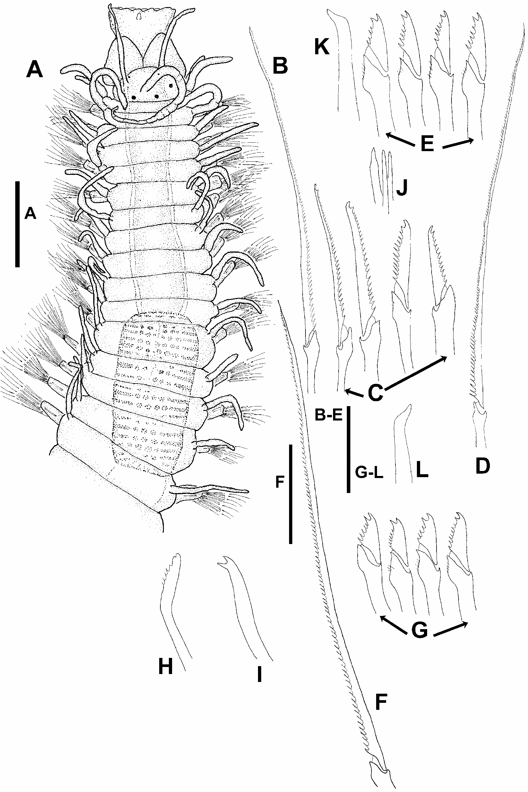
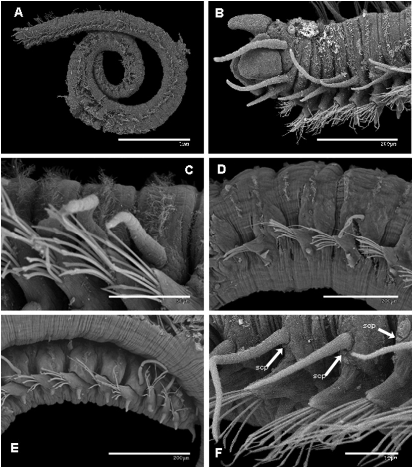
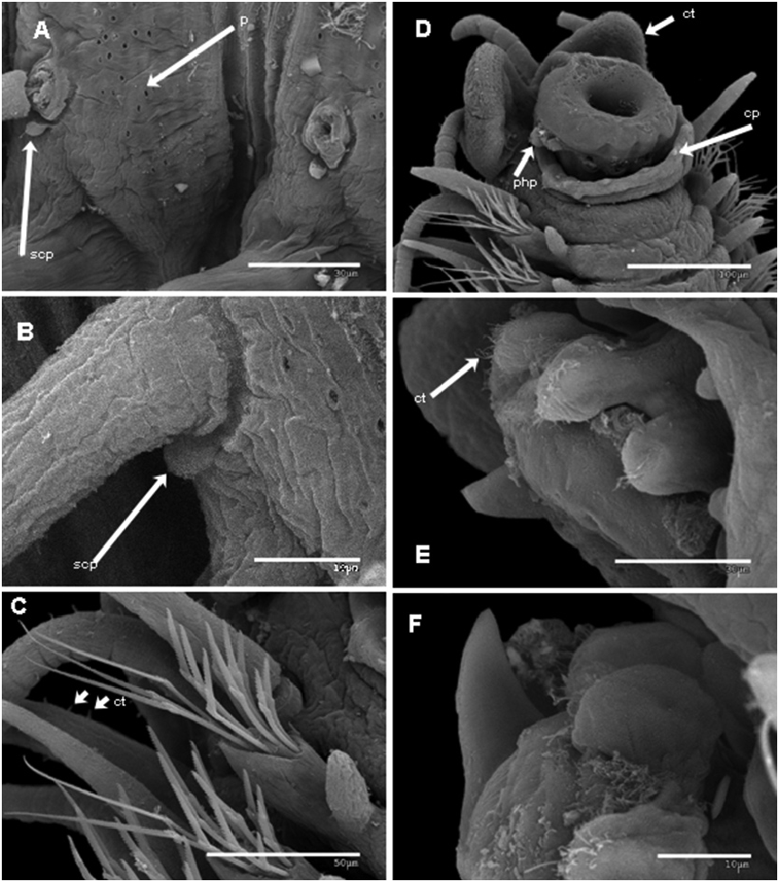
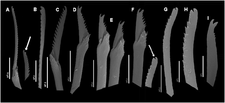
Description. Body slender, elongate, holotype 3.52 mm long, 0.3 mm wide, 60 chaetigers, lacking color; longest paratype 6 mm long, 67 chaetigers; some specimens with posterior part coiled ( Fig. 3A View FIGURE 3 ; preserved specimens) with minute pores on dorsal side ( Fig. 4A View FIGURE 4 ). Prostomium oval to semicircular, with four small eyes in open trapezoidal arrangement and two minute, anterior ocular spots ( Fig. 2A View FIGURE 2 ). Median antennae inserted almost on middle of prostomium, between posterior eyes, somewhat longer than prostomium and palps together, slightly pseudo-articulated distally; lateral antennae about half long as median one, inserted in front of anterior eyes, smooth or slightly pseudo-articulated depending on specimen ( Figs 2A View FIGURE 2 , 3A–B View FIGURE 3 ). Palps large, triangular, longer than prostomium, fused at bases with a distinct groove ( Figs 2A View FIGURE 2 , 3B View FIGURE 3 , 4D View FIGURE 4 ) and two ventro-lateral rows of small ciliary tufts ( Fig. 4D View FIGURE 4 ). Peristomium shorter than subsequent segments; dorsal tentacular cirri similar in length to lateral antennae, ventral tentacular cirri slightly shorter ( Figs 2A View FIGURE 2 , 3B View FIGURE 3 ). Anterior segments each with a single dorsal, transversal ciliary band ( Fig. 3B, C View FIGURE 3 ), double in midbody segments ( Fig. 3D View FIGURE 3 ), then single again on posterior segments ( Fig. 3E View FIGURE 3 ). Dorsal cirri slender, elongated, alternating long and short all along body, smooth ( Figs 2A View FIGURE 2 , 3F View FIGURE 3 ), or slightly pseudo-articulated distally on most anterior ones ( Figs 2A View FIGURE 2 , 3A, B View FIGURE 3 ). Subcirral papillae below anterior dorsal cirri, small, rounded ( Figs 3F View FIGURE 3 , 4A, B View FIGURE 4 ). Ventral cirri short, digitiform ( Figs 3D, E View FIGURE 3 , 4C, D View FIGURE 4 ). Anterior parapodia ( Figs 3B View FIGURE 3 , 4C, D View FIGURE 4 ) each with 3–5 compound chaetae with long, filiform, spiniger-like blades, 81 μm long, distally indistinctly bifid, with short spines on margin ( Figs 2B View FIGURE 2 , 4C View FIGURE 4 , 5A View FIGURE 5 ); and 7–9 falcigers with elongated, bidentate blades ( Figs 2C View FIGURE 2 , 5C View FIGURE 5 ), most dorsal 42 μm long, most ventral 20 μm, with both teeth of similar size, with short spines on margin ( Figs 2C View FIGURE 2 , 4C View FIGURE 4 , 5C View FIGURE 5 ). Number of spiniger-like and falcigers per parapodium decreasing posteriorly. Midbody parapodia with 1–2 (3), spiniger-like chaetae, similar to anteriormost parapodia, but with 95 μm long blades ( Fig. 2D View FIGURE 2 ), apparently unidentated under light microscope, slightly bidentate under SEM ( Fig. 5B View FIGURE 5 ), and 4–6 falcigers, about 17 μm long, with both teeth similar in size ( Figs 2E View FIGURE 2 , 5D View FIGURE 5 ). Posterior parapodia with chaetae similar to those at midbody, 1–2 spiniger-like chaetae 100 μm long ( Fig. 2F View FIGURE 2 ), and 4–5 falcigers 16 μm long, with both teeth similar or proximal slightly longer than distal, slightly curved ( Figs 2G View FIGURE 2 , 5E, F View FIGURE 5 ). Dorsal simple chaetae on posterior segments (also in anterior or midbody segments in some specimens), slightly curved, distally blunt and slightly bidentate, both teeth broad ( Figs 2H View FIGURE 2 , 5G–H View FIGURE 5 ); ventral simple chaetae only on posterior segments, markedly bidentate, both teeth acute, proximal slightly longer than distal one, apparently smooth under light microscope ( Fig. 2I View FIGURE 2 ), finely serrated under SEM ( Fig. 5I View FIGURE 5 ). Anterior parapodia each with three slender aciculae ( Fig. 2J View FIGURE 2 ); with decreasing posteriorly to only one distally oblique at late midbody and posterior parapodia ( Fig. 2K, L View FIGURE 2 ). Pharynx long and slender, longer than proventricle, through about 10 segments, with a small pharyngeal tooth located on anterior margin, surrounded by a crown of ten soft, ciliated papillae ( Figs 4D–F View FIGURE 4 ); pharynx inside a sheath with distal small papillae ( Fig. 5D View FIGURE 5 ). Proventricle rectangular, through about five segments, with about 18 muscle cell rows ( Fig. 2A View FIGURE 2 ). Pygidum small, triangular, with two long, smooth anal cirri.
No known copyright restrictions apply. See Agosti, D., Egloff, W., 2009. Taxonomic information exchange and copyright: the Plazi approach. BMC Research Notes 2009, 2:53 for further explanation.
|
Kingdom |
|
|
Phylum |
|
|
Class |
|
|
Order |
|
|
Family |
|
|
Genus |