
Leuceruthrus cf. ksepkai
|
publication ID |
https://doi.org/10.1645/22-36 |
|
DOI |
https://doi.org/10.5281/zenodo.7753990 |
|
persistent identifier |
https://treatment.plazi.org/id/039987AB-2164-FFF8-16D9-48D0AF3F92C5 |
|
treatment provided by |
Felipe |
|
scientific name |
Leuceruthrus cf. ksepkai |
| status |
|
Leuceruthrus cf. ksepkai View in CoL View at ENA
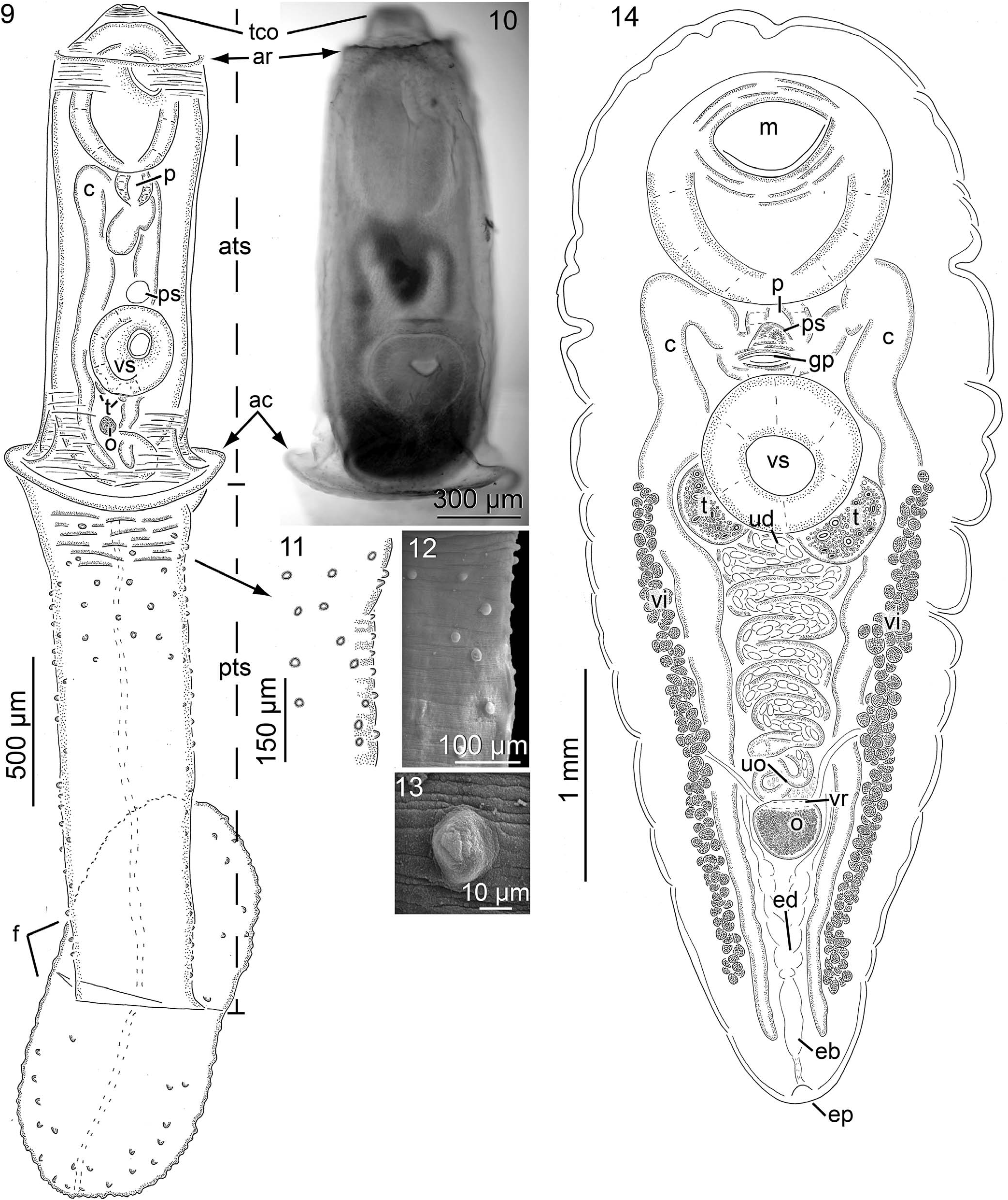
( Fig. 14 View Figures 9–14 ; Table II)
Diagnosis of adult (based on light microscopy of 5 stained, wholemounted adult specimens) ( Fig. 14 View Figures 9–14 ): Body of adult, 3,840 6,260 (4,904, 5) long or 1.7 3.0 (2.4, 5) X longer than wide, width at level of oral sucker 1,800 2,060 (1,928, 5) width at level of ventral sucker 1,800 2,500 (2,064, 5) ( Fig. 14 View Figures 9–14 ); forebody 1,400 2,460 (1,888, 5) long or 33 42% (38%, 5) of overall body length; hindbody 1,550 3,004 (2,381, 5) long or 38 58% (48%, 5) of overall body length, 0.90 1.6 (1.3, 5) X greater than forebody length; tegument unarmed, smooth, approximately 20 40 (28, 5) thick. Excretory system difficult to trace anteriorly branches joining at 81 91% (87%, 5) of body length from anterior body margin, forming excretory bladder ( Fig. 14 View Figures 9–14 ); excretory bladder 280 360 (340, 5) long, 70 160 (98, 5) wide, becoming confluent with diminutive excretory duct posteriorly; excretory duct 110 280 (184, 5) long, communicating excretory bladder and terminal excretory pore ( Fig. 14 View Figures 9–14 ). Nervous system not evident. Oral sucker 1,030 1,340 (1,150, 5) long or 21 27% (24%, 5) of body length or 54 74% (62%, 5) of forebody length ( Fig. 14 View Figures 9–14 ), 1,110 1,340 (1,222, 5) wide or 55 67% (62%, 5) of body width, anterior margin 4 8% (5%, 5) of body length from anterior body end, posterior margin 160 920 (494, 5) from anterior margin of ventral sucker ( Fig. 14 View Figures 9–14 ). Ventral sucker in anterior half of body, 690 860 (772, 5) long or 14 20% (16%, 5) of body length, 740 880 (797, 5) wide or 36 43% (40%, 5) of body width at level of ventral sucker, 64 74% (67%, 5) of oral sucker length, 60 69% (65%, 5) of oral sucker width ( Fig. 14 View Figures 9–14 ). Mouth opening ventrally ( Fig. 14 View Figures 9–14 ). Pharynx ovoid, 300 370 (334, 5) long or 5 9% (7%, 5) of body length, 230 350 (316, 5) wide ( Fig. 14 View Figures 9–14 ). Esophagus extending posteriad from mouth approximately 175 435 (341, 13), esophagus bifurcation and esophageal branches difficult to discern from surrounding tissue (stylized in illustration [ Fig. 14 View Figures 9–14 ]); dextral cecum 3,760 4,160 (3,950, 3) or 78 91% (84%, 3) of body length, prececal space, 860–1,260 (1,138, 4) or 22 31% (25%, 4) [18%] of body length from anterior end of body, postcecal space, 270 500 (384, 5) or 5 12% (8%, 5) of body length from posterior end of body; sinistral cecum 3,900 4,050 (3,975, 3) or 77 96% (84%, 3) of body length, prececal space 1,000 –1,300 (1,163, 4) or 23 31% (26%, 4) of body length from anterior end of body, postcecal space 300 480 (374, 5) or 5 10% (8%, 5) of body length from posterior end of body.
Testes oblique to askew, always posterior of horizontal midline of ventral sucker, oval to suboval ( Fig. 14 View Figures 9–14 ); dextral testis 350 400 (375, 2) long or 8% (2) of body length, 370 380 (375, 2) wide or 18% (2) of body width, pretestis space, 2,040 –2,200 (2,120, 2) from anterior end of body or 40 53% (47%, 2) of total body length, posttestis space, 2,560 2,640 (2,600, 2) from posterior end of body or 52 62% (57%, 2) of total body length; sinistral testis 350 470 (393, 3) long or 7 9% (8%, 3) of body length, 320 410 (353, 3) wide or 15 20% (18%, 3) of body width, pretestis space 2,060 2,340 (2,163, 3) from anterior end of body or 41 50% (45%, 3) of total body length, posttestis space 2,520 2,760 (2,603, 3) from posterior end of body or 48 67% (55%, 3) of total body length. Vasa efferentia not evident. Prostatic sac oval appearing densely filled with glandular cells, anterior margin 0 550 (232, 5) from posterior margin of oral sucker, 250 320 (286, 5) long, 290 330 (264, 5) wide ( Fig. 14 View Figures 9–14 ). Fine features of terminal male genitalia (i.e., seminal vesicle, pars prostatica, ejaculatory duct, sinus organ) difficult to delineate within prostatic sac; seminal vesicle present. Hermaphroditic pore directed ventrally, at 33 39% (36%, 3) of body length from anterior end of body. Genital atrium circular in outline, with seemingly strongly muscularized rim, 90 280 (186, 4) in diameter, 1 of 5 (20%) specimens containing ~ 35 uterine eggs. Genital pore near level with, or slightly posterior of midline of prostatic sac, opening ventrally at 34 41% (37%, 4) of total body length from anterior end of body ( Fig. 14 View Figures 9–14 ).
Ovary 200 360 (298, 5) long or 5 9% (6%, 5) of body length, 200 390 (313, 4) wide or 11 19% (16%, 4) of body width, postovary space 880 1,100 (1,028, 5) or 17 27% (17%, 9) [22%] of body length ( Fig. 14 View Figures 9–14 ). Fine features of the female genitalia (i.e., oviduct, Laurer’s canal, ovovitelline duct, and oötype) difficult to discern, putative highly glandular Mehlis’ complex present anterior to ovary ( Fig. 14 View Figures 9–14 ). Uterus comprising a field 1,840 1,940 (1,890, 2) long or 36 47% (42%, 2) of body length, proximal end extending anteriad from ovary, looping laterally between ceca posterior to testes, extending through space between testes ( Fig. 14 View Figures 9–14 ), distal portion of uterus difficult to delineate from surrounding tissue, synthesis with metraterm not evident; uterine seminal receptacle not evident; metraterm not evident. Vitellarium extending from near level of, or slightly posterior of, horizontal midline of ventral sucker, to near posterior end of body ( Fig. 14 View Figures 9–14 ), maximum distance between fields 1,000 1,440 (1,214, 5) or 53 70% (62%, 5) of body width; dextral vitelline field 1,910 2,920 (2,378, 5) long or 44 57% (49%, 5) of body length, terminating anteriorly at 41 54% (49%, 4) of body length, terminating posteriorly at 87 95% (90%, 4) of body length, 58 62% (60%, 3) of dextral cecum length; sinistral vitelline field 1,730 3,200 (2,492, 5) long or 45 66% (51%, 5) of body length, terminating anteriorly at 42 54% (47%, 5) of body length, terminating posteriorly at 88 96% (92%, 5) of body length, 58 68% (63%, 5) of sinistral cecum length; primary vitelline collecting ducts nearly symmetrical, extending posteromediad from respective vitelline field before becoming confluent and forming vitelline reservoir; dextral vitelline collecting duct 350 720 (535, 2) long, proximal end branches from vitellarium at 38 57% (47%, 2) of dextral vitelline field length; sinistral vitelline collecting duct 450 550 (500, 2) long, proximal end branches from vitellarium at 50 54% (52%, 2) of sinistral vitelline field length; vitelline reservoir dorsal to ovary ( Fig. 14 View Figures 9–14 ). Uterine eggs present in specimens greater than 4 (mm) in total body length, pyriform, varying in size from approximately 50 to 60 (55, 5) X 30 to 40 (37, 5) to approximately 70 (5) X 30 40 (37, 5).
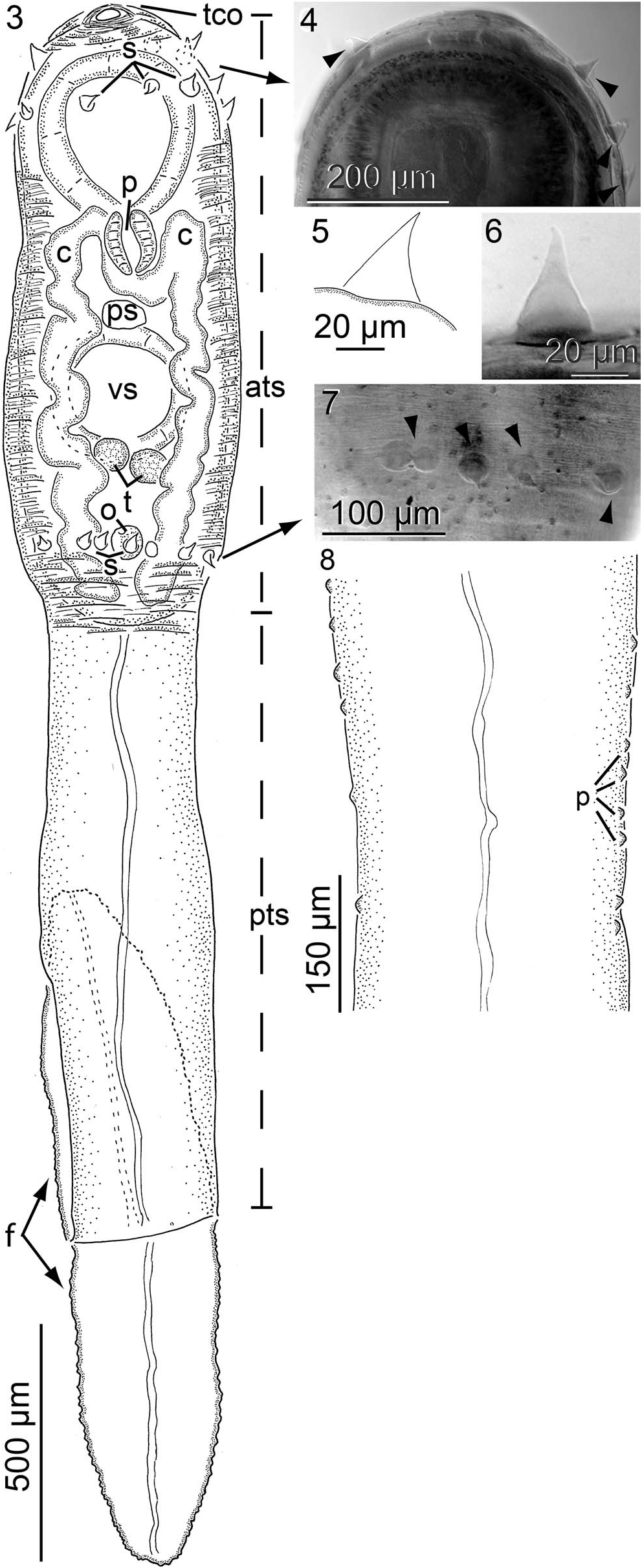
Diagnosis of young adult (based on light microscopy of 6 stained, whole-mounted specimens): Body 2,260 3,340 (2,960, 6) long or 2.2 3.2 (2.9, 6) X longer than wide, width at level of oral sucker 1,040 1,300 (1,156, 6) wide, width at level of ventral sucker 820 1,180 (1,040, 6); forebody 1,060 1,560 (1,322, 6) long or 39 50% (45%, 6) of overall body length; hindbody 770 1,455 (1,223, 6) long or 34 45% (40%, 6) of overall body length, typically less than forebody length; tegument unarmed, smooth, approximately 20 60 (34, 6) thick, 2 of 6 specimens enveloped with a thicker, yet distinct layer of tegument (¼possibly tail stem cavity of cercaria). Excretory system not evident anteriorly, branches joining at 87 93% (90%, 6) of body length from anterior body end, forming excretory bladder; excretory bladder 155 250 (185, 5) long, 30 50 (40, 4) wide, becoming confluent with diminutive excretory duct posteriorly; excretory duct extending 105 140 (122, 4) from posterior margin of excretory bladder to terminal excretory pore. Nervous system not evident. Oral sucker 560 790 (712, 6) long or 22 29% (24%, 6) of body length, 540 820 (735, 6) wide or 66 76% (71%, 6) of body width, anterior margin 2 3% (2%, 6) of body length from anterior body end, posterior margin 260 665 (476, 6) from anterior margin of ventral sucker. Ventral sucker near horizontal midline of body, 370 535 (474, 6) or 14 20% (16%, 6) of body length, 400 530 (498, 6) or 45 50% (48%, 6) of body width, 62 68% (66%, 6) of oral sucker length 65 74% (68%, 6) of oral sucker width. Mouth opening ventrally. Pharynx ovoid, 150 230 (195, 6) long or 6 9% (7%, 6) of body length 170 230 (212, 6) wide ( Fig. 4 View Figures 3–8 ). Esophagus extending posteriad from mouth approximately 295 450 (388, 6), before bifurcating 20 30 (25, 6) posterior to pharynx; dextral esophageal branch 100 230 (154, 6) long, sinistral esophageal branch 90 200 (144, 6) long; dextral cecum 1,640 2,800 (2,368, 6) or 72 84% (77%, 6) of body length, prececal space 660 890 (762, 6) or 22 29% (25%, 6) of body length from anterior end of body, postcecal space 100 300 (200, 6) or 4 9% (6%, 6) of body length from posterior end of body; sinistral cecum 1,710 2,750 (2,354, 6) or 72 82% (76%, 6) of body length, prececal space 650 860 (757, 6) or 22 29% (26%, 6) of body length from anterior end of body, postcecal space 75 330 (194, 6) or 3 10% (6%, 6) of body length from posterior end of body.
Testes abreast, always posterior of horizontal midline of ventral sucker, oval; dextral testis 150 270 (223, 6) long or 6 10% (8%, 6) of body length, 100 190 (161, 6) wide or 12 18% (15%, 6) of body width, pretestis space 1,420 1,950 (1,734, 6) from anterior end of body or 51 63% (57%, 6) of total body length, posttestis space 660 1,380 (1,085, 6) from posterior end of body or 29 43% (36%, 6) of total body length; sinistral testis 140 300 (222, 6) long or 5 9% (7%, 6) of body length, 95 190 (155, 6) wide or 12 18% (15%, 6) of body width, pretestis space 1,420 1,950 (1,762, 6) from anterior end of body or 51 63% (58%, 6) of total body length, posttestis space 680 1,320 (1,081, 6) from posterior end of body or 30 41% (36%, 6) of total body length. Vasa efferentia indistinct in whole-mounted specimens. Prostatic sac 95 210 (155, 6) long, 60 165 (141, 6) wide, anterior margin 105 440 (319, 6) from posterior margin of oral sucker. Features of terminal male genitalia (i.e., seminal vesicle, pars prostatica, ejaculatory duct, sinus organ) appearing as anlagen, difficult to discern. Genital pore near level with, or slightly posterior of midline of prostatic sac, opening ventrally at 39 56% (46%, 6) of total body length from anterior end of body.
Ovary 95 180 (139, 6) long or 4 6% (5%, 6) of body length, 70 120 (100, 6) wide or 8 12% (10%, 6) of body width, postovary space 250 850 (640, 6) or 11 26% (21%, 6) of body length. Fine details of the female genitalia (i.e., oviduct, Laurer’s canal, ovovitelline duct, oötype, and Mehlis’ gland) appearing as anlagen, difficult to discern. Uterus extending in straight line anteriorly, from near anterior margin of ovary, passing between testes, becoming indistinct distally; uterine seminal receptacle not evident; metraterm not evident. Vitellarium not developed. Uterine eggs none present.
Taxonomic summary for Leuceruthrus cf. ksepkai
Host: Micropterus salmoides , largemouth bass.
Locality: Holmes Creek ( 30°36 ′ 25 ′′ N, 85°44 ′ 49 ′′ W), Florida GoogleMaps .
Specimens and sequences deposited: Vouchers (USNM 1593589 [juvenile]; USNM 1593590 and USNM 1593591 [adults]); ITS2 (adults, GenBank ON877230 View Materials , ON877231 View Materials ).
Site in host: Adults and juveniles in stomach.
Site in host: Adults and young adults in stomach.
Prevalence and intensity of infection: 6 of 6 (100%) largemouth bass from Holmes Creek ( 30°36 ′ 25 ′′ N, 85°44 ′ 49 ′′ W) were infected with 1 7 (3) specimens of L. cf. ksepkai GoogleMaps .
Taxonomic remarks on Leuceruthrus cf. ksepkai
Adults of L. cf. ksepkai and L. micropteri are morphologically similar but can be readily differentiated. Leuceruthrus cf. ksepkai has a proportionally large oral sucker (24% body length and 62% forebody length vs. 18% and 49%, respectively, in L. micropteri ) and a small ventral sucker (16% body length vs. 12% in L. micropteri ). The adults of L. ocalana and L. stephanocauda have not been described.
No known copyright restrictions apply. See Agosti, D., Egloff, W., 2009. Taxonomic information exchange and copyright: the Plazi approach. BMC Research Notes 2009, 2:53 for further explanation.
|
Kingdom |
|
|
Phylum |
|
|
Class |
|
|
SubClass |
Digenea |
|
Order |
|
|
Family |
|
|
Genus |