
Leuceruthrus cf. stephanocauda (Faust, 1921) Bullard, 2022
|
publication ID |
https://doi.org/ 10.1645/22-36 |
|
DOI |
https://doi.org/10.5281/zenodo.7753986 |
|
persistent identifier |
https://treatment.plazi.org/id/039987AB-2160-FFE2-16E4-4EC6A9C795B0 |
|
treatment provided by |
Felipe |
|
scientific name |
Leuceruthrus cf. stephanocauda |
| status |
|
Leuceruthrus cf. stephanocauda View in CoL View at ENA
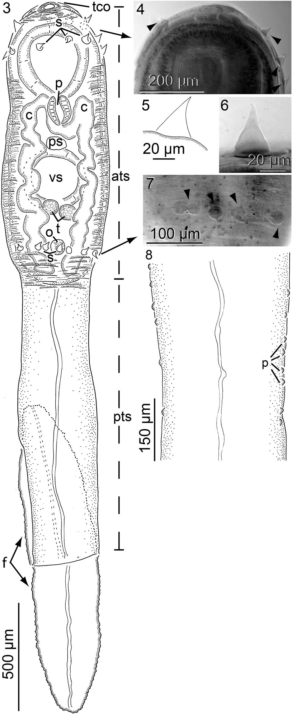
( Figs. 3 8 View Figures 3–8 ; Table II)
Diagnosis of cercaria (based on light microscopy of 2 stained, whole-mounted, naturally shed cercariae with withdrawn distome) ( Figs. 3 8 View Figures 3–8 ): Cercaria 3,320 3,580 (3,450, 2) long ( Fig. 3 View Figures 3–8 ). Tail stem lacking pigmentation, 2,600 2,900 (2,750, 2) long or 78 81% (80%, 2) of cercariae length, 510 660 (585, 2) wide or 4.4 5.1 (4.7, 2) X longer than wide, comprised of an anterior and posterior region ( Fig. 3 View Figures 3–8 ); anterior tail stem region (ATS) oblong, 1,320 1,460 (1,390, 2) long or 40 41% (40%, 2) of cercariae length, maximum width same as reported for tail stem, containing distome, tapering anteriorly, devoid of mammillae, armed with 2 rows of tail stem spines ( Figs. 3 8 View Figures 3–8 ); posterior tail stem region (PTS) dorsoventrally compressed, 1,280 1,440 (1,360, 2) long or 39 40% (39%, 2) of cercariae length, 360 340 (350, 2) wide, nearly uniform in width anteriorly to posteriorly, devoid of mammillae, lateral margins bearing many minute protuberances ( Figs. 3, 8 View Figures 3–8 ); PTS protuberances, minute, lacking pores, marginal ( Fig. 8 View Figures 3–8 ). Furcae lacking pigmentation, lanceolate, dorsoventrally compressed, margin bearing many protuberances, serrate ( Fig. 3 View Figures 3–8 ); furcal protuberances minute, pored, marginal; dorsal furca 660 700 (680, 2) long or 18 21% (19%, 2) of cercariae length, 320 380 (350, 2) wide or 1.7 2.2 X (2, 2) longer than wide; ventral furca 620 720 (670, 2) long or 17 22% (20%, 2) of cercariae length, 320 340 (330, 2) wide or 1.8 2.3 X (2, 2) longer than wide ( Fig. 3 View Figures 3–8 ). Tail cavity opening at anteromedial end of cercaria, directing anteriad ( Fig. 3 View Figures 3–8 ); tail stem cavity not evident. Mammillae not evident. Tail stem spines maximum length 40, maximum width 35 ( Fig. 5 View Figures 3–8 ), restricted to the anterior tail stem region, distributed in 2 concentric rows, anterior row encircling area near tail cavity opening ( Figs. 3, 4 View Figures 3–8 ), posterior row encircling area near synthesis of posterior tail stem ( Figs. 3, 7 View Figures 3–8 ). Excretory system with 1 primary excretory canal, extending posteriad along the medial axis of the posterior tail stem, bifurcating at the synthesis of the furcae, extending independently through each furca, opening via excretory pore at the distal end of each furca ( Fig. 3 View Figures 3–8 ).
Body of distome ( Fig. 3 View Figures 3–8 ) 1,220 1,780 (1,500, 2) long or 37 50% (43%, 2) of cercaria length, 470 580 (525, 2) wide or 2.6 3 (2.8, 2) X longer than wider, anterior margin 30 40 (35, 2) from tail cavity opening; forebody 630 900 (765, 2) long or 51 52% (51, 2) of overall body length; hindbody 350 600 (475, 2) long or 29 34% (31%, 2) of overall body length, 56 67% (61%, 2) of forebody length; tegument unarmed. Excretory system not evident. Nervous system not evident. Oral sucker 360 370 (365, 2) long or 21 30% (25%, 2) of body length, 340 370 (355, 2) wide or 64 72% (68%, 2) of body width, 60 30 (45, 2) or 2 3% (3%, 2) of body length from anterior body end, 850 1,360 (1,105, 2) or 70 76% (73%, 2) of body length from posterior body end ( Fig. 3 View Figures 3–8 ). Ventral sucker in posterior half of body, with anterior margin 630 900 (765, 2) or 51 52% (51%, 2) of body length from anterior body end ( Fig. 3 View Figures 3–8 ), 290 300 (295, 2) long or 24 25% (24%, 2) of body length, 290 300 (295, 2) wide or 50 64% (57%, 2) of body width, 78 83% (81%, 2) of oral sucker length, 78 88% (83%, 2) of oral sucker width ( Fig. 3 View Figures 3–8 ). Pharynx ovoid, 100 140 (120, 2) long or 6 11% (9%, 2) of body length, 110 135 (123, 2) wide ( Fig. 3 View Figures 3–8 ). Esophagus extending posteriad from mouth before bifurcating posterior to pharynx, esophageal branches arching posterolaterad before joining with intestinal ceca ( Fig. 3 View Figures 3–8 ); dextral cecum 1,050 1,518 (1,284, 2) or 82 85% (84%, 2) of body length, laterad cecum length 150 163 (157, 2), descending cecum length 900 1,355 (1,128, 2), prececal space 375 550 (463, 2) or 29 31% (30%, 2) of body length from anterior end of body, postcecal space 20 50 (35, 2) or 2% (2%, 2) of body length from posterior end of body; sinistral cecum 1,000 1,620 (1,310, 2) or 78 91% (85%, 2) of body length, laterad cecum length 150 (150, 2), descending cecum length 850–1,470 (1,160, 2), prececal space 375 510 (443, 2) or 29 30% (29%, 2) of body length from anterior end of body, postcecal space 25 50 (38, 2) or 2 3% (2%, 2) of body length from posterior end of body. Testes abreast, round to oval ( Fig. 3 View Figures 3–8 ); dextral testis 50 (50, 2) long or 2 4% (3%, 2) of body length, 65 90 (78, 2) wide or 1.3 1.8 (1.6, 2) X wider than long; sinistral testis 60 (60, 2) long or 3 5% (4%, 2) of body length, 60 75 (68, 2) wide or 1 1.3 (1.1, 2) X wider than long; pretesticular space 810 1,180 (995, 2) from anterior end of body or 63 66% (65%, 2) of total body length; posttesticular space 300 540 (420, 2) from posterior end of body or 23 30% (27%, 2) of total body length. Vasa efferentia not evident. Prostatic sac 55 75 (65, 2) long, 90 110 (100, 2) wide or 1.5 1.6 (1.5, 2) X wider than long ( Fig. 3 View Figures 3–8 ). Genital atrium circular in ventral view, 25 30 (28, 2) in diameter. Genital pore 650 840 (745, 2) of body length from anterior end of body or at 47 50% (49%, 2) of body length. Fine features of terminal male genitalia (i.e., seminal vesicle, pars prostatica, ejaculatory duct, sinus organ) not evident.
Ovary 140 210 (175, 2) of body length from posterior end of body, 65 70 (68, 2) long or 1.2 1.3 (1.2, 2) X longer than wide, 55 (55, 2) wide ( Fig. 3 View Figures 3–8 ). Fine features of terminal female genitalia (i.e., oviduct, Laurer’s canal, ovovitelline duct, oötype, and Mehlis’ gland) not evident. Uterus 430 655 (543, 2) long or 34 37% (35%, 2) of body length, 13 20 (17, 2) wide. Metraterm not evident. Vitellarium not developed in distome ( Fig. 3 View Figures 3–8 ).
No known copyright restrictions apply. See Agosti, D., Egloff, W., 2009. Taxonomic information exchange and copyright: the Plazi approach. BMC Research Notes 2009, 2:53 for further explanation.