
Lumbricillus nivalis, Klinth & Rota & Martinsson & Prantoni & Erséus, 2022
|
publication ID |
https://doi.org/ 10.1093/zoolinnean/zlab073 |
|
publication LSID |
lsid:zoobank.org:pub:3FB3FBB8-4112-463A-ADEF-35CD427C8AF4 |
|
DOI |
https://doi.org/10.5281/zenodo.6461097 |
|
persistent identifier |
https://treatment.plazi.org/id/D62E6765-ED4D-498D-A300-50D5232BDD14 |
|
taxon LSID |
lsid:zoobank.org:act:D62E6765-ED4D-498D-A300-50D5232BDD14 |
|
treatment provided by |
Plazi |
|
scientific name |
Lumbricillus nivalis |
| status |
|
LUMBRICILLUS NIVALIS View in CoL KLINTH,
ROTA & ERSÉUS SP. NOV.
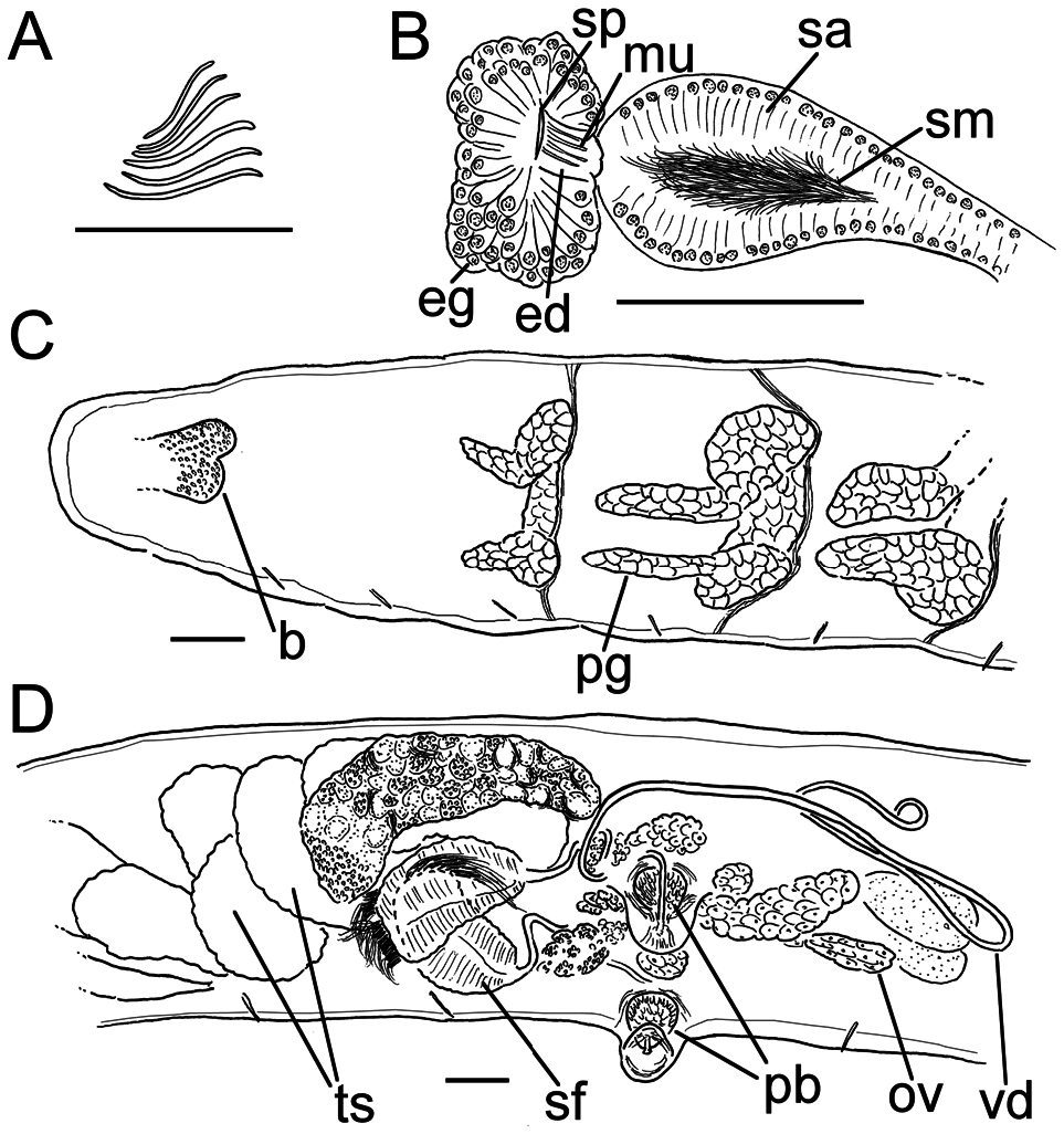
( FIG. 5 View Figure 5 )
Z o o b a n k r e g i s t r a t i o n: u r n: l s i d: z o o b a n k. org:act: D62E6765-ED4D-498D-A300-50D5232BDD14
Holotype: SMNH Type Coll. 9310 ( CE34644 ), a mature amputated specimen stained in Paracarmine and mounted on a slide. Leg. Karla Paresque, 15 January 2015. COI barcode, GenBank MZ 393952 View Materials ; accession numbers for additional genetic data are given in Table 1 View Table 1 and the Supporting Information ( Table S1 View Table 1 ).
Type locality: Snow Island , South Shetland Islands, Antarctica, from algae on rocks in intertidal zone, 62.7753 S, 61.2858 W GoogleMaps .
Etymology: The Latin adjective nivalis means snowy, alluding to the name of the type locality, Snow Island.
Diagnosis: This species, now differentiated by a unique COI barcode, is a member of the L. lineatus group, meaning that it has testis sacs forming clubshaped lobes arranged in a fan shape and spindleshaped spermathecae. It can be distinguished from other L. lineatus group members by: (1) its short sperm funnels, 1.5–2.0 times longer than wide; (2) vasa deferentia extending posterior to XII; (3) spermathecae with short ectal duct, rapidly widening into ampulla, with ampulla widest ectally, gradually tapering towards ental duct, which connects to oesophagus; and (4) clitellum ventrally absent in XII but present ventrally in (first half of) XIII.
Description: Length of first 25 segments 6 mm (fixed, amputated specimen); first 15 segments 3.7 mm long; width at clitellum 0.50 mm. Chaetae sigmoid ( Fig. 5A View Figure 5 ). Upper bundles dorsolateral (higher than the lateral line but closer to it than the ventral bundles), with three to five chaetae anterior and posterior of clitellum, at least to XXV. Ventral bundles with (three) four to six throughout to XXV. The longest measured chaetae 60 µm long, ~3–5 µm wide. Epidermis loosely covered with rows of pale gland cells. Clitellum with reticulate pattern of gland cells, extending over XII–1/2XIII, interrupted ventrally in XII but present ventrally in XIII.
Coelomocytes numerous, ~10–15 µm long; round, oval or spindle shaped; granulated with distinct nucleus. Paired pharyngeal glands ( Fig. 5C View Figure 5 ) in IV, V and VI; first two pairs narrowly connected dorsally, a trait not discernible in third pair owing to damaged specimen; ventral lobes elongated, possibly attributable to fixation. Dorsal vessel seemingly originating in XIII, with peristomial bifurcation. Nephridia ~130– 150 µm long, observed in 7/8, 8/9 and postclitellar segments. Anteseptale small, consisting of funnel only. Postseptale oval, tapering into posteroventral efferent duct. Brain with posterior incision.
Male genitalia paired ( Fig. 5D View Figure 5 ). Testes originating in anterior of XI, with testis sacs forming regular club-shaped lobes extending forwards into IX. Sperm funnels in XI, 240 µm long, 140 µm wide, making them 1.5–2.0 times longer than wide; funnels tapering towards vasa deferentia. Vasa deferentia irregularly coiled in XII–XIII, possibly reaching XIV, 15 µm wide. Penial bulbs round, 85 µm in diameter. Ovaries in XII. No mature eggs observed.
Spermathecae ( Fig. 5B View Figure 5 ) in V, pear or spindle shaped, with short ectal duct covered in musculature and rapidly widening into ampulla. Ampulla widest ectally and gradually tapering towards ental duct, which connects to oesophagus. Sperm in lumen of ectal part of ampulla; heads of spermatozoa embedded in wall of ampulla. Spermathecae 255 µm long, 20 µm wide at the ectal duct, expanding to 80 µm at widest part of ampulla. Gland cells surrounding ectal duct as a compact mass, 100 µm in diameter at its widest part. No midventral subneural glands observed.
Geographical distribution: Known from the South Shetland Islands only.
Remarks: Genetically and morphologically, this species is most closely related to the specimens we identified as L. antarcticus , which were found at the same locality on Snow Island. As such, this new species can be separated from other members of the L. lineatus group in the same way as described in the remarks for L. antarcticus above, where primarily the short sperm funnels help to distinguish this species from many somewhat similar Lumbricillus species from the Subantarctic. Owing to their likeness, we had to consider which of our two species was the true L. antarcticus . The clitellum is saddle-shaped in L. antarcticus , but in our single specimen of L. nivalis it is also present ventrally in the anterior of XIII. Moreover, L. nivalis has smaller penial bulbs and lacks subneural glands (possibly attributed to it not being fully mature; no mature eggs were observed). Finally, L. nivalis can be separated by its vasa deferentia extending all the way back to segment XIII or possibly XIV, whereas the vasa are consistently confined to XII in our and Stephenson’s material of L. antarcticus .
| SMNH |
Department of Paleozoology, Swedish Museum of Natural History |
| MZ |
Museum of the Earth, Polish Academy of Sciences |
No known copyright restrictions apply. See Agosti, D., Egloff, W., 2009. Taxonomic information exchange and copyright: the Plazi approach. BMC Research Notes 2009, 2:53 for further explanation.
|
Kingdom |
|
|
Phylum |
|
|
Class |
|
|
Order |
|
|
Family |
|
|
Genus |