
Lumbricillus macquariensis, BENHAM, 1905
|
publication ID |
https://doi.org/ 10.1093/zoolinnean/zlab073 |
|
publication LSID |
lsid:zoobank.org:pub:3FB3FBB8-4112-463A-ADEF-35CD427C8AF4 |
|
DOI |
https://doi.org/10.5281/zenodo.6461123 |
|
persistent identifier |
https://treatment.plazi.org/id/039DC377-FFBE-FFC4-4DBF-FA05FEA95D56 |
|
treatment provided by |
Plazi |
|
scientific name |
Lumbricillus macquariensis |
| status |
|
‘ LUMBRICILLUS’ CF. MACQUARIENSIS BENHAM, 1905 View in CoL
( FIG. 8 View Figure 8 )
? Lumbricillus macquariensis Benham, 1905: 295–297 View in CoL , pl. XIV, figs 8, 11–13; Benham, 1915: 189–191; Benham, 1922: 6; Stephenson, 1932: 254–255, fig. 7.
? Lumbricillus intermedius Benham, 1909: 261–262 View in CoL , pl. X, figs 8–11.
? Pachydrilus intermedius – Michaelsen, 1924: 197–199.
Type material: No information ( Reynolds & Wetzel, 2019). The material is probably located in the Otago Museum , New Zealand, because Benham mentions material being deposited and registered in the Museum by a Professor Parker, who was curator of the Otago museum collections at the time. Our inquiry did not receive any response .
Type locality: Macquarie Island, brackish pools, with planarians and Siphonaria View in CoL limpets .
Material examined: SMNH198150 View Materials ( CE12483 ), one mature specimen, and SMNH198151–198153 View Materials ( CE12484 –CE12486), three immature or semimature specimens, all collected in 2010 from South Georgia. For details of collection and GenBank accession numbers for COI barcodes and other gene sequences, see Table 1 View Table 1 and the Supporting Information ( Table S1 View Table 1 ) .
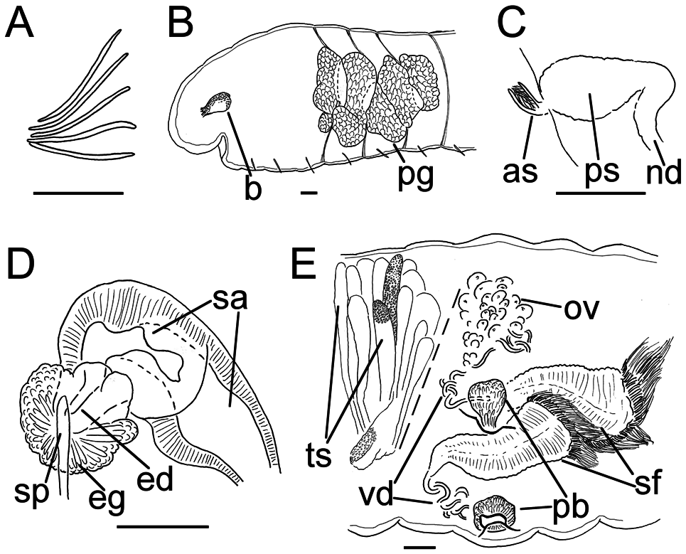
Description: Length of first 15–48 segments 2.5– 7.1 mm (fixed, amputated specimens); first 15 segments 2.5–3.0 mm long; width at clitellum 0.85– 1.10 mm. Chaetae slightly sigmoid ( Fig. 8A View Figure 8 ). Upper bundles dorsolateral (closer to lateral line than the ventral bundles), with four to six chaetae anterior to clitellum, and three to four chaetae in postclitellar segments, at least to segment XLVIII. Ventral bundles with (four) five to seven chaetae anterior to clitellum, and four to six chaetae posteriorly. The longest measured chaetae of each worm 135–145 µm long, ~6–7 µm wide. Epidermis loosely covered with rows of pale gland cells. Clitellum not fully developed. Head pore not observed.
Coelomocytes numerous, 15–30 µm long; round, oval or spindle shaped; granulated with distinct nucleus. Paired pharyngeal glands ( Fig. 8B View Figure 8 ) present in IV, V and VI, with third pair extending back into VII; each pair converging dorsally, but connections not discernible; dorsal lobes of about equal size, ventral lobes in IV–VI, increasing in size from IV to VI, absent in VII. Dorsal vessel originating in XVI–XVII, with peristomial bifurcation. Nephridia ( Fig. 8C View Figure 8 ) ~110– 190 µm long, observed in 7/8–9/10 and postclitellar segments. Anteseptale small, consisting of funnel only. Postseptale oval, tapering into posteroventral efferent duct. Brain with posterior incision.
Male genitalia paired ( Fig. 8E View Figure 8 ). Testes originating in anterior of XI, with testis sacs forming regular lobes extending forwards into X, but these lobes are fingerlike, thats is, much thinner than those of most species of Lumbricillus View in CoL . Sperm funnels in XI, in one specimen extending backwards into XII (see Fig. 8E View Figure 8 ), 425 µm long, 155 µm wide, making them about three times as long as wide; funnels tapering towards vasa deferentia. Most of vasa irregularly coiled in XII, 25 µm wide. Penial bulbs round or pear shaped, 125 µm in diameter, discharging into deep invagination of body wall ( Fig. 8E View Figure 8 ). Ovaries in XII. No mature eggs observed.
Spermathecae ( Fig. 8D View Figure 8 ) in V, pouch shaped, with short ectal duct gradually widening into ampulla. Ampulla with thicker epithelium in the ectal parts, which transitions to thinner epithelium in the more ental parts. Ental part seemingly connected with oesophagus. No sperm observed. Spermathecae 340 µm long, 55 µm wide at the ectal duct, 120 µm wide at widest part of ampulla. Gland cells surrounding ectal duct, forming compact mass, glandular body 125 µm in diameter at its widest part. In the mature specimen, we observed only one midventral subneural gland, 190 µm long, in XIV, but said specimen was amputated and ended in this segment.
Geographical distribution: Our specimens were collected from South Georgia Island, where L. macquariensis View in CoL was recorded by Stephenson (1932). However, the species was originally described from specimens that came from Macquarie Island ( Benham, 1905) and since then has also been reported from the Campbell and Auckland Islands ( Benham, 1922), Heard and McDonald Islands ( Lee, 1968) and Bishop Island ( Davies et al., 1997).
Remarks: Our specimens are similar to those identified as L. macquariensis View in CoL by Stephenson (1932) from South Georgia Island. In the original description from Macquarie Island (which lies south of Australia and New Zealand), Benham (1905) illustrated a spermatheca with a narrow ental duct connecting the sac-like ampulla with the oesophagus. Benham (1909) described L. intermedius (from Auckland Island), which he considered as intermediate between Lumbricillus maximus ( Michaelsen, 1888) View in CoL and L. verrucosus View in CoL , and which has a spermatheca with a small pore connecting the ampulla to the oesophagus without any narrow ental duct. However, Benham (1915) revisited his two species ( L. macquariensis View in CoL and L. intermedius ) and concluded that they were, in fact, the same, making L. intermedius a junior synonym of L. macquariensis View in CoL . He also concluded that in both samples the spermatheca did not have a narrow ental duct, but a pore connecting it directly to the oesophagus. The spermatheca illustrated by Benham (1909) is similar to that described by Stephenson (1932); both authors showed a thicker epithelium in the ectal part, much like in our specimens. This change in height of the duct epithelium and its expansion to merge with the ampulla are also reminiscent of the spermathecae of the re-examined types of L. maximus View in CoL ( Rota, 2001: fig. 1f). Furthermore, Stephenson and Benham noted pharyngeal glands as far back as VII, which is similar to what we observe and which distinguishes L. macquariensis View in CoL from most other Lumbricillus View in CoL species, except L. maximus ( Rota, 2001) View in CoL . Such posteriad extension of the third pair of glands represents a distinct situation from the development of an extra pair of glands in VII, a character distributed erratically also in other genera otherwise characterized by pharyngeal glands in IV–VI, for example, Fridericia Michaelsen, 1889a View in CoL ( Rota, 2001, 2015; Schmelz & Collado, 2010).
Another peculiar trait of our species is the structure of the testis sacs, which seemed at first sight to fall in the regularly lobed arrangement seen in most Lumbricillus species. However, the lobed sacs here are much thinner. This could perhaps be an indication that the sacs were not fully developed, but the developing testis sacs of, for example, L. sp. ‘Marion Is.’, from Marion Island (reported above) look completely different. It is possible that both Stephenson and Benham saw this structure but did not consider it deviant and therefore made no remarks upon it.
Our specimens might well belong to the same species as those studied by Stephenson from South Georgia and, like Stephenson, we found a good correspondence with Benham’s descriptions from Macquarie Island, particularly in the deep invagination of the body wall where the penial bulb discharges. A circumpolar distribution of the species would not be implausible, because a corresponding pattern has been observed for Lumbricillus View in CoL species in the Northern Hemisphere. The other possibility is that these are two or three separate but closely related species.
Genetically, our ‘ Lumbricillus ’ cf. macquariensis View in CoL is sister to Grania View in CoL in our phylogeny, but although its morphology is dissimilar in many ways to that of most Lumbricillus View in CoL (mentioned above), it is by no means closer to that of Grania View in CoL . Species of Grania View in CoL have a much more slender body, only one chaeta per ‘bundle’ and most often lack chaetae in several segments, and have masses of developing sperm cells and oocytes enveloped in septal distensions (seminal vesicles and ovisacs, respectively) extending backwards. In order to retain the well-defined genera Grania View in CoL and Lumbricillus View in CoL s.s., ‘ Lumbricillus ’ cf. macquariensis View in CoL needs to be transferred to another, probably new, genus. However, owing to the unresolved taxonomy and limited number of mature specimens we leave this to future studies.
No known copyright restrictions apply. See Agosti, D., Egloff, W., 2009. Taxonomic information exchange and copyright: the Plazi approach. BMC Research Notes 2009, 2:53 for further explanation.
|
Kingdom |
|
|
Phylum |
|
|
Class |
|
|
Order |
|
|
Family |
|
|
Genus |
Lumbricillus macquariensis
| Klinth, Mårten J., Rota, Emilia, Martinsson, Svante, Prantoni, Alessandro L. & Erséus, Christer 2022 |
Lumbricillus macquariensis
| Benham WB 1905: 297 |