
Capitella multibranchiata, Magalhäes & Hilliard, 2022
|
publication ID |
https://doi.org/ 10.11646/zootaxa.5125.2.7 |
|
publication LSID |
lsid:zoobank.org:pub:39562CC8-42D1-4CC6-930A-6F6F923A975C |
|
DOI |
https://doi.org/10.5281/zenodo.6425677 |
|
persistent identifier |
https://treatment.plazi.org/id/03A2627F-FFB3-D150-13F9-406E3A04C798 |
|
treatment provided by |
Plazi |
|
scientific name |
Capitella multibranchiata |
| status |
sp. nov. |
Capitella multibranchiata View in CoL sp. nov.
Figures 4–5 View FIGURE 4 View FIGURE 5
Capitella sp. nov.: Judge & Barry, 2016: Table 3 (in part).
Material examined. Holotype: Monterey Bay, California, “Deadwood 2” site, 36° 15.6768′ N, 122° 40.6790′ W, associated with deployed fragments of Spicebush ( Calycanthus occidentalis Hook. & Arn. ), Sta. WB20 , deployed on GoogleMaps October 18, 2011 and retrieved on October 26–28, 2013 by a benthic elevator and ROV Doc Ricketts on an MBARI cruise aboard the R / V Western Flyer , 3,100 m ( FMNM 15940 ). Paratypes same locality, date, collector and wood type as holotype (2 complete, FMNM 15941 ; 4 complete, FMNM 15942 ). Additional non-type material examined: Same locality, date and collector as type series, associated with Spicebush ( Calycanthus occidentalis ), Sta. WB19 (8) , Sta. WB20 (3 af) , and Sta. WB21 (4) ; Lyon , Sta. WB31 (2) , Sta WB32 (118) , Sta. WB33 (2c) ; yew ( Torreya californica Torr. ), Sta. WB34 (1) , Sta. WB35 (87) and Sta. WB36 (57) ; fern genus Cyathea Sm., Sta. WB 37 (2). Monterey Canyon off of coast of Monterey, Energetics Cruise Trawl #Oct 3000-4, sunken wood, 8 Oct. 2009, 3,000 –3,150 m, coll. J.C. Drazen (3, FMNH 14711 About FMNH ) GoogleMaps
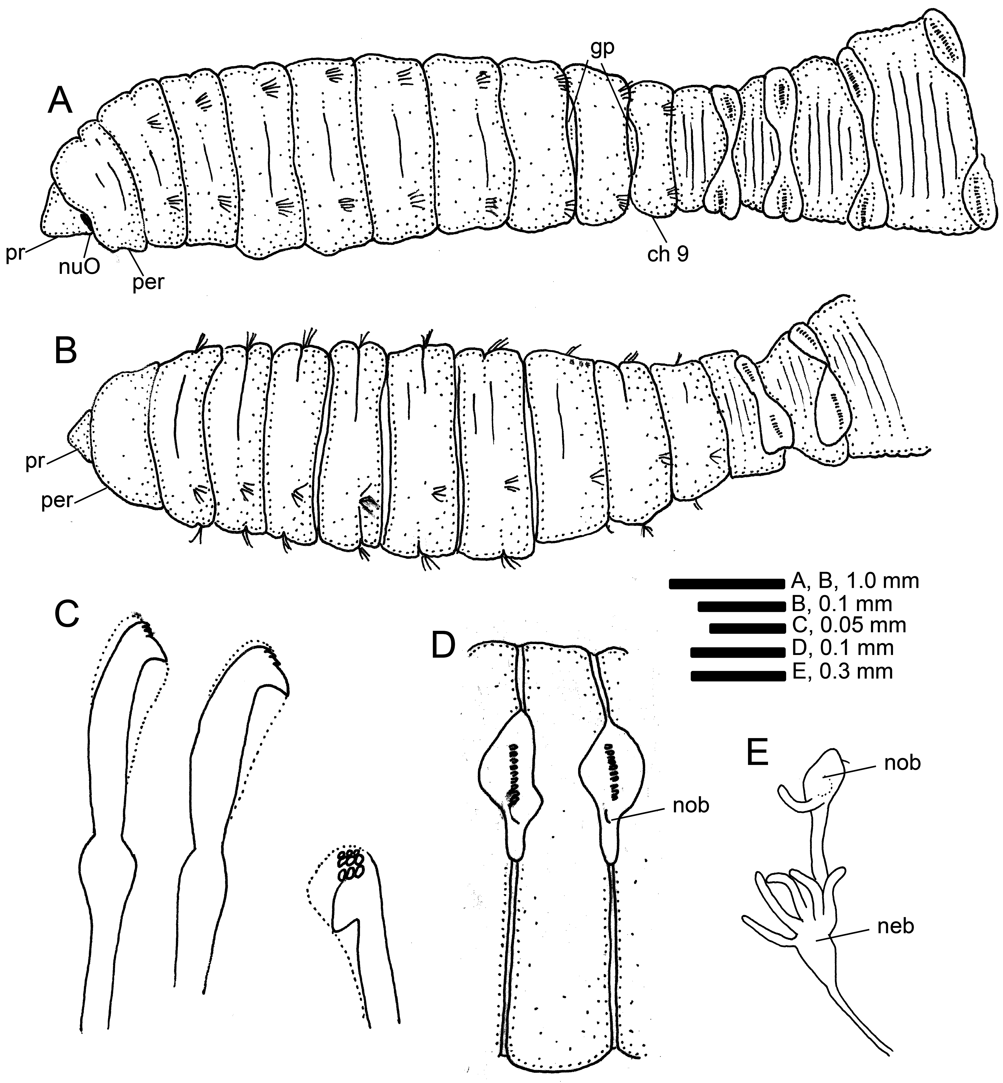
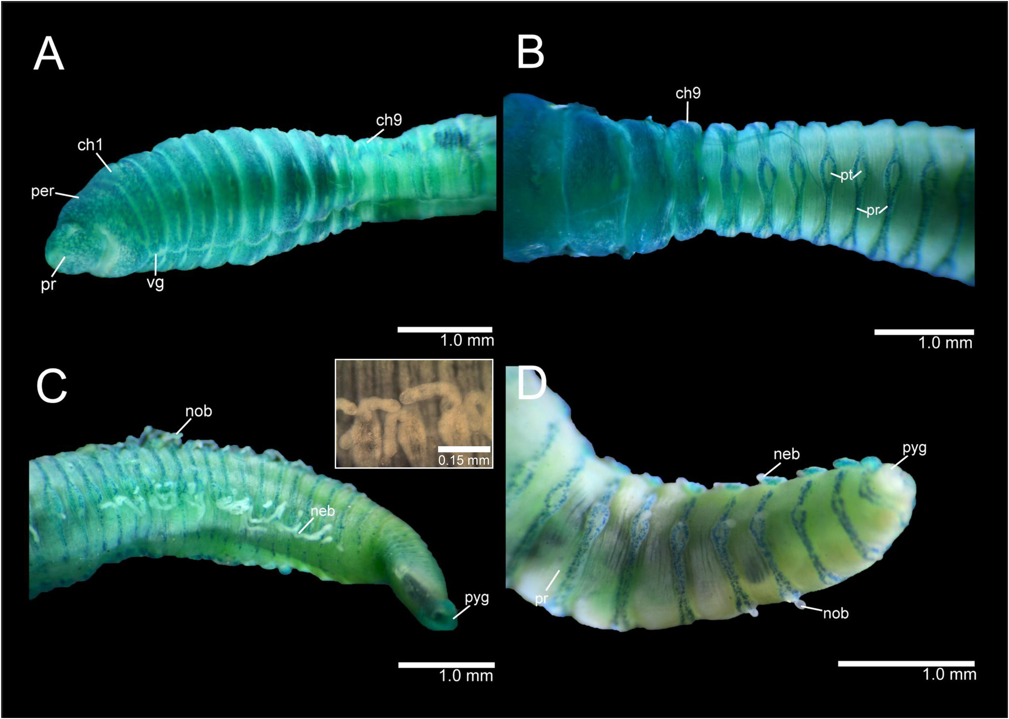
Description. All specimens with female external characteristics; holotype complete, 22 mm long, 2.2 mm wide for 64 chaetigers. Additional specimens including paratypes ranging from 15–26 mm long, 1.0– 2.2 mm wide for 52–94 chaetigers. Body elongate, wider on mid-thoracic chaetigers, narrowing on abdomen; body rounded dorsally and with ventral groove beginning from chaetiger 1 and deeper on abdominal segments; lateral groove absent. Color in alcohol pale yellow to dark brown.
Prostomium shorter than peristomium, broadly rounded ( Figs 4A View FIGURE 4 ; 5A View FIGURE 5 ); eyespots absent, nuchal organ depression postero-laterally in junction between prostomium and peristomium ( Fig. 4A View FIGURE 4 ). Peristomium distinct from prostomium, longer than chaetiger 1, forming a complete ring ( Figs 4A, B View FIGURE 4 ; 5A View FIGURE 5 ). Proboscis not observed.
Thorax with nine segments, epithelium smooth, not distinctly biannulated ( Figs 4A, B View FIGURE 4 ; 5A View FIGURE 5 ); chaetigers 1–8 or 1–9 having two rows of 18–20 unilimbate capillaries each. Notopodia of chaetiger 9 with only capillaries or mix of capillaries and hooded hooks; when mix of chaetae present, capillaries on lower part and hooded hooks on superior part of bundle. Neuropodia of chaetiger 9 with only hooded hooks (holotype, about 20 hooded hooks), or with only capillaries, or mix of capillaries and hooded hooks. Genital spines absent. Genital pore between chaetigers 7/8 and 8/9 ( Fig. 4A View FIGURE 4 ).
Abdominal segments distinctly multiannulated throughout, wider than long and slightly shorter than thoracic segments ( Fig. 5B–D View FIGURE 5 ); short hooded hooks present throughout abdomen, 15–20 hooks per fascicle, decreasing in number posteriorly. Noto- and neuropodia with well-separated glandular tori ( Fig. 5C View FIGURE 5 ); notopodial tori dorso-lateral and neuropodial tori ventro-lateral; parapodial tori connected by parapodial ridge ( Fig. 5B View FIGURE 5 ). Hooded hooks similar on thorax and abdomen—with moderate shaft, hoods not extending beyond main fang, with distinct node on shaft and, 3–4 rows of teeth above main fang ( Fig. 4C, D View FIGURE 4 ).
Notopodial and neuropodial branchiae present on posterior abdomen ( Fig. 5C, D View FIGURE 5 ). Notopodial branchiae from chaetigers 32–40, single to multiple filaments (up to five) emerging from lower region of tori ( Fig. 5C, D View FIGURE 5 ). Neuropodial branchiae from chaetigers 35–45 with up to seven filaments emerging from upper region of parapodial tori; branchiae retractile, isolated filaments, not branched, present to posterior end; when retracted, leaving a bulbous parapodial tori ( Fig. 4D View FIGURE 4 ).
Pygidium simple, anus terminal with a nondescript saclike projection present in few specimens, likely due to preservation conditions.
Methyl Green Staining Pattern. Staining clearly distinguishing thoracic and abdominal segments ( Fig. 5A– D View FIGURE 5 ). Thoracic segments staining with a solid green, leaving intersegmental areas and ventral groove unstained as well as the nuchal organ depression ( Fig. 5A View FIGURE 5 ). Prostomium stained with distinct speckles ( Fig. 5A View FIGURE 5 ). Abdominal segments with distinct stain on parapodial tori and ridges, forming a complete circle whereas segmental region remained unstained ( Fig. 5B–D View FIGURE 5 ). Pygidium staining with a light green ( Fig. 5C View FIGURE 5 ).
Etymology. The combination of ‘multi’ and ‘branchiata’ reflects the presence of multiple branchiae on all abdominal tori, a unique feature of this species.
Remarks. The presence of branchiae on abdominal notopodial and neuropodial segments is a unique feature of Capitella multibranchiata sp. nov. Capitella singularis ( Fauvel, 1932) was also described as having branchial filaments on abdominal segments. Fauvel (1932) reported on one or two short branchial filaments from chaetiger 80, inserted on the inner end of the dorsal tori. Magalhães & Bailey-Brock (2012) identified a branchiate Capitella from Hawaii as C. singularis having branchiae from neuropodia instead of notopodia and discussed that the deformation of abdominal segments on preserved specimens may have led previous authors to misinterpret the origin of branchiae. Capitella multibranchiata sp. nov. is clearly distinct from C. singularis by the presence of branchiae on both noto- and neuropodia, thoracic chaetal formulae and types of chaetae, distinct MGSP on thoracic region and absence of a lateral groove.
Both studied populations of branchiate Capitella (in India by Fauvel 1932 and in Hawaii by Magalhães & Bailey-Brock 2012) were from shallow waters and included only specimens with external male morphology. Conversely, only specimens with external female morphology of Capitella multibranchiata sp. nov. were collected. Dissections did not reveal any internal male anatomy. All branchiate Capitella have a distinct peristomial ring and abdominal tori taking up green stain and forming a ring around the segments.
Distribution. Type locality is Monterey Bay, off California, U.S. in 3,100 m.
| R |
Departamento de Geologia, Universidad de Chile |
| V |
Royal British Columbia Museum - Herbarium |
No known copyright restrictions apply. See Agosti, D., Egloff, W., 2009. Taxonomic information exchange and copyright: the Plazi approach. BMC Research Notes 2009, 2:53 for further explanation.
|
Kingdom |
|
|
Phylum |
|
|
Class |
|
|
Family |
|
|
Genus |