
Haplosyllis cf. spongicola
|
publication ID |
https://doi.org/ 10.11646/zootaxa.2552.1.1 |
|
persistent identifier |
https://treatment.plazi.org/id/03A487A3-FFA1-FF8B-2D99-FDBEB436FD05 |
|
treatment provided by |
Felipe |
|
scientific name |
Haplosyllis cf. spongicola |
| status |
|
Haplosyllis cf. spongicola View in CoL
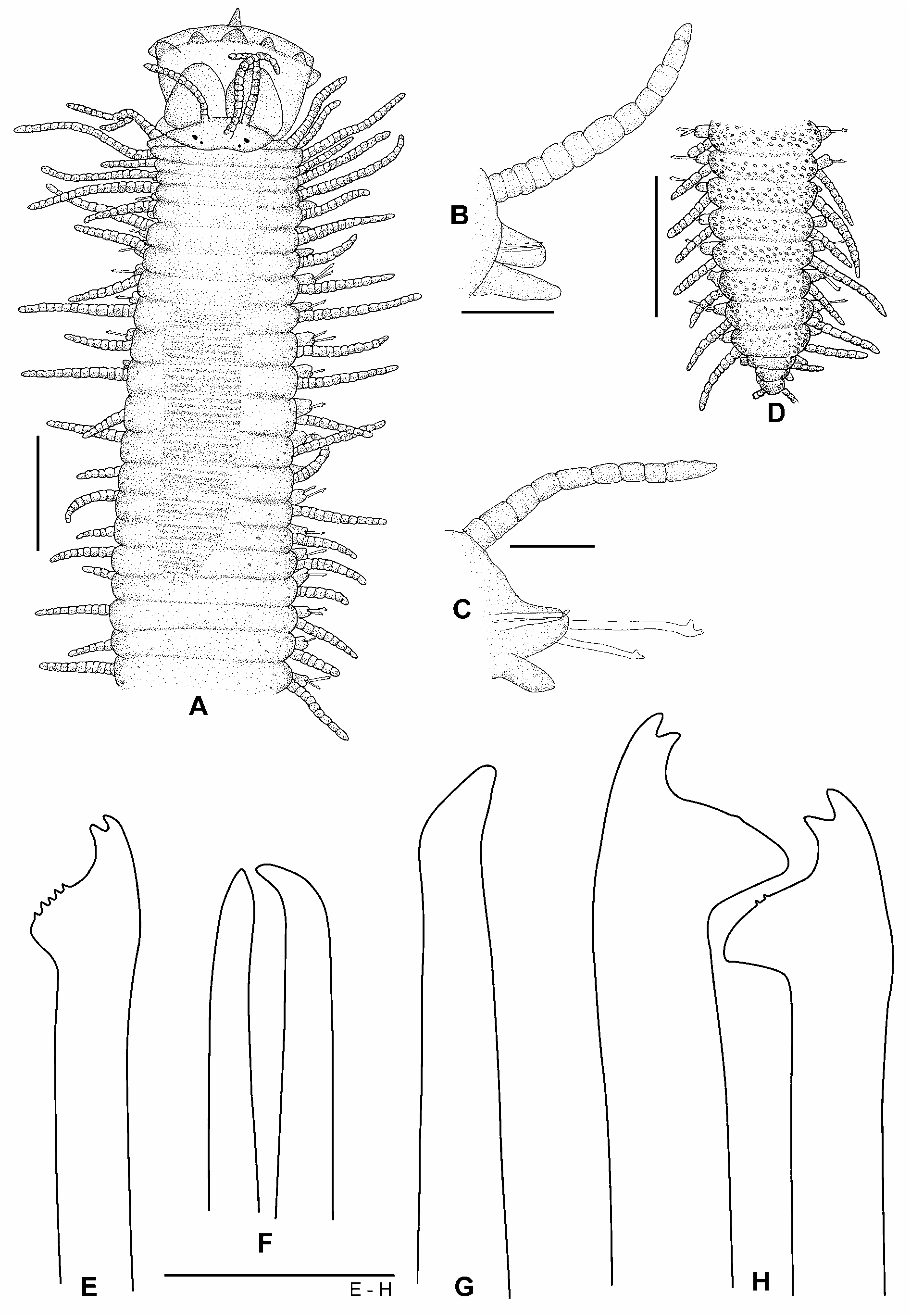
Figs 16A–H View FIGURE 16 , 17A–F View FIGURE 17
Examined material. AUSTRALIA. WESTERN AUSTRALIA: 3 specimens AM W36565 (plus 2 specimens mounted for SEM), off Jetty adjacent to Fisheries Hut, Beacon Island , 28º25'30''S 113º47'E, 12 m, dead coral, plate-like Acropora montipora , 23 May 1994 GoogleMaps . 1 specimen AM W36511, Goss Passage, Beacon Island , 28°25'30''S 113°47'E, 8 m, dead plates of Acropora , covered in coralline algae, coll. P.A. Hutchings, 19 May 1994 GoogleMaps . 9 specimens AM W36566, of south end of Long Island, Beacon Island , 28°28'48''S 113°46'18''E, 5 m, dead coral substrate covered in coralline algae, coll. P.A. Hutchings, 25 May 1994 GoogleMaps . NEW SOUTH WALES: 1 specimen AM W 36567, 100 m south of Split Solitary Island , 30º15'S 153º10'30''E, 16 m, sponge attached to rocky bottom, coll. R. Springthorpe, 23 June 1992 GoogleMaps . 1 specimen AM W36569, south-west side of South Solitary Island, Jervis Bay , 30º12'S 153º16'E, 18 m, coral rubble, coll. R. Springthorpe, 24 June 1992 GoogleMaps . 2 specimens AM W36568, Elizabeth Reef , south-west outer slope, 29°57'42"S 159°02'48"E, coll. P.A. Hutchings, 11 December 1987 GoogleMaps .
Description. Body robust, widest at proventricular segments, gradually tapering towards posterior end ( Fig. 17A View FIGURE 17 ); small to long-sized, 5–12 mm long, for 46–80 segments; pale yellow when preserved. Small, round dorso-lateral granules present, more abundant in posterior than in anterior and median regions ( Fig. 16A, D View FIGURE 16 ). Prostomium subpentagonal, wider than longer, with two pairs of small red eyes in trapezoidal arrangement ( Fig. 16A View FIGURE 16 ). Median antennae inserted on middle of prostomium, between posterior eyes (13–24 articles); lateral antennae inserted on anterior prostomial margin (10–21 articles). Palps longer than prostomium, broadly triangular, separated all along their length but in close contact at the base. Groups of cilia make up the sensory organs on ventral-upper side of palps ( Fig. 17C View FIGURE 17 ); nuchal organs not seen. Pharynx orange, broad, extending through 7–10 segments; with long triangular anterior tooth, crown of about 10 soft papillae and inner ring of cilia ( Fig. 17B View FIGURE 17 ). Ventral, upper side of papillae with groups of cilia making up the sensory organs. Proventricle cylindrical, long, extending through 7–14 segments, dark-brown, as broad as pharynx, with 33–61 muscle cell rows. Peristomium well defined, shorter than subsequent segments, difficult to see with pharynx everted. Dorsal tentacular cirri slightly longer (17–26 articles) than ventral ones. Antennae, tentacular cirri and dorsal cirri similar. Anterior dorsal cirri with well-defined length pattern, longest ones not exceeding body width: first cirri longer than remaining ones (18–35 articles), second short (9–15 articles), third and fourth long (11–16 and 12–17 articles, respectively), fifth short (9–12 articles), sixth long (14–24 articles). Thereafter, alternating with long (10–20 articles) and short (4–14 articles), gradually decreasing in length towards posterior end (2–10 articles) ( Fig. 16D View FIGURE 16 ). Ventral cirri digitiform, anterior ones broad, longer than parapodial lobes ( Fig. 16B View FIGURE 16 ); median and posterior ones gradually shorter, smaller than parapodial lobes ( Fig. 16C View FIGURE 16 ). Chaetae all bidentate, 2–3 per parapodia; anterior ones with LMF similar in length to SW, with numerous short spines on US of MF ( Figs 16E View FIGURE 16 ; 17D–E View FIGURE 17 ). Median and posterior chaetae with LMF longer than SW; MJP short and curved; US of MF with few, short denticles ( Figs 16H View FIGURE 16 , 17F View FIGURE 17 ). Apical teeth equal in length, with wide angle. Aciculae straight or with upward-directed curved tip; two in anterior and median parapodia ( Fig. 16F View FIGURE 16 ) one (sometimes two) in posterior parapodia ( Fig. 16G View FIGURE 16 ). Pygidium with two long cirri (about 10 articles).
Reproduction. Haplosyllis cf. spongicola reproduces by acephalous stolons; two female specimens (AM W36565) were observed in reproductive stage, with the last 9 (of a total of 46 body segments) with pairs of black parapodial ocular spots, capillary (i.e. swimming) chaetae, and 4–5 chaetae similarly to anterior ones. These reproductive segments, as well as the normal ones up to proventricular region contained numerous oocytes.
Remarks. The Australian specimens resemble the Mediterranean H. spongicola in chaetal characteristics and body shape. However, their cirri are much shorter (not longer than body width), with a marked alternation length pattern type (long and short), while Mediterranean species have longer cirri, all similar in size. Moreover, type of H. spongicola lack dorsal granules and has aciculae with curved tip bent at 90º. Therefore, the Australian specimens are here referred as Haplosyllis cf. spongicola , and future molecular studies seems to be the only way try to solve the identity of this morphotype.
Haplosyllis cf. spongicola most resembles Haplosyllis imajimai sp. nov., but has short, curved MJP (long, straight in the latter). Additionally, H. imajimai sp. nov., has a smaller, strongly curved acicula, long cirri and a long, robust body.
Distribution. AUSTRALIA (Western Australia and New South Wales)

| AM |
Australian Museum |
| R |
Departamento de Geologia, Universidad de Chile |
No known copyright restrictions apply. See Agosti, D., Egloff, W., 2009. Taxonomic information exchange and copyright: the Plazi approach. BMC Research Notes 2009, 2:53 for further explanation.
|
Kingdom |
|
|
Phylum |
|
|
Class |
|
|
Order |
|
|
Family |
|
|
Genus |