
Hydropsyche columnata Martynov 1931
|
publication ID |
https://doi.org/10.11646/zootaxa.4374.1.1 |
|
publication LSID |
lsid:zoobank.org:pub:6DDFE314-126F-4C82-936F-33C1C28EAC6E |
|
DOI |
https://doi.org/10.5281/zenodo.5987718 |
|
persistent identifier |
https://treatment.plazi.org/id/03A5866E-C119-7E25-D4E9-FA823C37F9D6 |
|
treatment provided by |
Plazi |
|
scientific name |
Hydropsyche columnata Martynov 1931 |
| status |
|
Hydropsyche columnata Martynov 1931
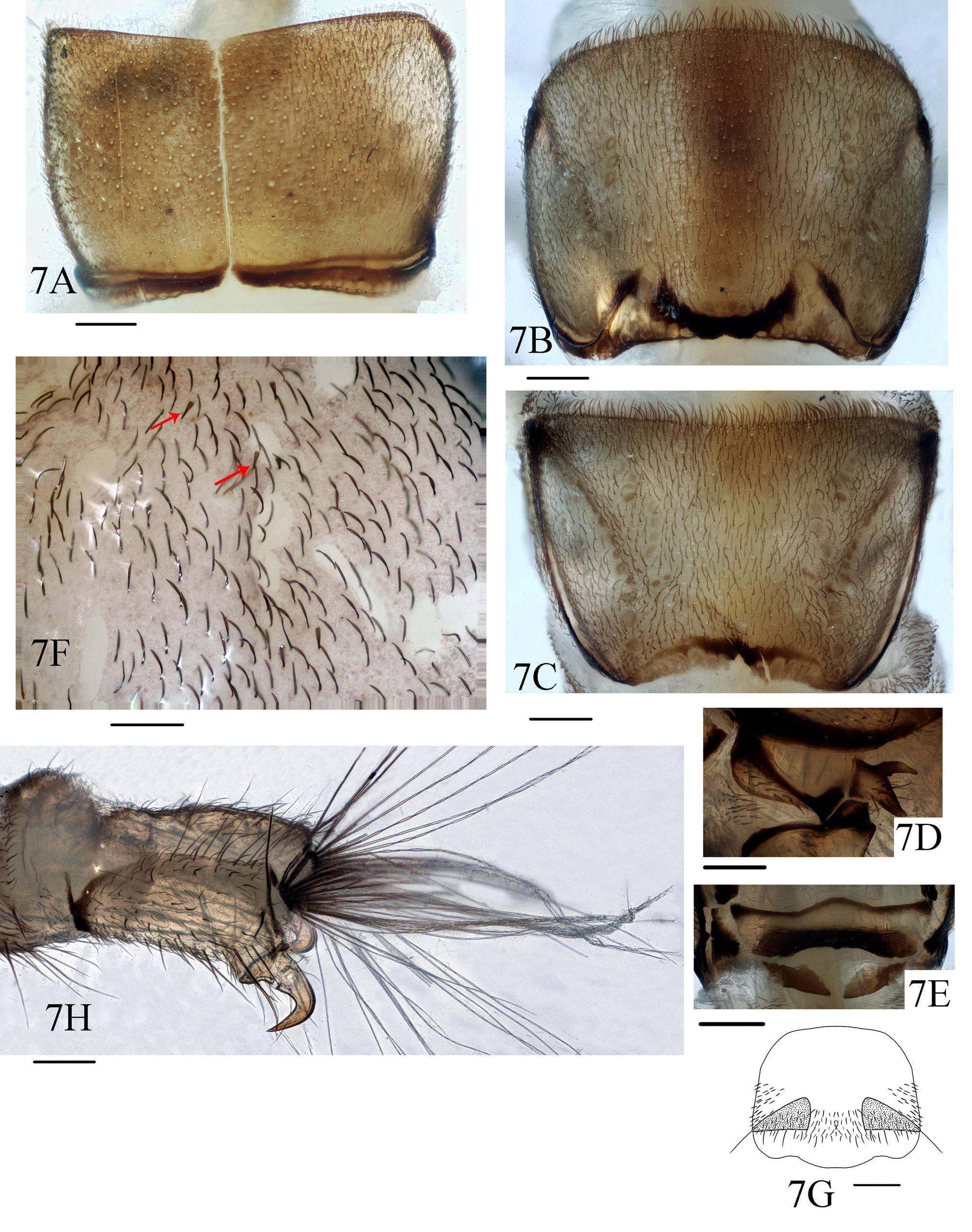
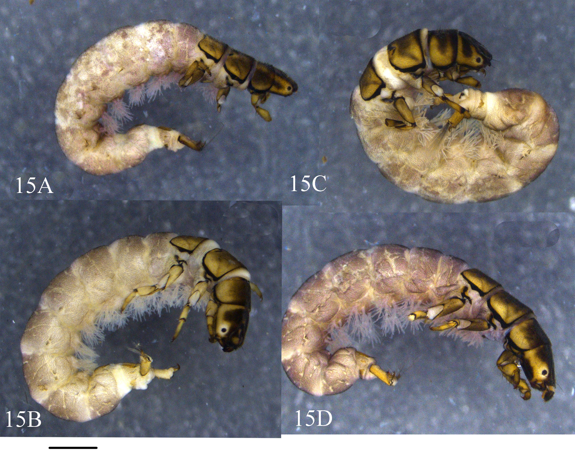
( Figs. 6–8 View FIGURE 6 View FIGURE 7 View FIGURE 8 , 15 View FIGURE 15 )
Description. Larvae (5th instar). Body length 10–15 mm (n = 5). Head. Head capsule ( Fig. 6A View FIGURE 6 ) subquadrate in dorsal view, 1.3 mm long, 1.1 mm wide at midlength. Overall coloration dark brown, with 3 yellow patches longitudinally arranged in frontoclypeal apotome in dorsal view, of which anterior one transverse and largest, middle and posterior ones relatively small and oval; in dorsal view parietals with oblique yellow stripes along posterior margins; in lateral view each stripe and yellow area around each eye fused into U-shaped patch collectively resembling shoulders, neck, and head of bird; muscle scars indistinct in postgenae ( Fig. 6C View FIGURE 6 ). Eyes oval, black. Frontoclypeal apotome ogival, with many protuberances on straight anterior margin. Dorsum of head with hair-like setae and semi-transparent truncate peg-like setae Labrum yellow, with anterior margin round and hair brush at each anterolateral corner. Mandibles ( Fig. 6D View FIGURE 6 ) yellow, triangular in ventral view, each with 5 apical teeth; left mandible with about 20 setae at basal half of lateral side and with brush of about dozen stiff hairs at middle of inner side; right mandible with more than 20 setae at basal half of lateral margin. Maxillae each with cardo transverse, sub-rectangular, dark brown ( Figs 6B,6F View FIGURE 6 ); stipes transverse and triangular, palpifer with about 22 setae, palp 5-segmented, maxillary lobe adjacent to mesal margin of palp ( Fig. 6G View FIGURE 6 ). Labium ( Fig. 6G View FIGURE 6 ) triangular and short in ventral view. Submentum ( Fig. 6E View FIGURE 6 ) with basal 2/3rds somewhat trapezoidal and distal 1/3rd incised mesally, forming two lobes; posterior margin slightly convex posteriorly; each anterolateral corner with 3–5 long, strong setae and many short setae; and each posterolateral corner bearing stout, brown setae. Anterior ventral apotome ( Fig. 6B View FIGURE 6 ) nearly triangular, brown, with anterior border slightly concave and anterolateral angles rounded.
Ventral ecdysial line more than twice as long as anterior ventral apotome. Posterior ventral apotome tiny, triangular, brown. Thorax. Pronotum ( Fig. 7A View FIGURE 7 ) subrectangular and brown in dorsal view, covered with slender, short, black hair -like setae; short, transparent truncate peg-like setae; and acuminate peg-like setae. Three types of setae alternatively arranged along anterior margin, with 2 types of peg-like setae shorter than hair-like setae. Prosternal plate ( Fig. 7E View FIGURE 7 ) dark brown in posterior half, with its width about 4 times its length, anterior margin sinuous, convex anteromesally, and posterior margin slightly concave; lateral piece and median piece behind each end of posterior prosternal sclerite fused into rhombic piece on each side, each with anterolateral and posteromesal corners acute.
Mesonotum ( Fig. 7B View FIGURE 7 ) mostly light-colored, U-shaped posterior mark black; pair of lateral fusiform marks dark brown, each with posterior half suddenly narrowed, extending to posterolateral corner of tergite; muscle scars distinct, darker than background, longitudinally arranged; dense hair-like setae and sparse truncate peg-like setae scattered over whole surface of notum. Metanotum ( Fig. 7C View FIGURE 7 ) lighter than pro- and mesonota in color, with posterior margin slightly incised; anterior margin with many hair-like setae and few truncate or acuminate peg-like setae; muscle scars darker than background, longitudinally arranged.
Legs. Legs yellow to brown. Forelegs much more stout than mid- and hind legs ( Figs. 8A–8C View FIGURE 8 ). Each foretrochantin bifurcate ( Fig. 7D View FIGURE 7 ), two branches divergent at angle of about 70°, with about 10 setae. Forecoxae somewhat conical; those of mid- and hind legs cylindrical. Trochanters triangular and 2-segmented, basal segment subtriangular and shorter than subtriangular apical segment, ventral trochanteral brush present on forelegs, with more than 30 spike-like setae. Forefemora in lateral view pentagonal, each with dorsal margin angulate and lower margin possessing dense long-slender setae and spike-like setae, mid- and hind femora cylindrical, each with lower margin bearing spike-like setae. Each tibia and tarsus more slender than its femur. Tarsal claw of each foreleg slightly shorter than its tarsus, conical, slightly curved downwards to acute apex, basal seta slender; claws of midlegs each about 2/3rds as long as its tarsus, its base thick and then suddenly narrowed to acute apex, slightly curved downwards, basal seta stout; claws of hind legs similar to those of midlegs in shape, but each about half as long as its tarsus, its basal seta stout.
Abdomen. Abdominal segments I–IX densely covered with hair-like setae and sparsely scattered and flattened scale -hair setae ( Fig. 7F View FIGURE 7 , arrows). Dorsolateral regions (sa 2) of segments I–VII each with 3 long setae. Tergum IX with pair of triangular tergites covered by hair-like setae and each with about 4 long-slender setae at posterior margin ( Fig. 7G View FIGURE 7 ); sterna of segments VIII–IX each with pair of ventral plates, covered with spike-like setae; apical margins of ventral plates on sternum VIII each with indentation. Anal prolegs slightly sclerotized, with anal claws yellow, hook-like, angled 90° ( Fig. 7H View FIGURE 7 ).
Diagnosis. The larva of H. columnata is very similar to the larva of H. trifora in the cephalic color patterns in dorsal view, and the overall shape and the color of pro-, meso-, and metanota, but differs from the latter in that (1) the yellow mark on the anterior margin of the frontoclypeal apotome is stripe-like and transverse on H. columnata , but it is oval on H. trifora ; (2) translucent acuminate peg-like setae occur on the genae of H. columnata , but stout, brown, truncate peg-like setae occur on those of the latter; (3) the anteromesal incision of the submentum is deeper on H. columnata than on that of H. trifora ; (4) the two branches of each foretrochantin are divergent about 90° on H. columnata , but divergent about 70° in H. trifora .
Materials examined. CHINA, Zhe-jiang Province , An-ji County, Hu-zhou : 5 larvae, Xiao-feng Town, Baiyang, 30.68°N, 119.50°E, alt. 139 m, 6 May 2015 GoogleMaps ; 14 larvae, Di-pu Town , Zi-xi-du, 30.67°N, 119.68°E, alt. 15 m, 6 May 2015 GoogleMaps ; 49 larvae, Di-pu Town , Lu-xi, 30.72°N, 119.80°E, alt. 85 m, 6 May 2015 GoogleMaps ; 3 larvae, Zhang-wu Town , Da-wu, 30.74°N, 119.50°E, alt. 205 m, 7 May 2015 GoogleMaps ; 12 larvae, Tian-jin-tang, 30.53°N, 119.25°E, alt. 478 m, 7 May 2015; 3 larvae, Bao-wu town , Shi-ling, 30.45°N, 119.50°E, alt. 375 m, 8 May 2015 GoogleMaps ; 13 larvae, Zhang-cun town , Chang-tan, 30.44°N, 119.39°E, alt. 213 m, 8 May 2015 GoogleMaps ; 5 larvae, Hang-gai Town , Leng-keng, 30.51°N, 119.37°E, alt. 421 m, 8 May 2015 GoogleMaps ; 3 males, Tian-huang-ping Town, Chi-wu-li, 30.50°N, 119.60°E, alt. 236 m, 12 May 2015. Lin-an, Hu-zhou: 387 larvae, Shi-bao-wu, 30.22°N, 119.60°E, alt. 114 m, 5 May 2015. Hangzhou, Yu-hang District : 112 larvae, Pai-lou-shan-cun, 30.45°N, 119.83°E, alt. 62 m, 11 May 2015. All materials were collected by Ji-hua Xu, Si-wen He, & Shu-zhao Gao GoogleMaps .
Known distribution. China (Bei-jing, Guang-dong, Gui-zhou, He-nan, Jiang-xi, Shaan-xi, Si-chuan, Yun-nan, Zhe-jiang).


No known copyright restrictions apply. See Agosti, D., Egloff, W., 2009. Taxonomic information exchange and copyright: the Plazi approach. BMC Research Notes 2009, 2:53 for further explanation.
|
Kingdom |
|
|
Phylum |
|
|
Class |
|
|
Order |
|
|
Family |
|
|
Genus |