
Amythas membranifera Benham, 1921
|
publication ID |
https://doi.org/ 10.5852/ejt.2021.733.1227 |
|
publication LSID |
lsid:zoobank.org:pub:1AAE62AF-ABD9-4930-B1DE-2C05F66BEC4A |
|
DOI |
https://doi.org/10.5281/zenodo.4531811 |
|
persistent identifier |
https://treatment.plazi.org/id/03A8045E-F710-FFDD-517B-F465FEEC6E07 |
|
treatment provided by |
Plazi |
|
scientific name |
Amythas membranifera Benham, 1921 |
| status |
|
Amythas membranifera Benham, 1921 View in CoL
Figs 4 View Fig , 5 View Fig A–E, J–L
Amythas membranifera Benham, 1921: 102–105 View in CoL , pl. 10, figs 124–132.
Amythas membranifera View in CoL – Monro 1939: 141–142, fig. 24.
Material examined
ANTARCTICA • 1 ♂ (BL = 55 mm, 20 AU); Terra Nova Bay ; -74.67597º S, 164.24592º E; depth 400 m; 30 Jan. 2014; Schiaparelli leg.; station: DR5; XXIX PNRA expedition (2013-2014); MNA- 07940 GoogleMaps • 1 ♂ (BL = 55 mm, 19 AU); same collection data as for preceding; MNA-07939 GoogleMaps • 1 ♂ (BL = 13 mm, 17 AU); same collection data as for preceding; MNA-07935 GoogleMaps • 1 ♂ (BL = 45 mm, 18 AU); same collection data as for preceding; MNA-07934 GoogleMaps • 1 spec., undetermined sex; Princess Elizabeth Land ; -67.05000º, 74.48333º; depth 437 m; station: 103; BMNH 1941.3.3.126–127 .
Description
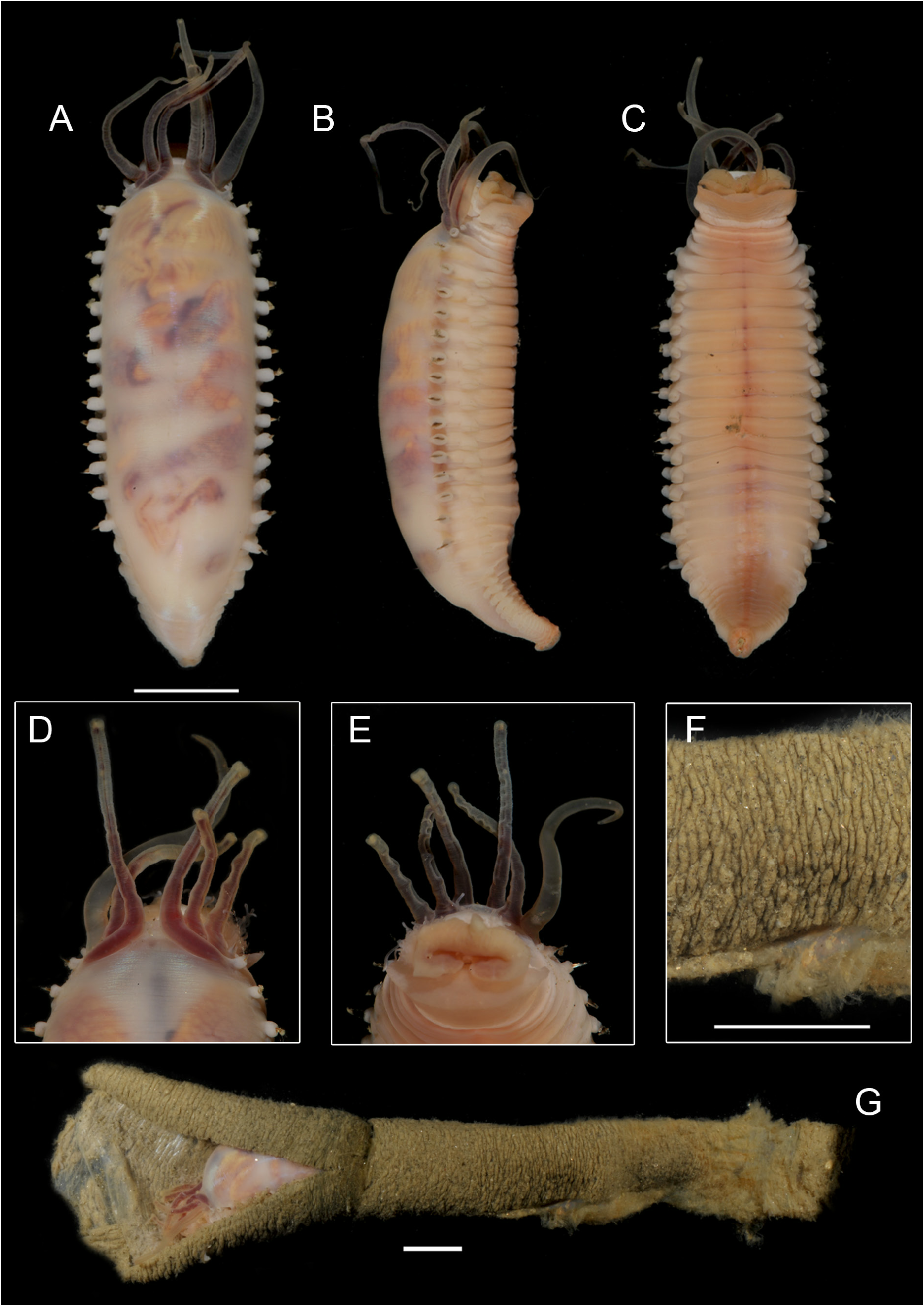
BODY. Body ( Fig. 4 View Fig A–C) short and thick. Length 13–55 mm, width 9–20 mm; girth of body spindleshaped, increasing to the middle of thorax, decreasing thereafter.
PROSTOMIUM. Prostomium trilobed, middle lobe anteriorly broadly rounded; any additional structures (nuchal organs, ridges, horns etc.) absent.
BUCCAL TENTACLES. Buccal tentacles numerous, short, smooth, attached behind a large-folded membrane. Lower lip broad, embracing mouth laterally and slightly latero-dorsally, covered by warts. Paleae totally absent.
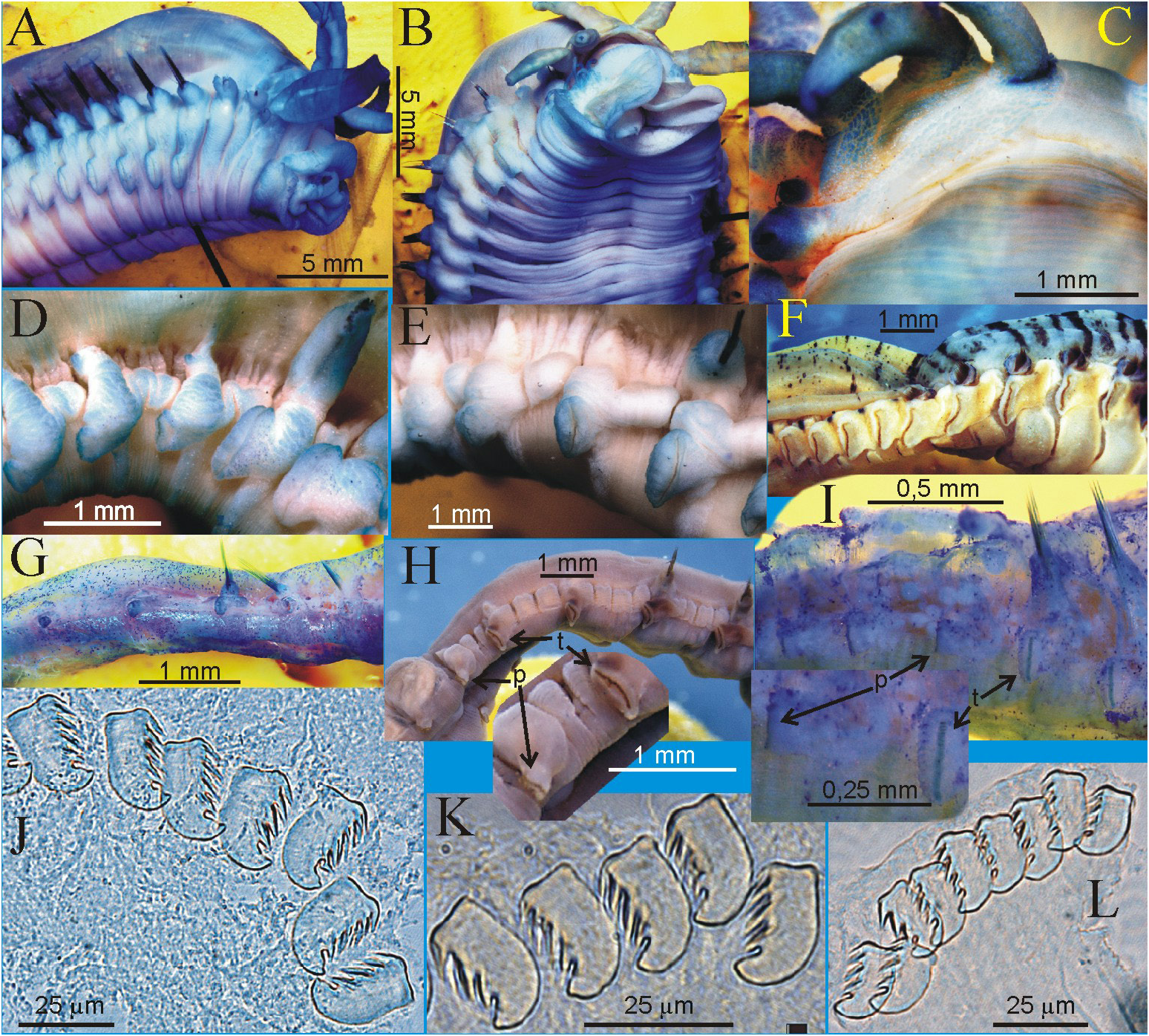
BRANCHIAE. Three pairs of branchiae, the outermost one originating from TC1, the innermost one from TC2, and the middle one from S2 ( Fig. 5C View Fig ). Branchostyles cirriform, short, organized in a straight line and showing a wide middle gap. Branchostyles and branchophores covered with warts. There are no visible nephridial papillae or nephropores.
NOTOPODIA AND NEUROPODIA. 17 TC, notopodia with capillary chaetae from S3; anterior notopodia small, increasing in size from first to third pair; elevated or modified notopodia absent; 14 TU, all thoracic neuropodia well developed, with almost equal size, middle ones slightly bigger than anterior and posterior ones. Ventral shields distinct anteriorly and completely disappearing before the end of thorax. Subdivision of thorax absent. 17–20 AU; shape of neuropodia gradually changing throughout in most specimens, from tori to pinnula with marked size reduction on the thorax/abdomen transition; uncini at the margin of neuropodia. All neuropodia without cirri; typically rudimental abdominal notopodia absent.
UNCINI. Uncini pectinate ( Fig. 5 View Fig J–L) with 4 teeth in double row, similar from TU1 to last AU; prow absent.
PYGIDIUM. Anal cirri absent.
TUBE ( Fig. 4 View Fig F–G). Tubes of this species appear to be formed by progressive additions of small quantities of homogeneous fine-grained sediment to form an irregular banded pattern ( Fig. 4F View Fig ). The interior part of
the tube has a homogeneous and transparent membranous lining that isolates the body of the worm from the outer layer and that can easily be peeled off from it ( Fig. 4G View Fig ).
Distribution
This species was reported from fjords along the West Antarctic Peninsula (WAP) ( Grange & Smith 2013) and in several stations from the Ross Sea ( NZ NIWA “IPY-CAML” Voyage TAN0802; records retrieved from GBIF, last accession 2020 Feb. 20).
Remarks
Benham in the description of the new genus Amythas , stated that there were no buccal tentacles and that these were replaced by a folded membrane ( Benham 1921). Hartman (1966) and Fauchald (1977) followed Benham.As it has already been stated by Monro (1939), buccal tentacles are present in Amythas as in all other Ampharetidae . The whole construction of buccal tentacles shows perfect resemblance to Terebellidae Johnston, 1846 and, at the same time, to Ampharetidae with an everted pharynx (see Jirkov 2016: fig. 3) which provides an indication of their homology and prostomial origin. The position of the uncini at the margin of the neuropodia was found to vary according to the individuals (e.g., Fig. 5 View Fig D–E). The smallest specimen examined (MNA-07935) has small rudimental abdominal notopodia and AU neuropodia more pinnuli-like ( Fig. 5D View Fig ). The morphology of the tube of this species was documented and described to occur in extant Sabellidae as well as in the ichnofossil Caprascolex antarcticus Schweitzer et al., 2005 from the Antarctic Eocene of La Meseta formation (Seymour Island, Antarctica) ( Schweitzer et al. 2005: figs 3b, 4). In situ images of tubes of this species are available in Grange & Smith (2013: fig. 2c).
| BMNH |
United Kingdom, London, The Natural History Museum [formerly British Museum (Natural History)] |
No known copyright restrictions apply. See Agosti, D., Egloff, W., 2009. Taxonomic information exchange and copyright: the Plazi approach. BMC Research Notes 2009, 2:53 for further explanation.
|
Kingdom |
|
|
Phylum |
|
|
Class |
|
|
Order |
|
|
Family |
|
|
Genus |
Amythas membranifera Benham, 1921
| Schiaparelli, Stefano & Jirkov, Igor A. 2021 |
Amythas membranifera
| Monro C. C. A. 1939: 141 |
Amythas membranifera
| Benham W. B. 1921: 105 |