
Petta pusilla Malmgren, 1866
|
publication ID |
https://doi.org/ 10.11646/zootaxa.4614.2.3 |
|
publication LSID |
lsid:zoobank.org:pub:3053533C-BDDE-4321-95B2-D557F3CF048D |
|
persistent identifier |
https://treatment.plazi.org/id/03A887E7-D42D-A033-60CE-DE743697F87B |
|
treatment provided by |
Plazi |
|
scientific name |
Petta pusilla Malmgren, 1866 |
| status |
|
Petta pusilla Malmgren, 1866 View in CoL
Figs 2–5 View FIGURE 2 View FIGURE 3 View FIGURE 4 View FIGURE 5 , Table 2 View TABLE 2
Petta pusilla Malmgren, 1866: 361 View in CoL , Tab. XVIII fig. 43 (Gullmarsfjord, west coast of Sweden, type locality).
Pectinaria pusilla — Levinsen 1883: 155 (Northern Europe); Wollebaek 1912: 35, pl. III, fig. 1–8 (Western Norway); Crawshay 1912: 346 (description, English Channel); Dauvin et al. 2003: 85 (name only, English Channel).
Petta View in CoL pusilla— LoBianco 1893: 48 (Naples, Italy); Nilsson 1912: pl. III, fig. 3–4 (anatomy of nervous system, Kristineberg, Sweden); Fauvel 1914: 279, pl. XXV, fig. 22–26 (Azores, Portugal); Hessle 1917: 83–84 (West coast of Sweden); Fauvel 1927: 224 (description, Plymouth, Ireland, Azores, Irish Sea, Marseille, Naples, North Sea, Arctic Ocean); Nilsson 1928: 83–85, Fig. 30 (Arctic Ocean, Atlantic Ocean and Mediterranean Sea); Holthe 1977 (zoogeography, Atlantic-boreal); Holthe 1986: 27–28, Fig. 7 View FIGURE 7 , map 6 (description, Norway); Castelli & Valentini 1995: 52–53 (name only, Italy); Méndez & Cardell 1996: 138 (name only, Catalonia, Spain); Arvanitidis 2000: 95 (name in checklist only, Greece); Orrhage 2001 (anatomy of nervous system, west coast of Sweden); Simboura & Zenetos 2002: 100 (name only, Mediterranean Sea); García-Diez et al. 2005 (name only, Azores); Zaâbi et al. 2010: 11 (name only, north-east coast of Tunisia), Jirkov & Leontovich 2013: 220 (name in key, Arctic Ocean); Çinar et al. 2014: 751 (name in checklist, Turkey).
Material examined. Holotype, with tube, SMNH T361 View Materials ,Bohuslän,Gullmarsfjord, Sweden, 58º16.002´N 11º28.002´E, depth not stated, but apparently subtidal, coll. L. S. Lovén; AM W.50333 (donated by Gothenburg Natural History Museum ( GNM) Polychaeta 749) GoogleMaps , 2 specimens, Gullmarsfjord, Sweden, coll. July 1878; AM W.50334 (donated by GNM Polychaeta 14334), 1 specimen plus tube, Kattegatt , 56º52.65´N 12º13.422´E, 20 m, coll. Sep 2005. GoogleMaps
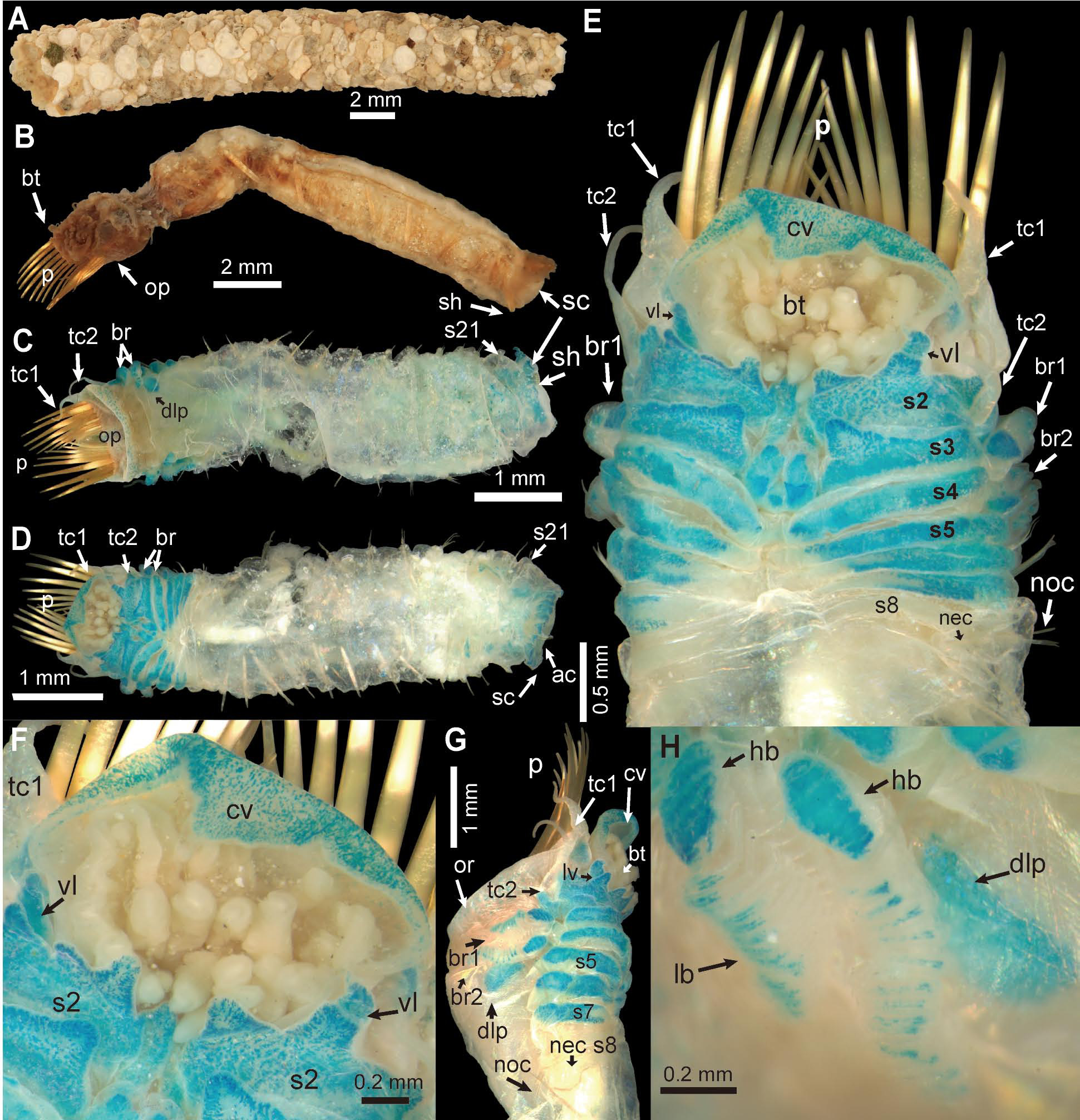
Description. Based on holotype and two specimens from Gullmarsfjord. Preserved specimens pale in colour. Body cylindrical, curved dorsally ( Fig. 3B, C View FIGURE 3 ). Body length 14.5–16.7 mm (15.2 mm), including paleae and scaphe, width at cephalic regions 2.8–3.2 mm (3.2 mm).
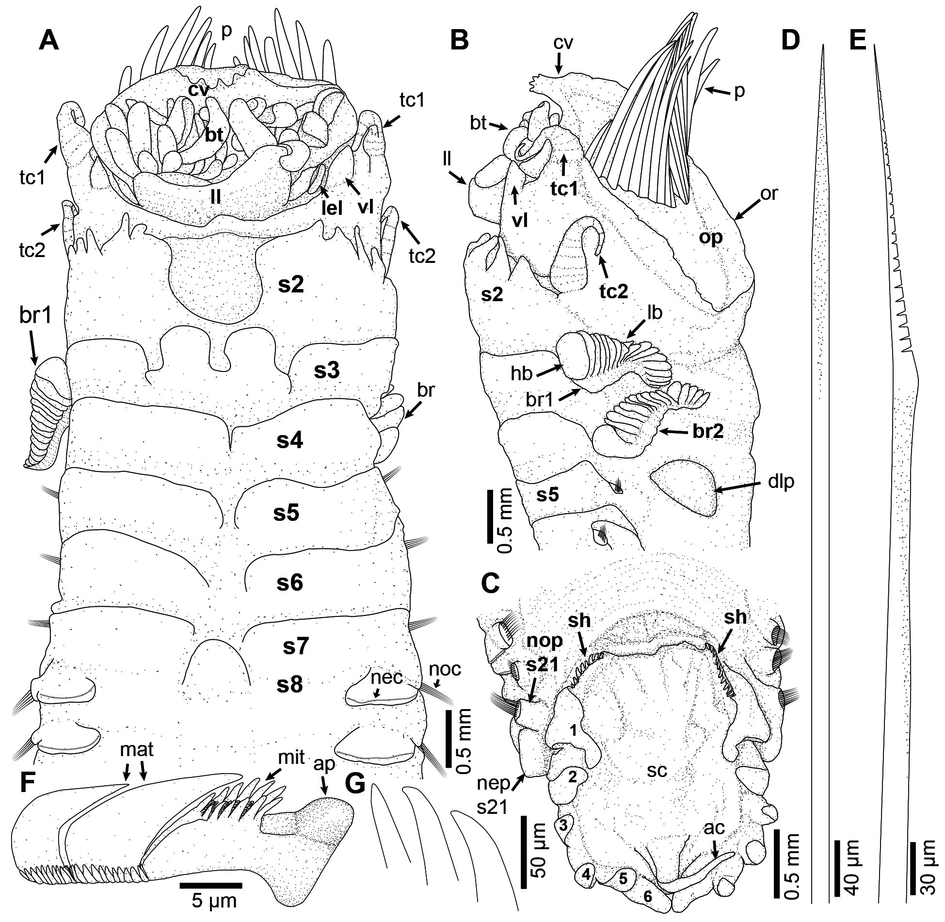
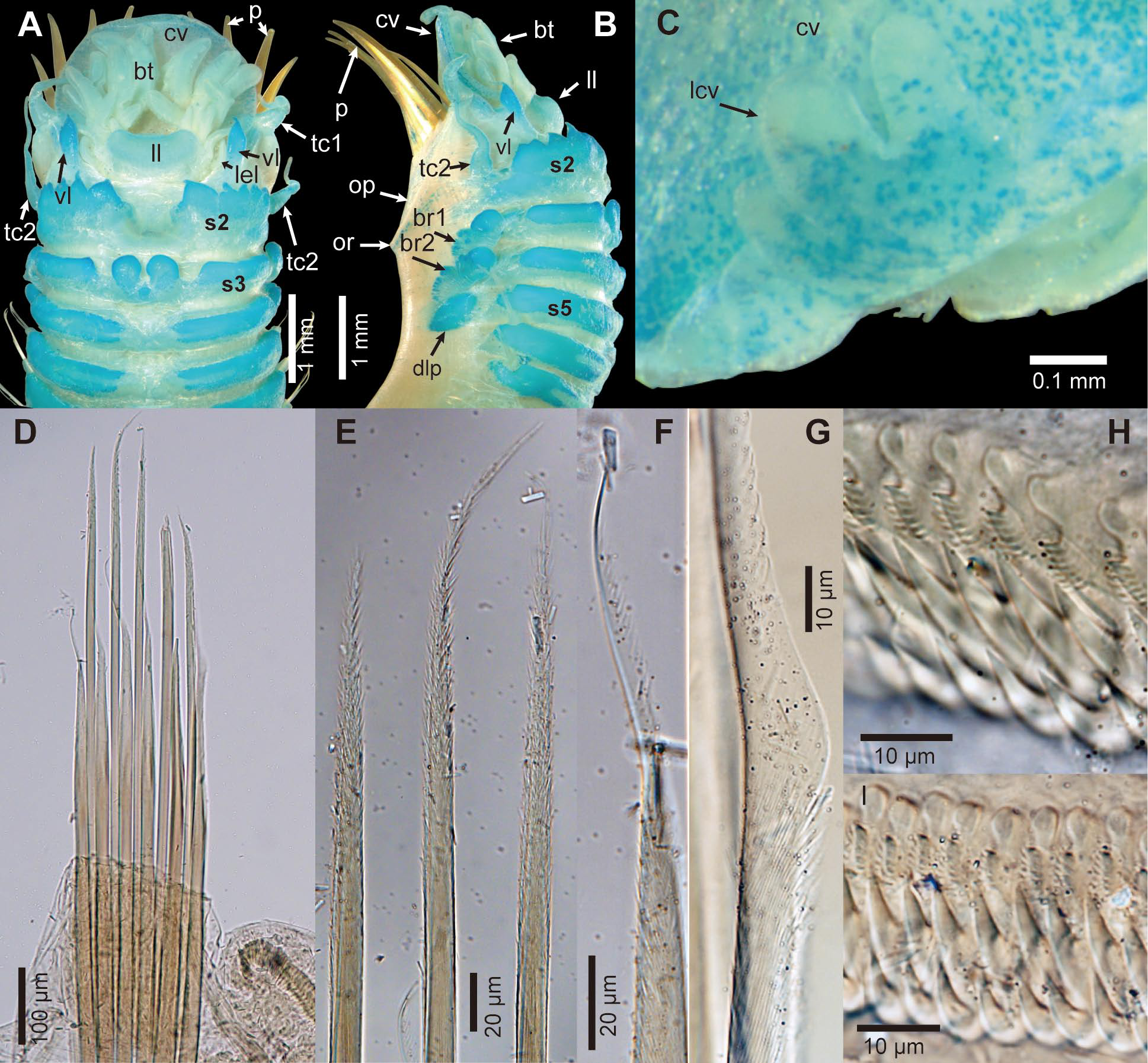
Cephalic veil leaf-shaped, free from operculum, with smooth lateral margins, distal margins smooth, except for a free and crenulated median extension with 3–4 (4) slightly distorted lappets (raised mounds) with crenulated margins ( Figs 2 View FIGURE 2 A–B; 4A, C–D; 5A–C). Pair of ventro-lateral ear-shaped lobes adjacent to dorsal side of cephalic veil ( Figs 2A View FIGURE 2 ; 4 View FIGURE 4 D–E; 5A). Buccal tentacles with deep longitudinal grooves, arising from around buccal cavity, posterior to cephalic veil ( Figs 2A View FIGURE 2 ; 4A View FIGURE 4 , C–D; 5A). Distinct broad ventral lobe (lower lip) between buccal cavity and segment 1 ( Figs 2A View FIGURE 2 ; 4A View FIGURE 4 , C–D; 5A).
Operculum semicircular; dorsal and lateral margins short and smooth; ventral margin (opercular ridge) with 10–11 (11) pairs of amber-coloured, stout notopodial paleae, curved dorsally, and ending in blunt tips ( Figs 2B View FIGURE 2 ; 3D View FIGURE 3 ; 5B View FIGURE 5 ).
First pair of tentacular cirri not extending beyond tips of paleae, slightly annulated with rounded cirri arising from inflated base between opercular margin and paleal ridge ( Figs 2 View FIGURE 2 A–B; 3D; 4A, C; 5B). Pair of sharp-tipped triangular ventral lappets present behind tentacular cirri, on segment 1, not covered by ventral lobes of segment 2 ( Figs 2 View FIGURE 2 A–B; 4A, C; 5A–B). Ventral region visible on segment 1 ( Figs 2A View FIGURE 2 ; 4A View FIGURE 4 ; 5A View FIGURE 5 ).
Second pair of tentacular cirri almost same length as first, but thinner and slightly annulated, its base more bulbous than that of 1 st pair of tentacular cirri, and slightly placed dorsally, on mid-lateral connecting ridge of segment 2 ( Figs 2 View FIGURE 2 A–B; 4C; 5A–B). Segment 2 with pair of broad and elongated ventro-lateral lobes separated from each other by a broad and deep mid-ventral groove; each lobe with 3–5 (left 5, right 4) continuous rows of triangular lappets ( Figs 2 View FIGURE 2 A–B; 4A, C; 5A–B).
Segments 3–4 with two pairs of almost equal sized comb-like branchiae, consisting of large basal hump and series of loose flat lamellae ( Figs 2B View FIGURE 2 ; 4B View FIGURE 4 ; 5B View FIGURE 5 ). First pair of branchiae on segment 3 inserted more ventrally than 2 nd pair on segment 4. Pair of dorso-lateral pads small and smooth, arising from dorsal side of notopodia on segment 5 ( Figs 2B View FIGURE 2 ; 3D View FIGURE 3 ; 5B View FIGURE 5 ).
Distinct ventral glandular lobes (pads) present on segments 2–7, becoming progressively more lateral and broader on segments 3–5 ( Figs 2A View FIGURE 2 ; 3C View FIGURE 3 ; 5 View FIGURE 5 A–B). Humps near branchiae absent ( Figs 2B View FIGURE 2 ; 4C View FIGURE 4 ; 5B View FIGURE 5 ). Segment 3 with a pair of smooth broad ventro-lateral lobes and a pair of mid-ventral lappets, separated by deep notches; ventro-lateral lobes without projection on ventral margin; mid-ventral lappets rounded, narrow about 1/4 width of ventro-lateral lobes, and more anterior than ventro-lateral lobes ( Figs 2A View FIGURE 2 ; 3C View FIGURE 3 ; 4A View FIGURE 4 ; 5A View FIGURE 5 ). Segments 4–6 with a pair of broad ventrolateral lobes separated from each other by a shallow median groove becoming progressively broader on segments 4–6. Segment 7 with a pair of broad ventro-lateral lobes separated from each other by a median swelling about 1/4 width of ventro-lateral lobes.
Notopodia of segment 1 with paleae, from segment 5 to 21 (17 pairs) notopodia with two rows of different chaetae. Anterior row bearing shorter chaetae with distal serrated wings, covered with numerous minute spines from below wing to about mid-basal portion of chaeta; posterior row with about 1.3 times longer capillary chaetae, straight and stout, tapering to acute tip, anterior surface covered with numerous spines from mid-length to tip ( Figs 2 View FIGURE 2 D–E; 3B–C; 5D–G). Neuropodia on segments 8–21 (14 pairs in total) with slightly raised torus bearing a transverse row of uncini; uncinus with a rounded anterior peg with a blunt tip embedded into torus, followed by several rows of minor teeth on a swelling and one longitudinal row of two major teeth, each covered with many small teeth basally ( Figs 2F View FIGURE 2 ; 5 View FIGURE 5 H–I). Neuropodia of segment 21 with enlarged posterior lobe ( Fig. 4 View FIGURE 4 H–I).
Scaphe long, ovoid, flattened dorsally, slightly separated from posterior segments. Lateral margins dorsally rolled, with six pairs of lobes; first pair largest and elongated, connected to dorsal margin of scaphe; posterior lobes narrow, triangular, almost same size; dorsal margin of scaphe smooth with two shallow notches on holotype ( Figs 2C View FIGURE 2 ; 3 View FIGURE 3 B–C; 4G–I). Anal flap vestigial with oblong swollen area distally bearing long anal cirrus ( Figs 2C View FIGURE 2 ; 4G View FIGURE 4 ). Anus located behind anal cirrus, between last pair of lateral lobes on scaphe. Scaphal hooks amber-coloured, 7–8 (8) pairs arising from both sides of dorsal margin of scaphe, with blunt tips slightly curved dorsally ( Figs 2G View FIGURE 2 ; 4 View FIGURE 4 F–G).
Tube slightly curved, robust, made of small gastropod shells ( Fig. 3A View FIGURE 3 ).
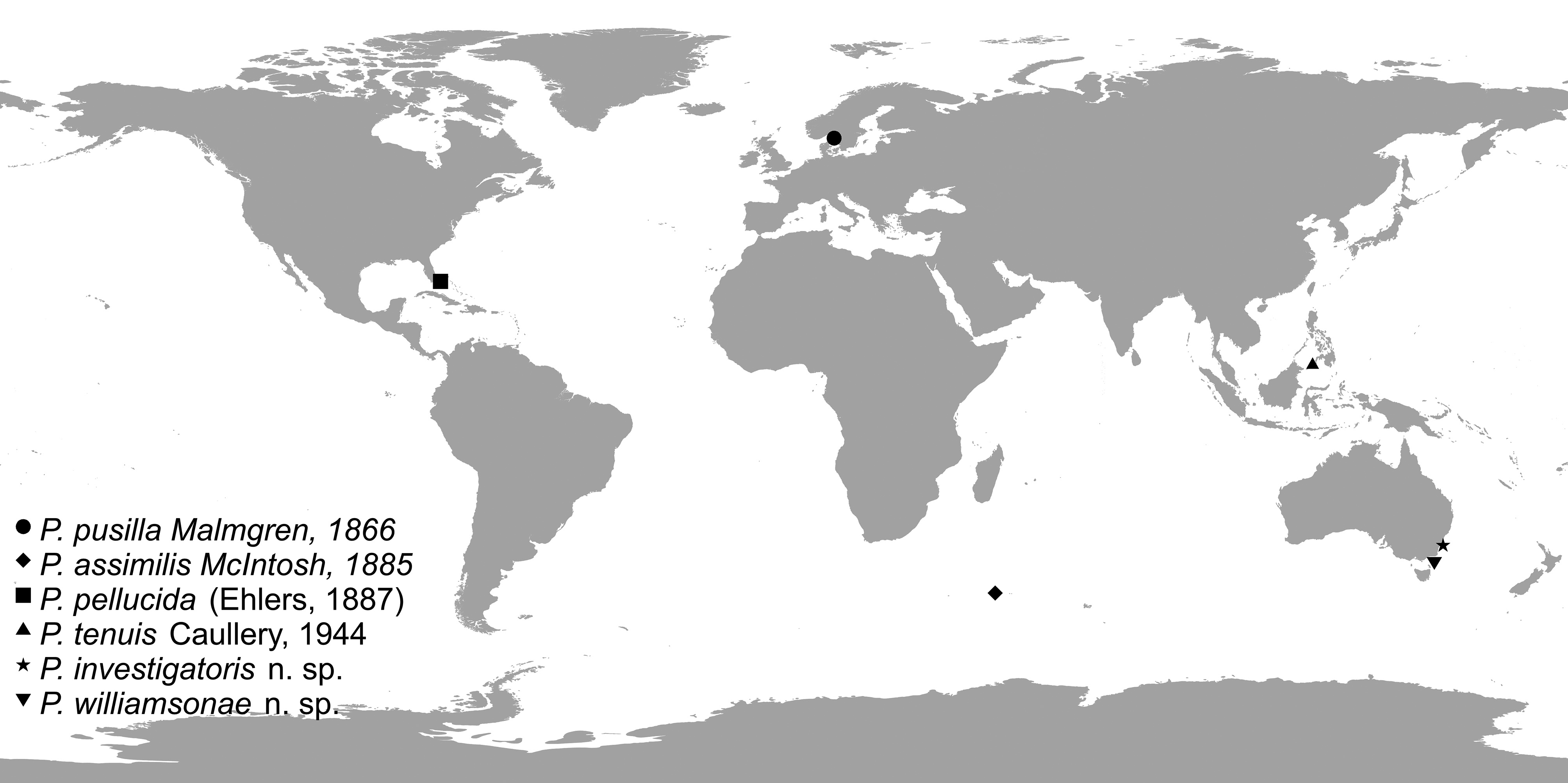
Distribution. Type locality Gullmarsfjord, Sweden ( Fig. 1 View FIGURE 1 ), Arctic Ocean: along Norwegian coast from Lofoten Islands to Swedish West Coast to northern part of Oresund, North Eastern Atlantic Ocean ( Faroe Islands, Irish Sea, Azores, the UK coasts) and Mediterranean Sea (coasts of France, Italy, Turkey, Greece, Tunisia, Spain). According to Jirkov & Leontovich (2013), this is a low boreal species, although only a few available records from northern areas suggest that this species is restricted to more southern regions.
Habitat. According to Holthe (1986), the species is found in Norwegian waters in sandy or mixed sediments, 15–200 m deep, while the record from 800 m in the Azores seems questionable.
Remarks. Considerable discrepancies in some morphological characters exist between our description of the holotype and the description of Malmgren (1866). For example, he reports smooth anterior tip of cephalic veil, while it actually bears 3–4 lappets. We also expanded the description with additional characters not mentioned in the original description, such as presence of broad ventral lobe (lower lip) between buccal cavity and segment 1, pair of pointed ventral lappets on segment 1, ventral region visible on segment 1, large basal hump on branchiae, a pair of dorso-lateral pads on segment 5, and uncini with a longitudinal row of two major teeth. Petta pusilla is distinguished from other species of this genus by having 3–4 lappets on anterior margin of cephalic veil, broad ventral lobe (lower lip) between buccal cavity and segment 1 and a pair of rounded mid-ventral lappets on segment 3 ( Table 2 View TABLE 2 ). Petta pusilla is similar to P. pellucida , P. tenuis and P. williamsonae n. sp. as all have smooth ventro-lateral lobes on segment 3 and a long anal cirrus. Petta pusilla differs from P. assimilis and P. investigatoris n. sp. because the last two species have a continuous row of lappets on ventro-lateral lobes of segment 3. Petta pusilla also differs from P. pellucida which has neurochaetae on segments 7–21, whereas P. pusilla has neurochaetae on segments 8–21.
Nilsson (1928) illustrated specimens of P. pusilla with three minute lappets on the anterior margin of cephalic veil and also a pair of dorso-lateral pads on segment 5, however the locality where his illustrated specimens came from is unclear, he reports a distribution in the Arctic Ocean, Atlantic Ocean and Mediterranean Sea. Nilsson (1928) believed that P. pusilla and P. pellucida from the Caribbean were synonymous, and the material from Mediterranean Sea, which has 12 pairs of paleae, and more spinose capillary chaetae, could be another species, but his material needs to be examined to confirm this. All records of this species from outside the type locality should be carefully checked against the expanded here re-description of the type as they may represent additional undescribed species.


No known copyright restrictions apply. See Agosti, D., Egloff, W., 2009. Taxonomic information exchange and copyright: the Plazi approach. BMC Research Notes 2009, 2:53 for further explanation.
|
Kingdom |
|
|
Phylum |
|
|
Class |
|
|
Order |
|
|
Family |
|
|
Genus |
Petta pusilla Malmgren, 1866
| Zhang, Jinghuai, Hutchings, Pat & Kupriyanova, Elena 2019 |
Pectinaria pusilla —
| Dauvin, J. - C. & Dewarumez, J. - M. & Gentil, F. 2003: 85 |
| Wollebaek, A. 1912: 35 |
| Crawshay, L. R. 1912: 346 |
| Levinsen, G. M. R. 1883: 155 |
Petta pusilla
| Malmgren, A. J. 1866: 361 |