
Perinereis latipalpa (Schmarda, 1861), 2019
|
publication ID |
https://doi.org/10.5252/zoosystema2019v41a24 |
|
publication LSID |
urn:lsid:zoobank.org:pub:9347D7C7-1D9D-4682-A9B9-BD7E11AF97B4 |
|
DOI |
https://doi.org/10.5281/zenodo.4439496 |
|
persistent identifier |
https://treatment.plazi.org/id/03B28785-F476-BE5C-A4DA-A4F8FDD171E0 |
|
treatment provided by |
Felipe |
|
scientific name |
Perinereis latipalpa (Schmarda, 1861) |
| status |
|
Perinereis latipalpa (Schmarda, 1861) View in CoL , reinst., n. comb.
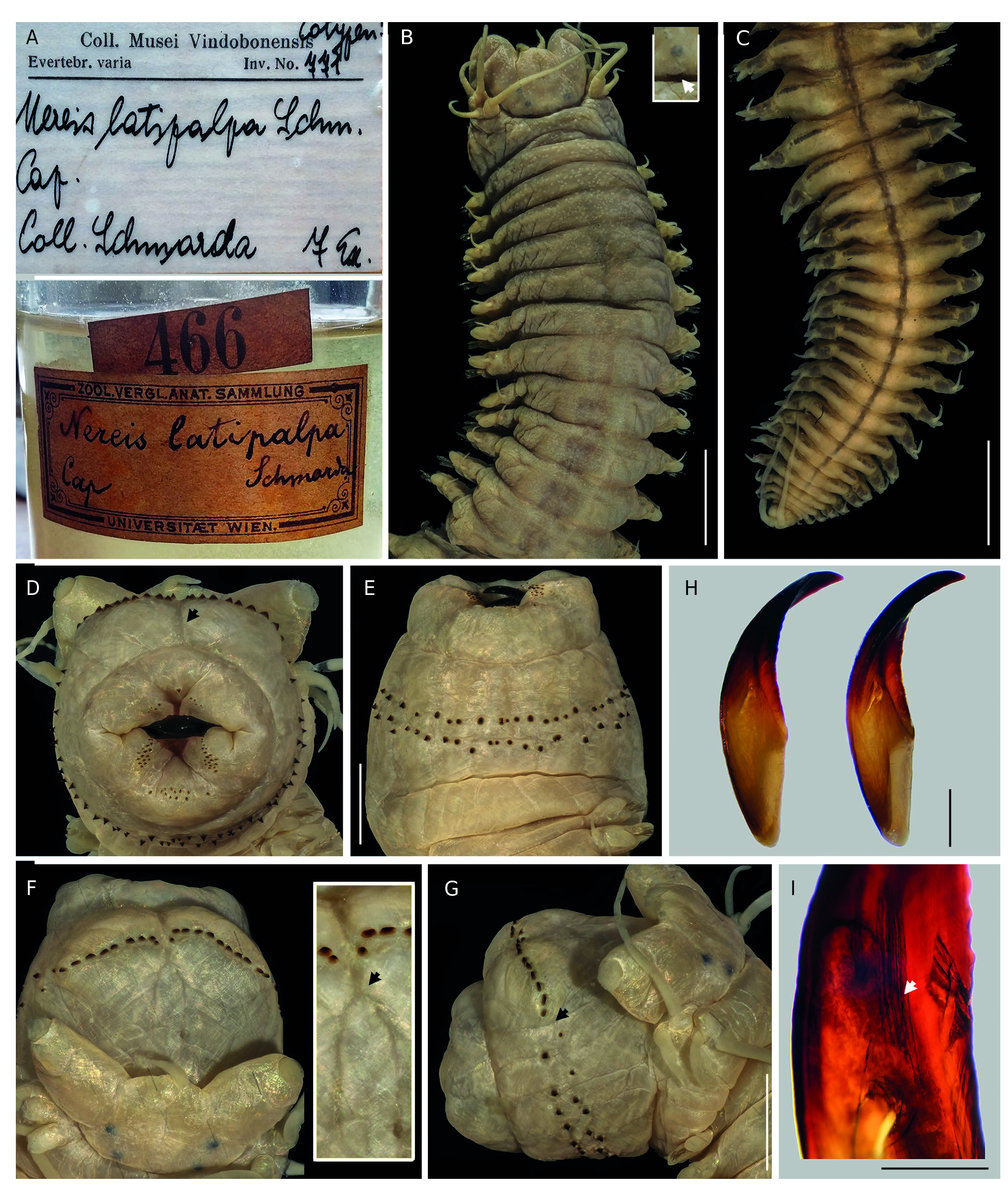
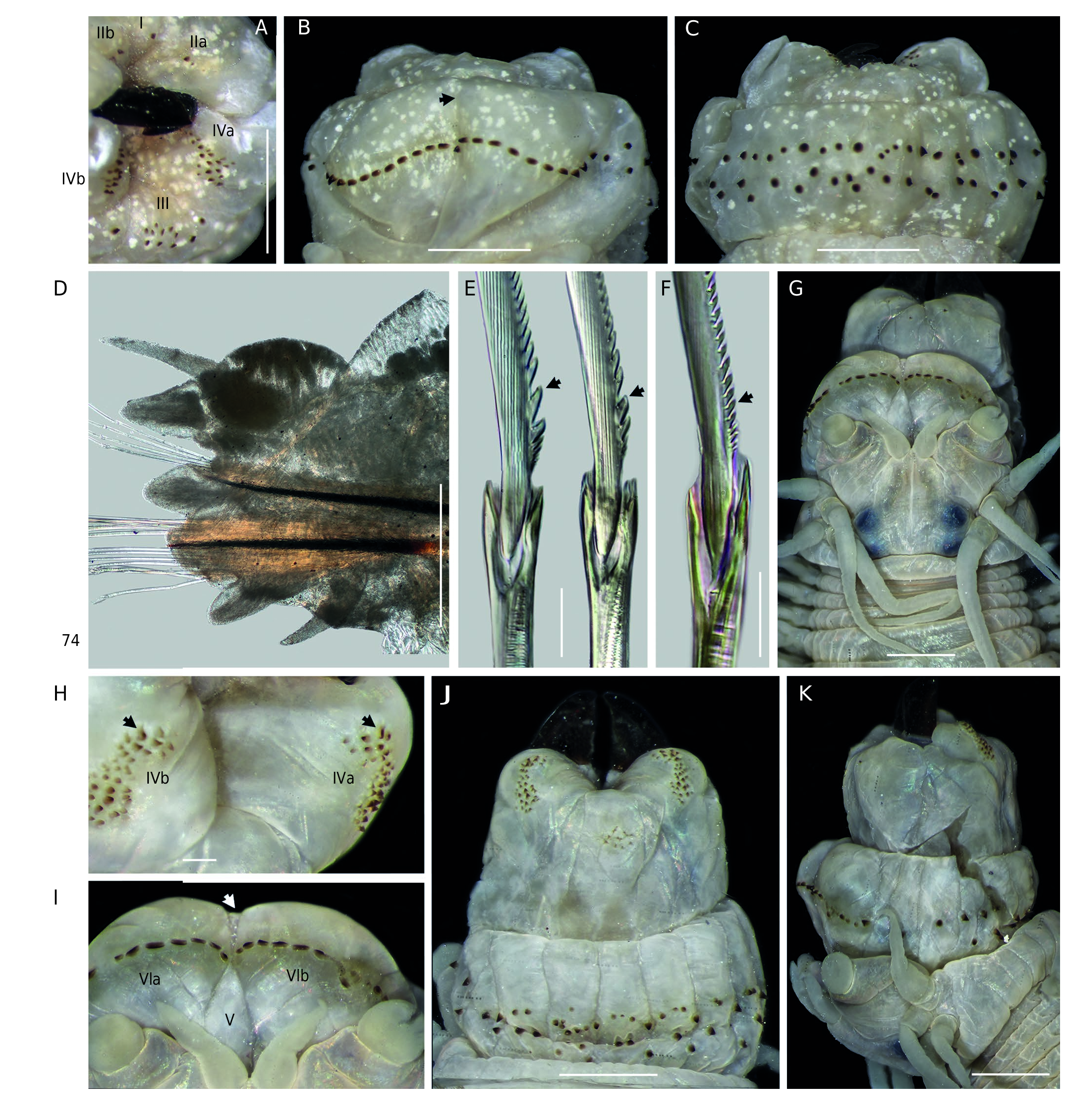
( Figs 1 View FIG A-C; 3A-I; 4A-L; 5A-L; 6A-O; 7A-E)
Nereis ( Nereis) latipalpa Schmarda, 1861: 104-105 View in CoL , textfig.A, B, K, a, b, pl. 31, fig. 244.
Neanthes latipalpa View in CoL – Kinberg 1865: 171. — von Marenzeller 1888: 6-7, fig. 2. n. syn.
Neanthes latipalpa typica View in CoL – Willey 1904: 260-261, pl. 13, fig. 9, pl. 14, figs 1, 2, 2a, b. n. syn.
Perinereis nuntia vallata View in CoL – Day 1967: 334, fig. 14.12.p-s (material from South Africa, partim, non Grube & Kröyer in Grube 1858).
Perinereis namibia Wilson & Glasby, 1993: 265-266 View in CoL , fig. 10a-k. n. syn.
TYPE MATERIAL. — 9 specimens of Nereis ( Nereis) latipalpa (originally labelled as “cotypen”) in three different lots. Herein, 1 designated as lectotype NMHW 769a and 8 as paralectotypes NMHW 769b, 770, 771. Collected by L. Schmarda, Table Bay, Cape Town (originally as “Tafelbai”, “Cap.”), South Africa, mud, under stones. Older label and catalog number attached on the outer surface of each jars which originally belonged to the “Zoologisch-vergleichend-anatomische Sammlung, Universität Wien” ( Fig. 3A View FIG ). Recent labels inside the jars with the NMHW imprint (as “Coll. Musei Vindobonensis, Evertebrata varia”) and handwritten ( Fig. 3A View FIG ) (see remarks for additional information). — Holotype of Neanthes latipalpa Kinberg, 1865 SMNH 37900, collected by Eugenie Expedition 1851-1853, sta. 1620-28, Green Point Lighthouse, Cape Town, South Africa, 9 Apr. 1854; jar with one original and two recent labels. One permanent slide of holotype’s left parapodium 27th SMNH 37900, housed at the NHMUK ( Fig. 5L View FIG ), apparently loaned to NHMUK for Arthur Willey . — Sixteen syntypes of Neanthes latipalpa typica Willey, 1904 NHMUK 1911.2.1.23-26, collected by W.F. Purcell, Green Point, Table Bay, Cape Town, South Africa, Nov. 1896 . One slide of a syntype NHMUK 1911.2.1.23-26a; Fig. 6N View FIG , right parapodia 8, 33 and 73 . One slide of a syntype NHMUK 1911.2.1.23-26b; Fig. 6O View FIG , small specimen, right parapodium 28 .
ADDITIONAL MATERIAL. — Seven specimens of Perinereis namibia Wilson & Glasby, 1993 ( ZMB 4109b), collected by L. Schultze, Lüderitz Bay, Namibia, Jul. 1903, identified by R. Wilson and C. Glasby . — One specimen of Perinereis vallata (Grube & Kröyer in Grube, 1858) ( ZMB 3666), collected by L. Plate, Puerto Montt, Los Lagos, Chile, identified by A.E. Grube and E. Ehlers . — One specimen of Perinereis akuna Wilson & Glasby, 1993 ( LACM-AHF Poly 10199), collected by S. J. Edmonds, Venus Bay inlet, Eyre Peninsula, South Australia, 33°13’00”S, 134°40’00”E, associated with Modiolus areolatus (Gould) clusters, no date, identified by T. Villalobos.
DIAGNOSIS. — Specimens with antennae separated, nuchal organs subequal to posterior eyes, postero-dorsal tentacular cirri reaching chaetigers 3-6. Jaws with 5-6 canals. Maxillary ring: AI = 1-2; AII = 2-10; AIII = 8-20, 1-2 lateral cones; AIV = 14-28, merged paragnaths absent. Oral ring: AVI-V-VI pattern, λ- shaped; AV = 0-2, paragnaths slightly behind AVI; AVI = 9-12, arc long, oblique, bars short, even; AVII-VIII = 48-66, 2-3 transverse rows, proximal with large cones. Gap between AVI and AVII-VIII narrow. Dorsal cirri 1.5-2 times longer than dorsal ligule, inserted posteriorly on three-quarters of it. Dorsal ligule barely uneven throughout body, subequal to median ligule; distal lobe bluntly conical posteriorly. Ventral cirri cirriform. Homogomph spinigers with proximal teeth notoriously thickened, separated; absent in subacicular neurochaeta. Heterogomph spinigers with proximal teeth barely thickened, evenly spaced; present throughout. Heterogomph falcigers with medium blades.
VARIATIONS. — LT = 48-145 mm, L15 = 8-24 mm, W15 = 1.7- 5.5 mm, 89-143 chaetigers. Antero-dorsal tentacular cirri reaching chaetiger 1-2. Postero-dorsal reaching chaetiger 3-6. Jaws with 4-8 denticles, 5-6 canals. Paragnaths: I =1-2, II =2-10, III =8-20, lateral cones 1-2, IV = 14-28, none merged paragnaths, V = 0-2, VI = 9-12, VII-VIII = 48-66, arranged in 2-3 transverse rows. Dorsal ligule expanding from parapodia 26-38. Notoacicular process developed in parapodia 5 to 14-22. Superior lobe developed in first 53-57 parapodia, projecting beyond end of neuracicular ligule in first 22-25 parapodia. Anal cirri as long as last 4-9 chaetigers. Falcigers with serrated region 0.43-0.52 total blade length.
HABITAT. — Mud beneath stones (Schmarda 1861), among rocks ( Willey 1904).
TYPE LOCALITY. — Table Bay, Cape of Good Hope, South Africa.
DISTRIBUTION. — Lüderitz Bay, Namibia and Table Bay, Cape Town, South Africa.
DESCRIPTION ( LECTOTYPE AND TWO PARALECTOTYPES) Lectotype, MPW 769 a, atoke, complete
Measurements. TL= 127 mm, L15 = 20.5 mm, W15= 5 mm, with 109 chaetigers. Paralectotypes atokes, longer female, complete, smaller lacking posterior region, TL= 95-137 mm, L15= 19-23.7 mm, W15= 4.5-4.6 mm, with 49-131 chaetigers.
Pigmentation. Brown, most intense anteriorly. Blackish glandular patches in ligules, cirrophore of ventral cirri and ventral ends of segments, most intense posteriorly ( Fig. 3C View FIG ); same glandulation running transversally on dorsum of posterior segments ( Fig. 3C View FIG ).
Head. Prostomium sub-pentagonal with broad anterior end ( Fig. 3B View FIG ), as long as wide; anterolateral sides 1.5 times wider than antennal diameter. Palpophores oval ( Fig. 3B View FIG ), massive, slightly longer than wide, equaling entire length of prostomium; one deep wrinkle placed obliquely in one-half of palpophore. Antennae separated, gap three-quarters of antennal diameter ( Fig. 3B View FIG ); conical, slender, extending backward to one-third of prostomium. Eyes in sub-trapezoidal arrangement, blackish, anterior and posterior pairs well separated (twice size of posterior; Fig. 3B View FIG ); lens barely visible, grayish. Anterior pair of eyes rounded, nearly subequal to antennal diameter; lens oval, located anterolaterally, touching margin of eye, covering 20%. Posterior pair of eyes rounded, similar-sized to anterior pair; lens rounded, located posterolaterally, not touching margin, covering 70%. Nuchal organs deeply embedded, slightly convex, subequal to posterior eyes ( Fig. 3B View FIG ).
Apodous anterior segment & tentacular cirri. Apodous anterior segment 4 times wider than long, 1.4 (1.4-1.5) times longer than chaetiger 1.Tentacular cirri pattern: Postero-dorsal cirri 1.5 times longer than antero-dorsal ones; anterior-ventral cirri slightly longer than postero-ventral one. Antero-dorsal cirri reaching chaetiger 1 (1-2); antero-ventral slightly smaller than palpophore. Postero-dorsal reaching chaetiger 3 (2-5); postero-ventral extending over prostomium to reach one-third of it. Dorsal cirrophores wrinkled, cylindrical; postero-dorsal cirrophores longest, 2.5 times length of postero-ventral ringshaped ones.
Pharynx. Not everted, previously dissected (based on lectotype, figures referring to a paralectotype with everted pharynx). Only one jaw available, blackish in distal quarter, then brownish; 7 (6-8) denticles,poorly developed, inner margin of fang flattened, equaling next 4 denticles; pulp cavity two-fifths (two-fifths to three-fifths) length of jaw ( Fig. 3H View FIG ), distal apex leveling third basal denticle; 5 (5-6) longitudinal canals emerging from pulp cavity ( Fig. 3I View FIG ). Maxillary ring ( Fig. 3D View FIG ): paragnaths conical, none worn, brownish amber colored, much smaller than those on oral ring. AI = 1 (1-2), longer than those on AII. AIIa =4 (4-5), AIIb= 5 (4-9), oblique irregular patch, two irregular rows. AIII= 9 (9-12), oval patch, two slightly regular transverse rows, one cone laterally isolated in each side. AIVa =18 (19-22), AIVb =23 (19-27), lemniscate-shaped patch, maxillary portion smaller than proximal one, outer cones smallest; merged paragnaths absent. Oral ring ( Fig. 3 View FIG E-G): AVI-V-VI pattern, λ- shaped; AVI overlapping proximally AV further behind arc of paragnaths ( Fig. 3F View FIG ). Paragnaths conical and shield-shaped bars, reddish amber colored.AV=1 (1-2), aligned to paragnaths of AVI. AVIa =9 (9-10), AVIb =11 (9-10), arc of paragnaths long, slightly oblique, shield-shaped bars; bars short, tip barely worn, similar in length; ridges prominent, broad, slightly wider than palpophore. AVII-VIII=53 (49- 66), conical paragnaths, bigger ones arranged in longitudinal wrinkles of ring ( Fig. 3E View FIG ); single band of two main rows, not increasing in number dorsoventrally, distal row slightly more regular than proximal one. Gap between AVI and AVII-VIII narrow, as wide as palpostyle ( Fig. 3G View FIG ).
Notopodia. Dorsal cirrus slightly longer than dorsal ligule in first parapodia, subequal to ligule or slightly longer in anterior and median parapodia, 1.5-2 times longer in posterior ones; cirri longer than length of proximal lobe of dorsal ligule in anterior parapodia ( Fig. 4A, B View FIG ), subequal to it from median ones ( Fig. 4 View FIG C-F); cirrus inserted on one-third in anterior parapodia ( Fig. 4A, B View FIG ), one-half in median ( Fig. 4C, D View FIG ), three-quarters in posterior ones ( Fig. 4E, F View FIG ). Dorsal ligule somewhat uneven throughout body, becoming wider from parapodia 35 (28-37), barely expanded from about parapodia 85 ( Fig. 4E, F View FIG ); ligule 2.5 (2.3-2.5) times width of median ligule in posterior parapodia ( Fig. 4E, F View FIG ). Distal lobe of dorsal ligule bluntly conical, longer than proximal one in anterior parapodia, two-thirds or three-fifths length of proximal in median, one-third or two-fifths length in posterior ones ( Fig. 4E, F View FIG ). Dorsal ligule subequal to median ligule in anterior parapodia, slightly shorter in median ( Fig. 4C, D View FIG ), slightly longer in posterior ones ( Fig. 4E, F View FIG ). Two main glandular patches in dorsal ligule, central and proximal ( Fig. 4F View FIG ); oval, similar-sized in anterior parapodia (covering 25-30% of total ligule area; Fig. 4B View FIG ); proximal patch 1.5 times bigger than central one in median parapodia (covering 40-50% of total ligule area; Fig. 4D View FIG ) and twice bigger in posterior ones (covering 60-70% of total ligule area; Fig. 4F View FIG ). Notoacicular process developed from parapodia 5 to 21 (14-22), digitiform, short.
Neuropodia. Neuracicular ligule subequal to ventral ligule or slightly longer throughout body; 1.5 times wider than ventral ligule in anterior parapodia, twice wider from median ones. Superior lobe digitiform ( Fig. 4C View FIG ), shorter than inferior lobe and projecting beyond end of neuracicular ligule in first 25 (22-24), reduced from parapodia 57 (54-58). Inferior lobe rounded ( Fig. 4A, B View FIG ), narrowing progressively from parapodia 28 (25-27). Ventral ligule digitiform, shorter than median ligule throughout body; two-thirds length of ligule in anterior parapodia, one-half length from median ones. Ventral cirri cirriform, smaller than ventral ligule throughout body, onehalf length of ligule, except in first 5 parapodia, three-quarters or nearly subequal to ligule.
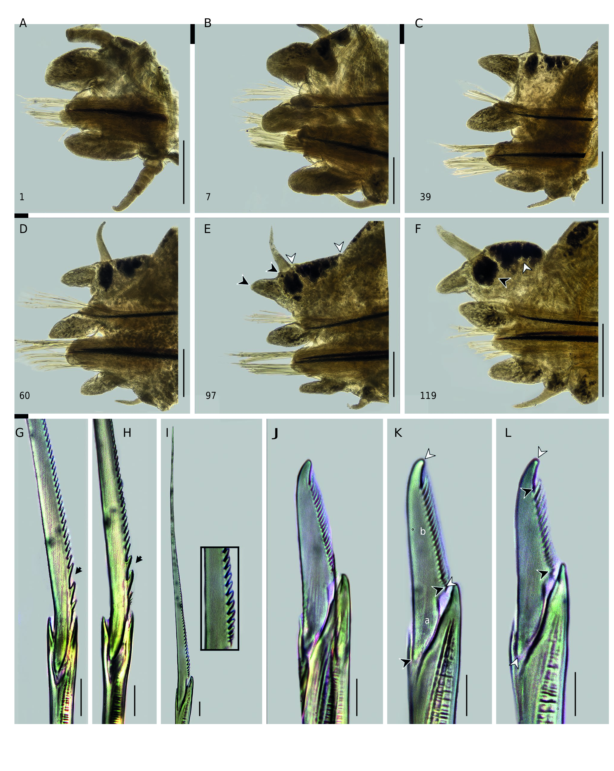
Chaetae. Notochaetae with homogomph spinigers, blade with few first proximal teeth thick, notoriously separated ( Fig. 4G, H View FIG ). Neurochaetal supracicular fascicle with homogomph spinigers and heterogomph falcigers, both present throughout; spinigers as notopodial ones, more numerous than falcigers in fascicle; falcigers with blade of medium length ( b/ a =1.27- 1.57 times), serrated region 0.50-0.52 of total blade length ( Fig. 4J View FIG ). Neurochaetal subacicular fascicle with heterogomph spinigers and heterogomph falcigers, both present throughout; spinigers with proximal teeth barely thickened, evenly spaced ( Fig. 4I View FIG ), less numerous than falcigers in fascicle; falcigers with blade of medium length ( b /a = 1.61-1.79 times), serrated region 0.49-0.51 of total blade length ( Fig. 4K, L View FIG ).
Pygidium. With slender anal cirri ( Fig. 3C View FIG ), as long as last 9 (7) parapodia, cirrophores well developed, broad.
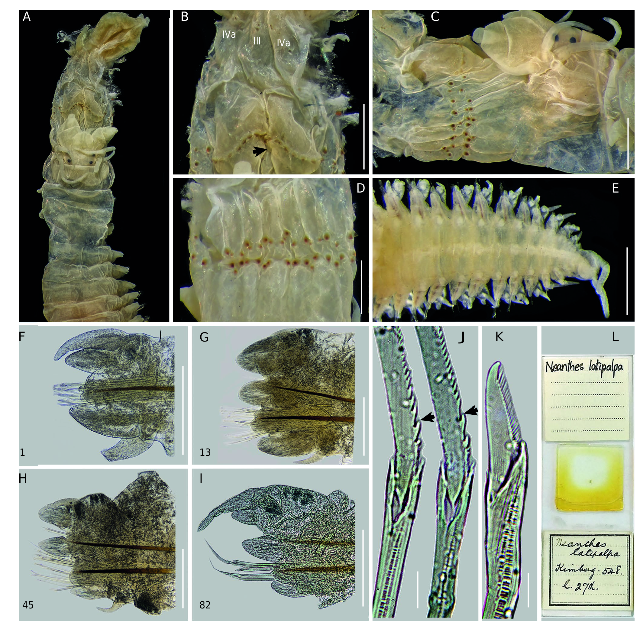
Holotype of Neanthes latipalpa Kinberg ( Fig. 5 View FIG A-L)
Body. Atoke, SMNH 37900, complete, TL = 50 mm, L15 = 10.8 mm, W15 = 2.4 mm, with 89 chaetigers. Body pigmentation absent, probably faded. Blackish glandular patches in ligules, cirrophore of ventral cirri and ventral ends of segments, most intense posteriorly.
Head. Prostomium sub-pentagonal, broad anterior end; barely longer than wide ( Fig. 5A View FIG ). Palpophores oval, massive, as long as wide ( Fig. 5A, C View FIG ), nearly equaling entire length of prostomium. Antennae separated ( Fig. 5A View FIG ), conical, slender, extending backward to one-quarter of prostomium. Eyes in sub-trapezoidal arrangement, blackish ( Fig. 5A, C View FIG ); anterior pair barely reniform due to lens, subequeal to antennal diameter, lens anterolateral, covering 30%; posterior pair rounded, slightly shorter than anterior, lens posterolateral, covering 60%.
Apodous anterior segment & tentacular cirri. Apodous anterior segment 3 times wider than long, 1.3 times longer than chaetiger 1. Antero-dorsal tentacular cirri reaching chaetiger 1, anteroventral one smaller than palpophore. Postero-dorsal reaching chaetiger 2 but incomplete, postero-ventral one extending over prostomium to reach one-third of it. Postero-dorsal cirrophores longest, 2 times length of postero-ventral ring-shaped ones.
Pharynx. Everted, damaged ( Fig. 5 View FIG A-C), paragnath areas still recognizable, jaws not present. Maxillary ring ( Fig. 5B View FIG ): paragnaths conical, small, brownish amber. AI=2, in longitudinal line. AIIa=4, AIIb=4, oblique row. AIII =8, rectangular patch, two slightly regular rows. AIVa =14, AIVb=16, spoonshaped patch, merged paragnaths absent. Oral ring ( Fig. 5 View FIG B- D): AVI-V-VI pattern, λ- shaped; AVI overlapping proximally AV further behind arc of paragnaths ( Fig. 5B View FIG ). Paragnaths conical, except shield-shaped bars on AVI, brownish amber. AV =1, aligned to paragnaths of AVI. AVIa = 11, AVIb = 11, arc of paragnaths long, oblique, bars short, similar in length, tip barely worn. AVII-VIII =56, single band of two main rows, irregular. Gap between AVI and AVII-VIII narrow ( Fig. 5C View FIG ).
Notopodia. Dorsal cirri slightly longer than dorsal ligule in first parapodia, subequal to ligule or slightly longer in anterior and median parapodia ( Fig. 5 View FIG F-H), 1.5-2 times longer in posterior ones ( Fig. 5I View FIG ); cirri inserted on one-third in anterior parapodia, one-half in median, three-quarters in posterior ones. Dorsal ligule becoming wider from parapodia 27, barely expanded from about parapodia 65 ( Fig. 5I View FIG ); subequal to median ligule in anterior and median parapodia ( Fig. 5 View FIG F- H), longer in posterior ones ( Fig. 5I View FIG ). Two main glandular patches in dorsal ligule, central and proximal, most intense in posterior parapodia. Notoacicular process developed from parapodia 5 to 14, digitiform, short.
Neuropodia. Neuracicular ligule subequal to ventral ligule or slightly longer.Superior lobe digitiform ( Fig. 5F, G View FIG ), projecting beyond end of neuracicular ligule in first 22 parapodia, reduced from parapodia 56. Ventral cirri smaller than ventral ligule.
Chaetae. Homogomph spinigers with blade bearing few first proximal teeth thickened and notoriously separated ( Fig. 5J View FIG ). Heterogomph spinigers present in all chaetigers, blade with proximal teeth barely thickened, evenly spaced. Falcigers in both fascicles with serrated region 0.49-0.51 total blade length ( Fig. 5K View FIG ).
Pygidium. With slightly thickened anal cirri ( Fig. 5E View FIG ), apparently incomplete, as long as last 4 parapodia; cirrophores well developed.
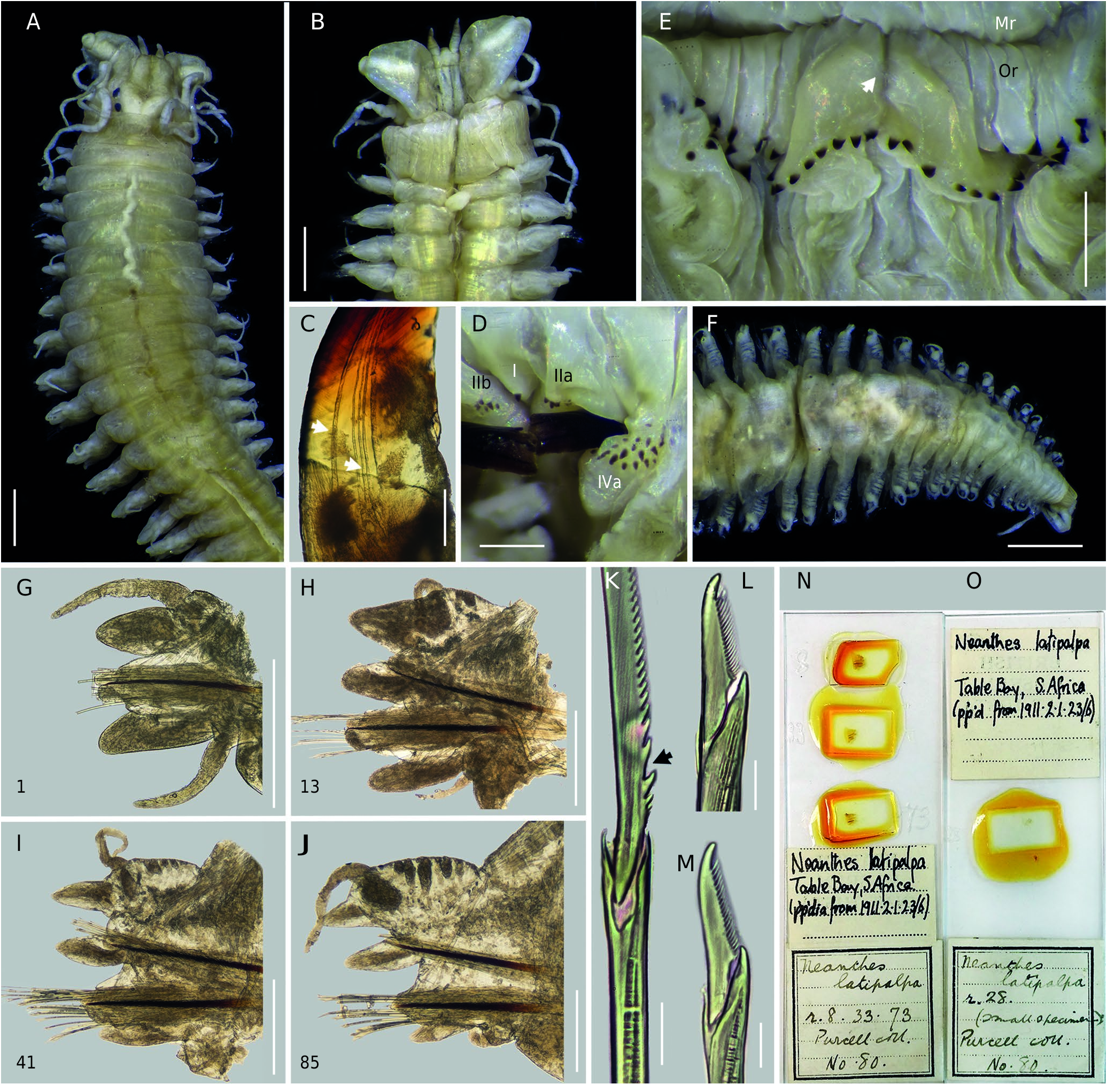
Syntype of Neanthes latipalpa typica ( Fig. 6 View FIG A-O)
Body. Atoke, NHMUK 1911.2.1.23-26,complete,TL= 48 mm, L15 = 8.3 mm, W15 = 1.7 mm, with 93 chaetigers. Body pigmentation on dorsal surface, faint, pale brown, most intense anteriorly. Brownish glandular patches in ligules, cirrophore of ventral cirri and ventral ends of segments.
Head. Prostomium sub-pentagonal, broad anterior end; barely longer than wide ( Fig. 6A View FIG ). Palpophores oval, massive, as long as wide ( Fig.6B View FIG ), equaling entire length of prostomium.Antennae separated ( Fig. 6A View FIG ), conical, slender, extending backward to one-quarter of prostomium. Eyes in sub-trapezoidal arrangement, blackish ( Fig. 6A View FIG ); anterior pair barely reniform due to lens, slightly wider than antennal diameter, lens anterolateral, covering 25%; posterior pair rounded, slightly shorter than anterior, lens posterolateral, covering 40%.
Apodous anterior segment & tentacular cirri. Apodous anterior segment 3.4 times wider than long, 1.4 times longer than chaetiger 1. Antero-dorsal tentacular cirri reaching chaetiger 2, antero-ventral one smaller than palpophore. Postero-dorsal reaching chaetiger 5, postero-ventral one extending over prostomium to reach one-half of it. Postero-dorsal cirrophores longest, 2 times length of postero-ventral ring-shaped ones.
Pharynx. Not everted. Jaws with 6 denticles, barely prolonged; pulp cavity two-fifths length of jaw, oblique distal apex, five longitudinal canals ( Fig. 6C View FIG ). Maxillary ring ( Fig. 6D View FIG ): paragnaths conical, small, dusky brown. AI =1, longer than cones on AII. AIIa =9, AIIb=10, irregular oblique patch. AIII=12, oval patch in three rows, one isolated cone. AIVa = 21, AIVb =21, spoon-shaped patch, merged paragnaths absent. Oral ring ( Fig. 6E View FIG ): AVI-V-VI pattern, λ- shaped; AVI overlapping proximally AV further behind arc of paragnaths. Paragnaths conical, except shield-shaped bars on AVI, dusky brown. AV= 1, aligned to paragnaths of AVI. AVIa =10, AVIb =10, arc of paragnaths long, oblique ( Fig. 6E View FIG ), bars short, similar length, tip barely worn. AVII-VIII= 52, single band of two main rows, irregular.Gap between AVI and AVII-VIII narrow.
Notopodia. Dorsal cirri longer than dorsal ligule in first parapodia, subequal to ligule or slightly longer in anterior and median parapodia ( Fig. 6 View FIG G-I), 1.5-2 times longer in posterior ones ( Fig. 6J View FIG ); cirri inserted on one-third in anterior parapodia, one-half in median, three-quarters in posterior ones.Dorsal ligule becoming wider from parapodia 29, barely expanded from about parapodia 63 ( Fig. 6J View FIG ); subequal to median ligule in anterior and median parapodia ( Fig. 6 View FIG G-I), longer in posterior ones ( Fig. 6J View FIG ). Two main glandular patches in dorsal ligule, central and proximal, most intense in posterior parapodia. Notoacicular process developed from parapodia 5 to 18, digitiform, short.
Neuropodia. Neuracicular ligule slightly longer than ventral ligule. Superior lobe digitiform ( Fig. 6G, H View FIG ), projecting beyond end of neuracicular ligule in first 24 parapodia, reduced from parapodia 54. Ventral cirri smaller than ventral ligule, except in first anterior parapodia, subequal to it.
Chaetae. Homogomph spinigers with blade bearing few first proximal teeth thickened and notoriously separated ( Fig. 6K View FIG ). Heterogomph spinigers present in all chaetigers, blade with proximal teeth barely thickened, evenly spaced. Falcigers in both fascicles with serrated region 0.47-0.49 total blade length ( Fig. 6L, M View FIG ).
Pygidium. With slightly thickened anal cirri ( Fig. 6F View FIG ), as long as last 6 parapodia, cirrophores well developed.
Topotype of Perinereis namibia ( Fig. 7 View FIG A-E)
Body & head. Atoke, ZMB 4109b, complete. Prostomium sub-pentagonal, broad anterior end. Palpophores oval, massive, as long as wide. Antennae separated, conical, slender, extending backward to one-third of prostomium. Eyes in subtrapezoidal arrangement; anterior pair rounded, subequal to antennal diameter, lens anterolateral, covering 20%; posterior pair rounded, slightly shorter than anterior pair, lens posterolateral, covering 30%. Apodous anterior segment 3 times wider than long, 1.3 times longer than chaetiger 1.
Pharynx. Everted. Jaws with 6 denticles, barely prolonged. Maxillary ring ( Fig. 7A View FIG ): paragnaths conical, small, dusky brown. AI= 1, barely longer than cones on AII. AIIa = 4, AIIb= 5, oblique row. AIII= 12, oval patch in three irregular rows, one isolated cone. AIVa =22, AIVb = 22, lemniscateshaped patch, merged paragnaths absent. Oral ring ( Fig. 7B, C View FIG ): AVI-V-VI pattern, λ- shaped; AVI overlapping proximally AV further behind arc of paragnaths ( Fig. 7B View FIG ). Paragnaths conical, except shield-shaped bars on AVI, dusky brown. AV=1, aligned to paragnaths of AVI. AVIa= 10, AVIb= 9, arc of paragnaths long, oblique ( Fig. 7B View FIG ), bars short, similar in length, tip barely worn. AVII-VIII = 54, single band of two main rows, irregular ( Fig. 7C View FIG ). Gap between AVI and AVII-VIII narrow.
Notopodia. Dorsal ligule becoming barely expanded from parapodia 32; subequal to median ligule in anterior parapodia, barely shorter in median, slightly longer in posterior ones ( Fig. 7D View FIG ). Two main glandular patches in dorsal ligule, central and proximal, most intense in posterior parapodia ( Fig. 7D View FIG ). Notoacicular process developed from parapodia 5 to 23, digitiform, short.
Neuropodia. Neuracicular ligule slightly longer than ventral ligule ( Fig. 7D View FIG ). Superior lobe digitiform, projecting beyond end of neuracicular ligule in first 26 parapodia, reduced from parapodia 56. Ventral cirri smaller than ventral ligule.
Chaetae. Homogomph spinigers with blade bearing few first proximal teeth thickened and notoriously separated ( Fig. 7E View FIG ). Heterogomph spinigers present in all chaetigers, blade with proximal teeth barely thickened, evenly spaced. Falcigers in both fascicles with medium blade ( b/a = 1.47-1.76 times), serrated region 0.44-0.53 total blade length.
Other characteristics well described and drawn in Wilson & Glasby (1993).
REMARKS
Schmarda (1861) described 16 new species of nereidids in two genera, Heteronereis Örsted, 1843 and Nereis . In the preface of his work, he simultaneously referred Mastigonereis Schmarda, 1861 and Nereilepas de Blainville, 1828 as subgenera of Nereis . Although they were not represented as such in the descriptions, it was clear that he considered subdivisions in Nereis . Therefore, I herein refer the subgeneric level for Schmarda’s new species, including N. ( Nereis) latipalpa .
Schmarda (1861) described N. ( Nereis) latipalpa using specimens collected by himself in Table Bay during his visit to Cape Town ( South Africa) in February-June 1854 (see details in Appendix). The type material of N. ( Nereis) latipalpa is still deposited at the NHMW. It consists of nine syntypes, which match the original description (Schmarda 1861); the best specimen preserved is here designated as lectotype (NMHW 769a), whereas the others as paralectotypes (NMHW 769b, 770, 771). The size and number of chaetigers of the lectotype do not fit the specimen described by Schmarda; nevertheless, it was the only specimen dissected among the type material, agreeing with dissected parts mentioned and illustrated by Schmarda: one jaw, and one anterior (chaet. 13) and posterior (chaet. 78) parapodia, which are lost.
De Quatrefages (1866) recognized N.latipalpa as a valid species. Kinberg (1865), based on a specimen collected in Cape Town and on the description of N. latipalpa by Schmarda, described a new species, Neanthes latipalpa , but he was not entirely sure whether it was the same species as Schmarda’s since the latter did not consider the paragnaths arrangement, a relevant feature used by Kinberg to distinguish nereidid genera and species.
Von Marenzeller (1888), after an examination of some polychaetes from Angra Pequena Bay (now as Lüderitz Bay), noticed the morphological similarities between the Namibian and Cape of Good Hope specimens. Further, he realized that N. ( Nereis) latipalpa by Schmarda and Neanthes latipalpa by Kinberg are the same species because of the shape of the parapodia. Thus, he synonymized N. latipalpa Kinberg but transferred N. ( Nereis) latipalpa Schmarda to Neanthes Kinberg, 1865 , the latter following the Kinberg’s classification of genera by using the pharyngeal arrangement of paragnaths.
Ehlers (1901), who certainly did not recognize as valid Neanthes and other Kinberg’s nereidid genera (see Ehlers 1868), examined the type material and some specimens of N. ( Nereis) latipalpa Schmarda from Angra Pequena, regarding the species as a junior synonym of Nereis vallata (currently in Perinereis sensu Hartman 1959 ). Later, Willey (1904) examined the type specimen of Neanthes latipalpa and confirmed the synonymy proposed by von Marenzeller (1888), but established a trinominal name, N. latipalpa typica , to emphasize that it is barely different to N. latipalpa brevicirris (currently as Perinereis brevicirris sensu Hartman 1959 ). Afterward, Augener (1918) reinforced Ehlers’ assumptions and considered N. latipalpa typica as identical to N. vallata , but placed in the subgenus Perinereis ( sensu Augener 1913) by having, among other characters, a continuous transverse line of paragnaths on AVI.
Fauvel (1919, 1921, 1932, 1953) grouped the species morphologically similar to Perinereis nuntia , mainly characterized by having an arc of paragnaths on AVI.Within the subspecies of this group, P. nuntia var. vallata was different from the others by having AI= 1-3, AV =1, most extended tentacular cirri reaching chaetigers 7-8, and dorsal cirri subequal to dorsal ligule or slightly smaller. Fauvel considered the species as having a wide distribution (the Red Sea, India, Madagascar, South Africa, Philippine Islands, New Zealand, Australia, and Chile) based on the records of the species under several names. Also, Fauvel (1919, 1921, 1932, 1953) still retained Schmarda, Kinberg and Willey’s N. latipalpa as a synonym of P. nuntia var. vallata ; and since then, the three latipalpa ’s names have not been recognized (e.g. Hartmann-Schröder 1962; Wesenberg-Lund 1962), except by Hartman (1949, 1959), who still retained the Kinberg’s species as valid in Neanthes , but details were not provided.
Hutchings et al. (1991) established three groups within Perinereis according to the number of bars on AVI; those valid species more related to P. nuntia were placed in the third group (AVI with numerous bars in an arc). A few years later, Wilson & Glasby (1993) carried out a relevant study of 20 species within the P.nuntia species complex.They restricted the morphology of the P. nuntia complex, redescribed some of the species using type, topotype or non-type materials, such as P. nuntia and P. vallata ; also, they recognized as valid some previously synonymized species, and described two new ones: Perinereis akuna ( New South Wales, Australia) and P. namibia (Lüderitz Bay, Namibia). Nevertheless, the authors overlooked N. latipalpa since it was not included in the source of synonyms of any species, particularly P. vallata , neither in the appendix at the end of the document.
After the comprehensive revision of the type material of N. Nereis ) latipalpa Schmarda , Neanthes latipalpa Kinberg and Neanthes latipalpa typica Willey , herein is confirmed that they belong to the same species. Furthermore, this species is here considered as valid since it is different the other species within the P. nuntia complex, particularly P. vallata . Therefore, it is reinstated as P. latipalpa (Schmarda, 1861) n. comb. Besides, P. namibia is regarded as a junior synonym of P. latipalpa n. comb., based on the original description and the revision of some topotypes formerly used by Wilson & Glasby (1993).
Perinereis latipalpa n. comb. and P. vallata are indeed closely related species since they have a similar parapodial pattern, as well as a similar number and arrangement of paragnaths on the ventral areas of the pharynx. Nevertheless, according to my revision on the type material of P. latipalpa n. comb. and a specimen from Chile ( Fig. 7 View FIG F-K), the description of P. vallata and its accurate posterior characterizations ( Ehlers 1901; Wesenberg-Lund 1962; Rozbaczylo & Castilla 1973; Bertrán 1980; Wilson & Glasby 1993; Sampértegui et al. 2013), the differences between these two species remain in the shape of the proximal teeth of homogomph spinigers and the AVI-V-VI pattern. In P. latipalpa n. comb., the first proximal teeth of homogomph spinigers are notoriously thick and separated ( Figs 4G, H View FIG ; 5J View FIG ; 6K View FIG ; 7E), whereas in P. vallata these are of similar size and equidistant from one another ( Fig. 7F View FIG ). Also, in P. latipalpa n. comb., the AVI-V-VI pattern is λ- shaped ( Figs 3D, F View FIG ; 5B View FIG ; 6E View FIG ; 7B), with the AVI overlapping AV proximally further behind the arc of paragnaths ( Figs 3F View FIG ; 5B View FIG ; 7B View FIG ); whereas in P. vallata , it is χ- shaped ( Fig. 7G, I View FIG ), with the AVI overlapping AV proximally at the level of the arc of paragnaths ( Fig. 7I View FIG ).
Perinereis latipalpa n. comb. also differs from other two closely related species of P. vallata : Perinereis gualpensis Jeldes, 1963 (based on the original description and characterizations of topotype specimens, e.g. Bertrán 1980; Sampértegui et al. 2013) and P. akuna Wilson & Glasby, 1993 . Perinereis gualpensis , who also shares the same type of blade of homogomph spiniger and the same pattern of AVI-V-VI as P. vallata , can be distinguished from P. latipalpa n. comb. because the latter has AIII =8-20 and AVII-VIII= 48-66, whereas P. gualpensis has AIII= 21-55 and AVII-VIII = 27-47. Likewise, the dorsal ligule is longer than median ligule and slightly expanded in posterior parapodia of P. latipalpa n. comb.; whereas in P.gualpensis , it is subequal to median ligule and not expanded in same parapodia. Furthermore, in P. latipalpa n. comb., the dorsal cirrus extends beyond the end of dorsal ligule, and it is placed on the distal quarter of the ligule, whereas in P. gualpensis the dorsal cirrus is subequal to dorsal ligule and it is located on one-half of the ligule.
On the other hand, although P. latipalpa n. comb. and P. akuna share the same λ- shaped pattern in AIV-V-IV, they differ in several respects. Perinereis latipalpa n. comb. has the first proximal teeth of homogomph spinigers notoriously thick and separated, whereas in P. akuna these are of similar size and equidistant from one another. The dorsal ligule is longer than median ligule and slightly expanded in posterior parapodia of P. latipalpa n. comb., whereas in P. akuna the dorsal ligule is subequal to median ligule and not expanded in same parapodia. Also, in P. latipalpa n. comb. the dorsal cirri extend beyond the end of dorsal ligule and it is placed on the distal quarter of the ligule; whereas in P. akuna , the dorsal cirri is subequal to dorsal ligule and it is located on one-half of the ligule. Likewise, P. latipalpa n. comb. has a lower number of paragnaths on AVII-VIII (48-66) which are arranged in 2-3 rows of small and large cones; whereas P. akuna has a larger number of paragnaths (76-191) which are arranged in more than four rows, with tiny cones in a distinct band. Finally, P. latipalpa n. comb. always lacks merged paragnaths on AIV, which are always present in P. akuna (numbering 1-5).
Wilson & Glasby (1993) regarded the occurrence of merged paragnaths on AIV as a relevant taxonomic feature to distinguish species within the P. nuntia complex; however, this character may be present or absent in individuals of the same species ( Hutchings et al. 1991; Wilson & Glasby 1993; Park & Kim 2007; Sampértegui et al. 2013). Concerning P. vallata, Sampértegui et al. (2013) considered this character as varying since it was only present in a few of their Chilean specimens. The specimens herein examined of P. latipalpa n. comb. lack this type of paragnath, but a small merged one was present in the examined individual P. vallata . It is uncertain if their presence/absence on AIV of the latter species is related to size or to a particular developmental stage. Nevertheless, it is noteworthy that any of the specimens of P. latipalpa n. comb. herein examined and many of those examined by Wilson & Glasby (1993) as P. namibia , have merged paragnaths. For this reason, this feature should be carefully used to distinguish species within the P. nuntia complex.

No known copyright restrictions apply. See Agosti, D., Egloff, W., 2009. Taxonomic information exchange and copyright: the Plazi approach. BMC Research Notes 2009, 2:53 for further explanation.
|
Kingdom |
|
|
Phylum |
|
|
Class |
|
|
Order |
|
|
Family |
|
|
Genus |
Perinereis latipalpa (Schmarda, 1861)
| Villalobos-Guerrero, Tulio F. 2019 |
Perinereis namibia
| WILSON R. S. & GLASBY C. J. 1993: 266 |
Perinereis nuntia vallata
| DAY J. H. 1967: 334 |
Neanthes latipalpa typica
| WILLEY A. 1904: 260 |
Neanthes latipalpa
| VON MARENZELLER E. 1888: 6 |
| KINBERG J. G. H. 1865: 171 |