
Anaptomecus yarigui, Galvis & Rheims, 2018
|
publication ID |
https://doi.org/ 10.11646/zootaxa.4521.2.11 |
|
publication LSID |
lsid:zoobank.org:pub:27C50B2D-FDD6-4AB3-A6A0-FF57F15DBE94 |
|
DOI |
https://doi.org/10.5281/zenodo.5990663 |
|
persistent identifier |
https://treatment.plazi.org/id/03BE4E5D-7E00-FFA8-FF31-FD29D202FED9 |
|
treatment provided by |
Plazi |
|
scientific name |
Anaptomecus yarigui |
| status |
sp. nov. |
Anaptomecus yarigui View in CoL sp. nov. ( Figs 1 View FIGURE 1 , 18–30 View FIGURES 18–24 View FIGURES 25–30 )
Type material. Holotype: GoogleMaps male, COLOMBIA: Santander: San Vicente de Chucurí, Serranía de los Yariquíes National Natural Park GoogleMaps , Vereda Cantagallo GoogleMaps , Finca El Prado GoogleMaps , 2138 m, 6.81833°N, 73.36274°W, 23–25 September 2015, J.A. Moreno-González leg. (ICN-Ar 10851).
Additional material examined. 1 subadult male, 2 immatures, same data as holotype (ICN-Ar 10852–10853) GoogleMaps ; 1 subadult male, same locality data and collector as holotype, 2200 m, 6.81727°N, 73.36155°W, 21–23 September 2015 (ICN-Ar 10854); 2 immatures, same municipality and National Natural Park as holotype, Vereda Centro, Fincal El Llanito, 1702 m, 6.84547°N, 73.38380°W, 8–10 October 2015, J.A. Moreno-González leg. (ICN-Ar 10855) GoogleMaps .
Etymology. The specific epithet honors the Yariguí people, an extinct indigenous group that inhabited the region and the particular location where the species was found, the Serranía de los Yariquíes National Natural Park. It has been said that they committed mass suicide instead of submitting to Spanish colonial rules; noun in apposition.
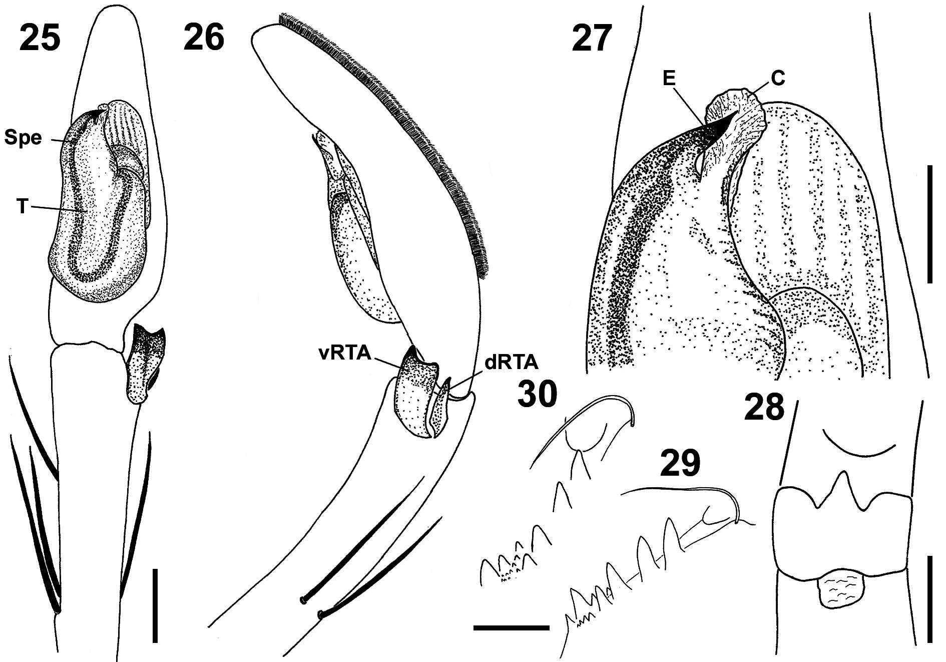
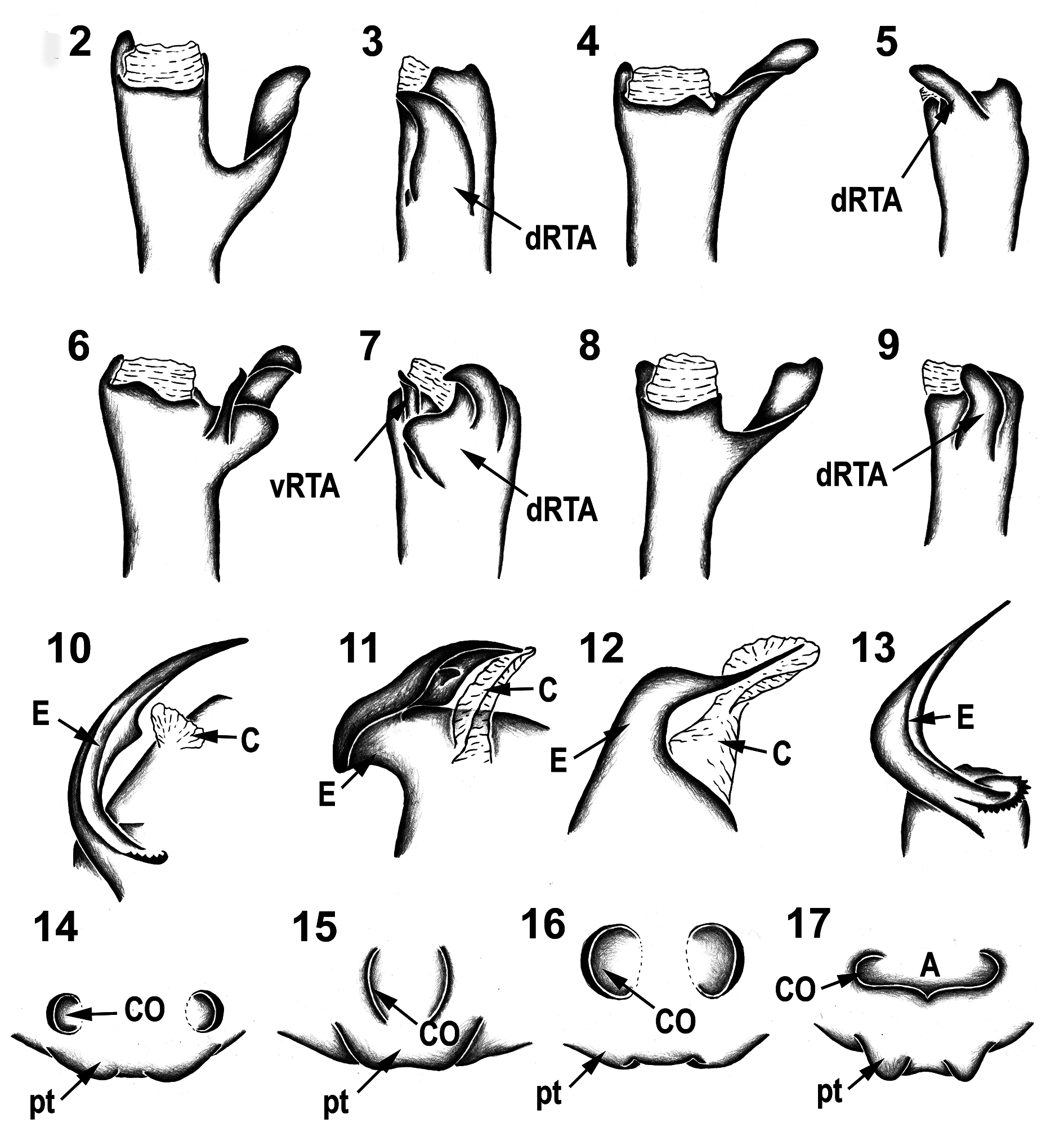
Diagnosis. Males of A. yarigui sp. nov. resemble those of A. suni in having a vRTA and an untwisted embolus. They are distinguished from the latter by the vRTA laminar, wide and distally indented ( Figs 23–26 View FIGURES 18–24 View FIGURES 25–30 ; slender, prong-like in A. suni , Figs 6–7 View FIGURES 2–17. 2–9 ) and by the embolus with a short tip ( Figs 21–22 View FIGURES 18–24 , 25, 27 View FIGURES 25–30 ; long and slender in A. suni , Fig. 12 View FIGURES 2–17. 2–9 ).
Description. Male (holotype). Total length 13.35. Prosoma: 4.51 long, 3.50 wide. Opisthosoma: 8.84 long, 2.98 wide. Eyes in two recurved rows ( Fig. 18 View FIGURES 18–24 ), diameters: AME 0.22, ALE 0.23, PME 0.16, PLE 0.21; interdistances: AME– AME 0.16, AME–ALE 0.07, PME–PME 0.28, PME–PLE 0.21, AME–PME 0.26, ALE–PLE 0.26. Legs (2143): I: 38.17 (10.35, 2.26, 10.34, 11.80, 3.42); II: 39.85 (10.37, 2.41, 11.29, 12.50, 3.28); III: 25.06 (6.89, 1.82, 7.18, 7.35, 1.82); IV: 37.06 (9.22, 1.96, 8.30, 14.41, 3.17). Spination: palpal tibia I, p 3, d 1, r1; femora I, III: p 1–1–1–1, d 0–1–0, r 1–1–1; femur II: p 1–1–1, d 0–1–0, r 1–1–1; femur IV: p 1–1–1, d 1–0–0, r 0–0–1; tibiae I–II: p 0–1–0, d 1–0–1, r 0–1–0, v 2–2 – 2; tibia III: p 0–1–0, d 0–0–1, r 0–1–1, v 2 –0–2; tibia IV: p 0–1–1, d 0–0–1, r 0–1–1, v 2 –0–2; metatarsi I–II: p 1–0–0, r 1–0–0, v 2–2 –0; metatarsus III: p 1–1–0, r 1–1–0, v 2–2 –0; metatarsus IV: p 1–1–0, r 1–1–0, v 2 –0–0. Median lobe of trilobate membrane acuminate and longer than lateral projections ( Fig. 28 View FIGURES 25–30 ). Chelicerae with four anterior and five posterior teeth, and approximately 17 intermarginal denticles between them ( Figs 29–30 View FIGURES 25–30 ). Male palp: tibia long, slightly longer than cymbium; dRTA slender and conical; vRTA as in diagnosis; tegulum with U-shaped spermophor; embolus short with wide base, arising from tegulum at approximately 11:30 o´clock position; conductor hyaline, laminar, arising from tegulum at 12 o´clock position ( Figs 21–27 View FIGURES 18–24 View FIGURES 25–30 ). Coloration: General coloration pale brown. Dorsal shield of prosoma laterally with some brown narrow marks, six central small patches next to fovea and one longitudinal mark behind fovea. Legs with numerous brown spots distributed randomly and one irregular mark per spine base encircling it. Sternum, endites and labium pale brown. Opisthosoma light brown with four dorsal and anterior spots, and numerous bright guanine crystal spots under tegument ( Figs 18–20 View FIGURES 18–24 ).
Female: Unknown.
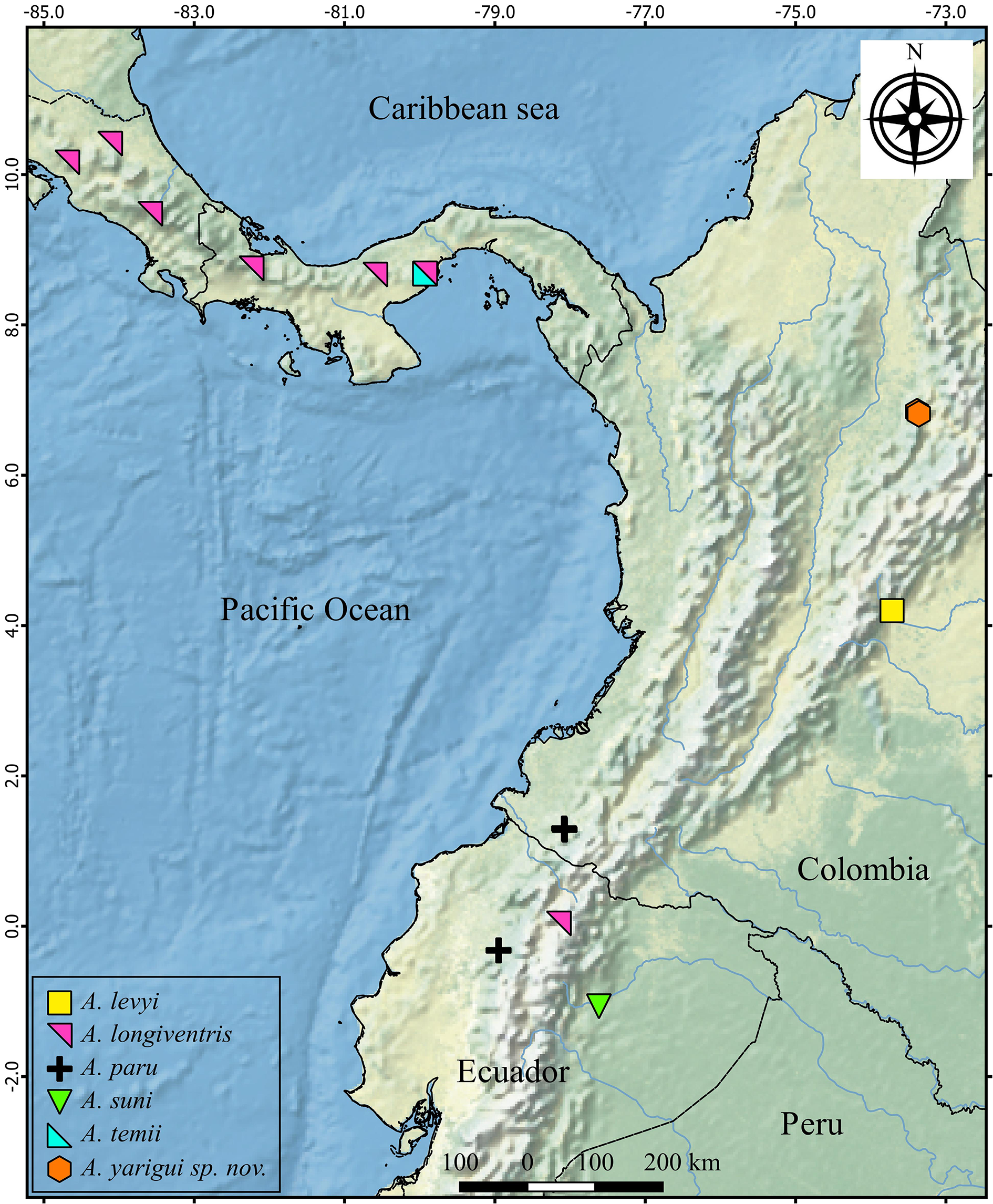
Distribution. Known only from the type locality, at the Serranía de los Yariquíes National Natural Park, in the Andean department of Santander, Colombia ( Fig. 1 View FIGURE 1 ).
Natural history. The male holotype was collected by beating low vegetation in a highly conserved wet Andean forest. Subadult males and juveniles were collected in the same type locality by beating low vegetation, and manually during the day, in both secondary and very-intervened ecosystems.
Comments. Anaptomecus yarigui sp. nov. is known solely from one male and there is another species from Colombia, A. levyi , known solely from a female. Nevertheless, we consider them separate species due to the fact that the type localities have distinctly different altitudes, and they are more than 300 km apart from each other. Both in a region, as northern Andes, known for its extremely high levels of diversity and endemisms in plants and animals ( Halffter 1992), including spiders ( Galvis 2017). Anaptomecus levyi was collected between 800–1200 m, probably in the Susumuco Mountain range in the department of Meta, just east of Bogotá [see Jäger et al. (2009) for discussion on the type locality], with altitudes reaching 1900 m , while A. yarigui sp. nov. was collected between 1700–2200 m, in the Serranía de los Yariguíes , in the department of Santander, with much higher altitudes, more than 3200 m .
In addition, both species show differences in coloration pattern, leg measurements and cheliceral dentition. While A. yarigui is generally pale brown, with a darker pattern on the dorsal shield of prossoma and opisthosoma completely covered by guanine crystal spots, A levyi is generally pale yellow with simple pale brown marginal bands on prossoma and fewer guanine spots on opisthosoma. Moreover, Anaptomecus yarigui has 6–7 retromarginal teeth on the chelicerae and leg formula 2143, while A. levyi has 5 retromarginal teeth and leg formula 1243.

No known copyright restrictions apply. See Agosti, D., Egloff, W., 2009. Taxonomic information exchange and copyright: the Plazi approach. BMC Research Notes 2009, 2:53 for further explanation.
|
Kingdom |
|
|
Phylum |
|
|
Class |
|
|
Order |
|
|
Family |
|
|
Genus |