
Anthothela echinata ( Kükenthal, 1915 ) Watling, 2020
|
publication ID |
https://doi.org/ 10.11646/zootaxa.4881.2.9 |
|
publication LSID |
lsid:zoobank.org:pub:37F1B897-6C25-4F8C-BCF1-1EFDDDFB6E59 |
|
DOI |
https://doi.org/10.5281/zenodo.4328164 |
|
persistent identifier |
https://treatment.plazi.org/id/03C087E4-FFFD-4D5E-FF39-F924FE7DDDA9 |
|
treatment provided by |
Plazi |
|
scientific name |
Anthothela echinata ( Kükenthal, 1915 ) |
| status |
comb. nov. |
Anthothela echinata ( Kükenthal, 1915) View in CoL n. comb.
( Figs. 1–4 View FIGURE 1 View FIGURE 2 View FIGURE 3 View FIGURE 4 )
Muricellisis echinata Kükenthal, 1915, p. 124 View in CoL ; Kükenthal, 1919, p. 627
Diagnosis. Colony membranaceous. Polyp cylindrical, often exsert or invaginated into calyx, crowded together with little coenenchyme space. Coenenchyme and calyx sclerites clubs, thorn-clubs, sticks and spindles; polyp sclerites sticks and spindles arranged as collaret and points. Tentacle sclerites short, flat tuberculate rods along the rachis with spatulate clubs in the pinnules. Pharyngeal sclerites not known.
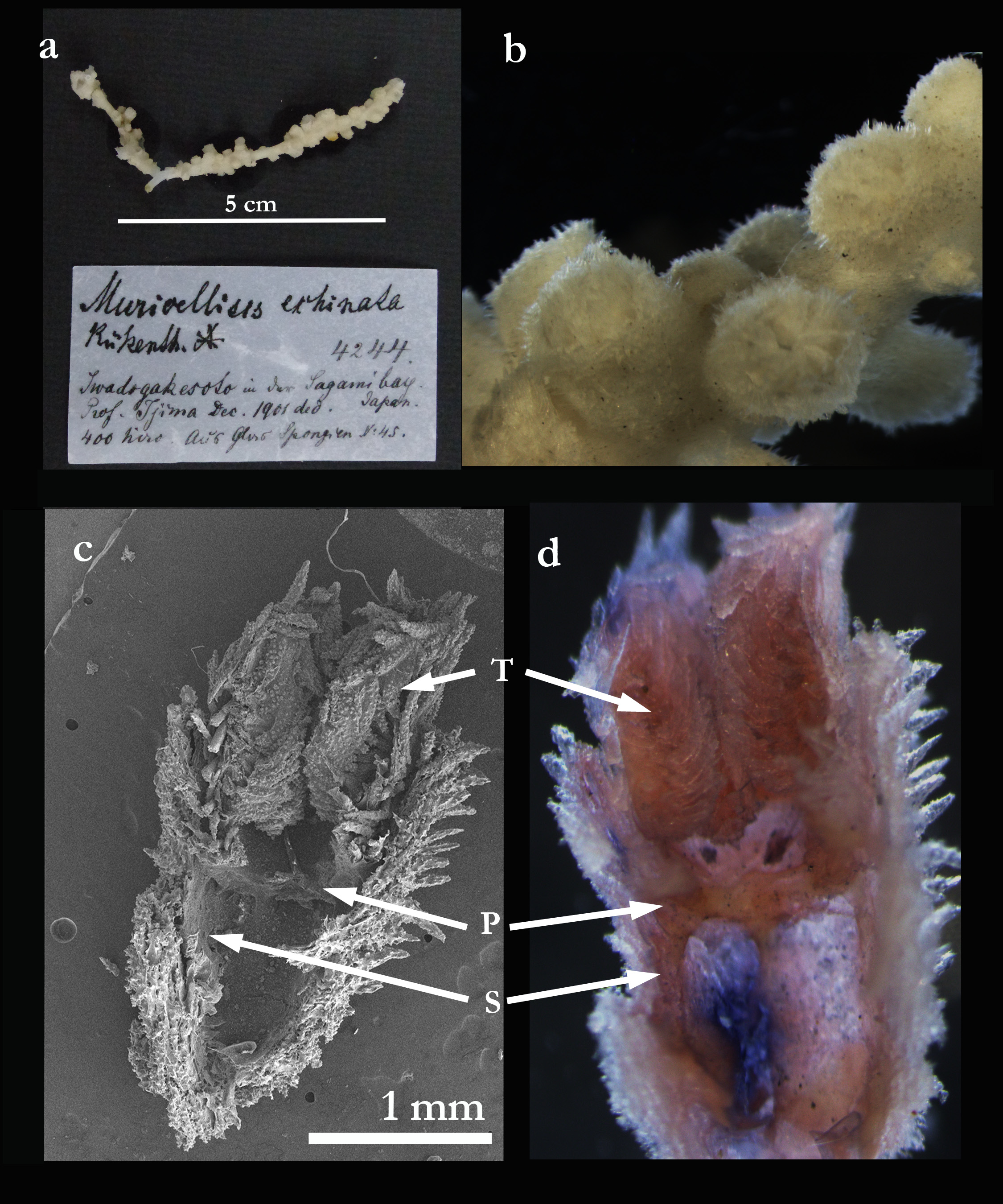
Type specimen. Museum für Naturkunde, Berlin, Germany
Type locality. Sagami Bay , 730 m.
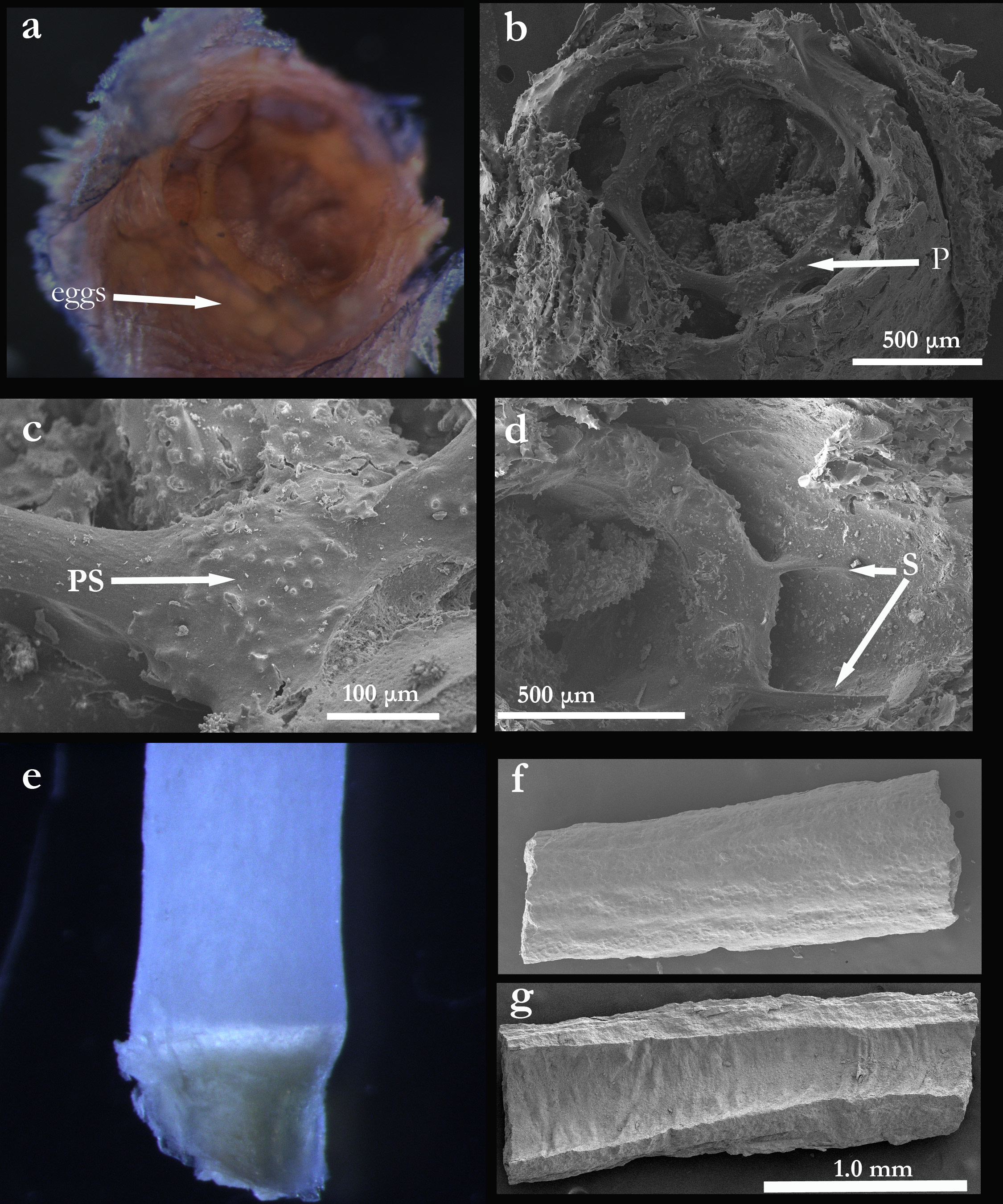
Description. The type consists of three fragments totaling 11 cm according to Kükenthal (1919). The following description applies to an 8 cm piece loaned from the Museum für Naturkunde ( Fig. 1a View FIGURE 1 ). The colony is membranaceous, overgrowing a piece of axis from a keratoisid bamboo coral ( Fig. 2 View FIGURE 2 e–g).
Polyp calyces are very closely arranged with little coenenchyme space between them ( Fig. 1b View FIGURE 1 ). The polyps are cylindrical, short, and mostly invaginated within the calyx, with the tentacles folded into the polyp ( Fig. 1c, d View FIGURE 1 ). The polyp does not appear to have a typical cylindrical pharynx, rather there is a thick ring of tissue (“pharyngeal ring”) attached at the proximal end of the polyp as it invaginates into the calyx ( Fig 1c, d, P View FIGURE 1 ). A few sclerites are visible under the tissue on the proximal (ventral) side of the pharyngeal ring ( Fig. 2b, c View FIGURE 2 ). Short septa (S) extend proximally to the calyx wall from the pharyngeal ring and seem to spread across the interior of the calyx ( Fig. 1c, d View FIGURE 1 , Fig. 2d View FIGURE 2 ). In one case, eggs appear to be present along the inner calyx wall ( Fig. 2a View FIGURE 2 ).
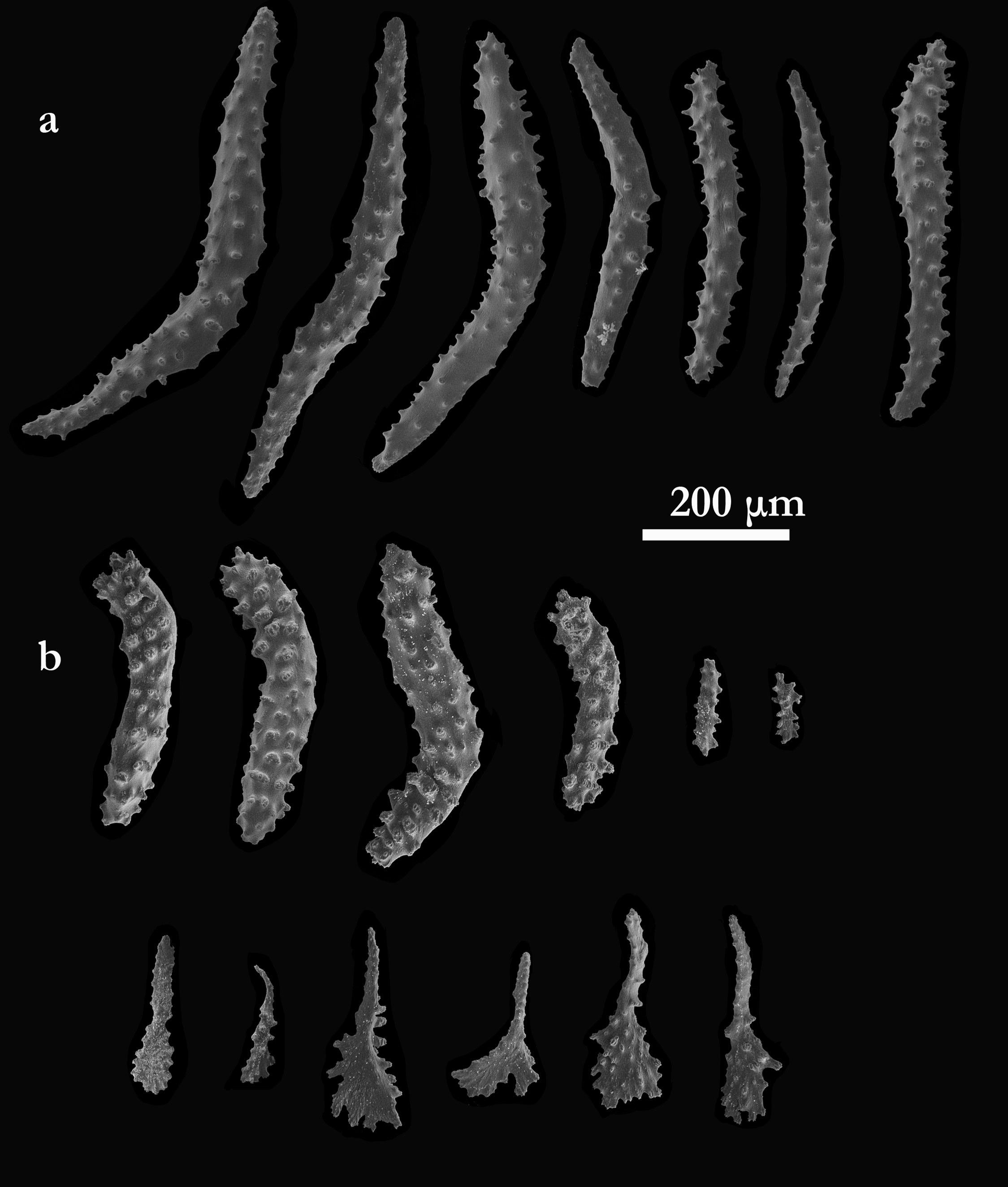
The sclerites of the coenenchyme consists of sticks and small clubs ( Fig. 3a View FIGURE 3 ). Most of the calyx sclerites are small clubs and thorn clubs of various sizes ( Fig. 3b View FIGURE 3 ). The polyp sclerites are spindles in a collaret (?) and points arrangement. Flattened tuberculate rods are common on the tentacle rachis and small rods and spatulate clubs are found in the pinnules.
Remarks. Kükenthal (1915, 1919) created the new genus Muricellisis for this species and added it to the Family Isididae as a new subfamily Muricellisidinae due to the presence of a calcareous axis with nodes and internodes underlying the polyps and coenenchyme. However, the curious form of the polyps and the spiny and tuberculate sclerites were unlike anything seen in the Isididae to that time. Kükenthal noted that had the calcareous axis not been present, he would have thought this species to be similar to Muriceides .
The sclerites of this species, in particular the thorn-clubs of the calyx wall and spatulate clubs in the pinnules, suggests that the species belongs to the Anthothelidae . In addition, the documentation by Moore et al. (2017) that some members of the genus Anthothela could exist as membranous colonies with no axis cortex and medulla, hinted at the possibility that the keratoisidid axis present here could merely be a substrate on which the colony was growing. In fact, the form of the axis is definitely keratoisid-like, but also seems to be considerably degraded (images not shown). However, there are no members of the Keratoisidinae that have sclerites in the form possessed by this species. The most parsimonious explanation for the disparity between the form of the axis and the nature of the polyps and sclerites is that the octocoral and the axis on which it is growing do not belong to the same species.
From the review of the Anthothelidae by Moore et al. (2017), there does not seem to be any known species in the genus Anthothela to which this species could belong. Several species have colonies with fully formed axes but as well are membranous growing on some unrelated substrate such as sponge spicules. Of the seven species of Anthothela , three have thorn clubs: in A. vickersi the thorn clubs are mostly rounded at the tips; those of A. aldersladei and A. tropicalis are most like those seen here in A. echinata . As with A. aldersladei , the thorn clubs are shorter (0.4–0.55 mm) than in A. tropicalis (0.33–0.78 mm). The calyces in A. aldersladei , however, are much taller than in A. echinata (1.5–2.5 mm in height vs. 1–1.5 mm, and 2–2.5 mm in width vs. 1.5–2 mm). Hockey stick sclerites were not observed in the few calyces and polyps of A. echinata that were examined. Their absence would further distinguish these two species.

No known copyright restrictions apply. See Agosti, D., Egloff, W., 2009. Taxonomic information exchange and copyright: the Plazi approach. BMC Research Notes 2009, 2:53 for further explanation.
|
Kingdom |
|
|
Phylum |
|
|
Class |
|
|
Order |
|
|
Family |
|
|
Genus |
Anthothela echinata ( Kükenthal, 1915 )
| Watling, Les 2020 |
Muricellisis echinata Kükenthal, 1915 , p. 124
| Kukenthal 1915: 124 |