
Anthothela aldersladei, Moore & Alderslade & Miller, 2017
|
publication ID |
https://doi.org/ 10.11646/zootaxa.4304.1.1 |
|
publication LSID |
lsid:zoobank.org:pub:3D557C94-0783-4C39-80C3-9C321DA94800 |
|
DOI |
https://doi.org/10.5281/zenodo.6015369 |
|
persistent identifier |
https://treatment.plazi.org/id/039B87ED-3E03-FFFE-FF4B-E63E7DD2D958 |
|
treatment provided by |
Plazi |
|
scientific name |
Anthothela aldersladei |
| status |
sp. nov. |
Anthothela aldersladei View in CoL sp. nov. Moore & Miller in Moore, Alderslade & Miller
http://zoobank.org/A55A7194-F698-4F8B-A9B9-E4FCC8759297 ( Figs. 42–55 View FIGURE 42 View FIGURE 43 View FIGURE 44 View FIGURE 45 View FIGURE 46 View FIGURE 47 View FIGURE 48 View FIGURE 49 View FIGURE 50 View FIGURE 51 View FIGURE 52 View FIGURE 53 View FIGURE 54 View FIGURE 55 )
Material examined. Holotype: WAM Z 31463, 190km NW of Karratha , Pluto Gas Field, Western Australia, SKM Pluto Gas Field Survey (PF06/S1–600/R2), 19.874°S, 115.166°E, depth 600 m, 7th December 2005. GoogleMaps
Paratypes: WAM Z13059 View Materials , North West Cape , Exmouth, Western Australia, AIMS North West Cape Survey II 2002, Fromont, J., Marsh, L.M. & Alderslade, P.N., stn. 0 4, 21.48°S, 113.966°E, depth 570 m, 20th March 2002 GoogleMaps ; WAM Z90585, Perth Canyon, SE Indian Ocean, Western Australia, CSIRO RV Southern Surveyor , stn. 73 (SS 200510 /073-020), 31.9650°S, 115.105°E, depth 1000 m, 30th November 2005 GoogleMaps .
Description:
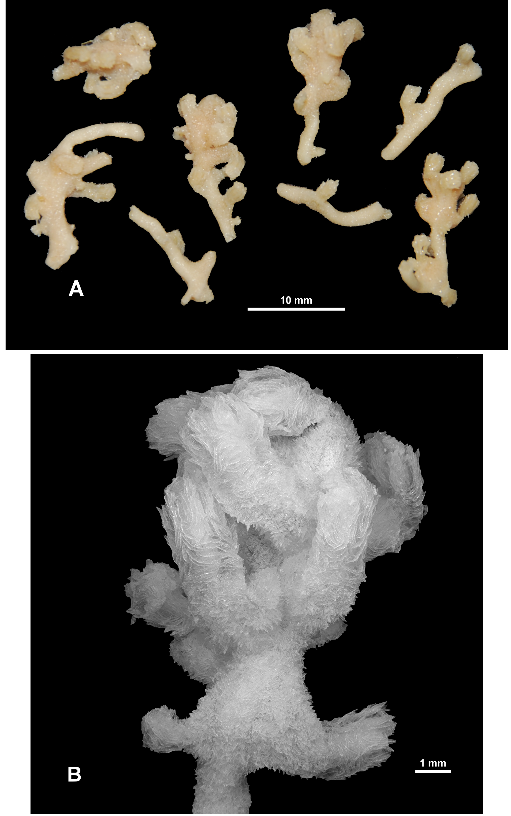
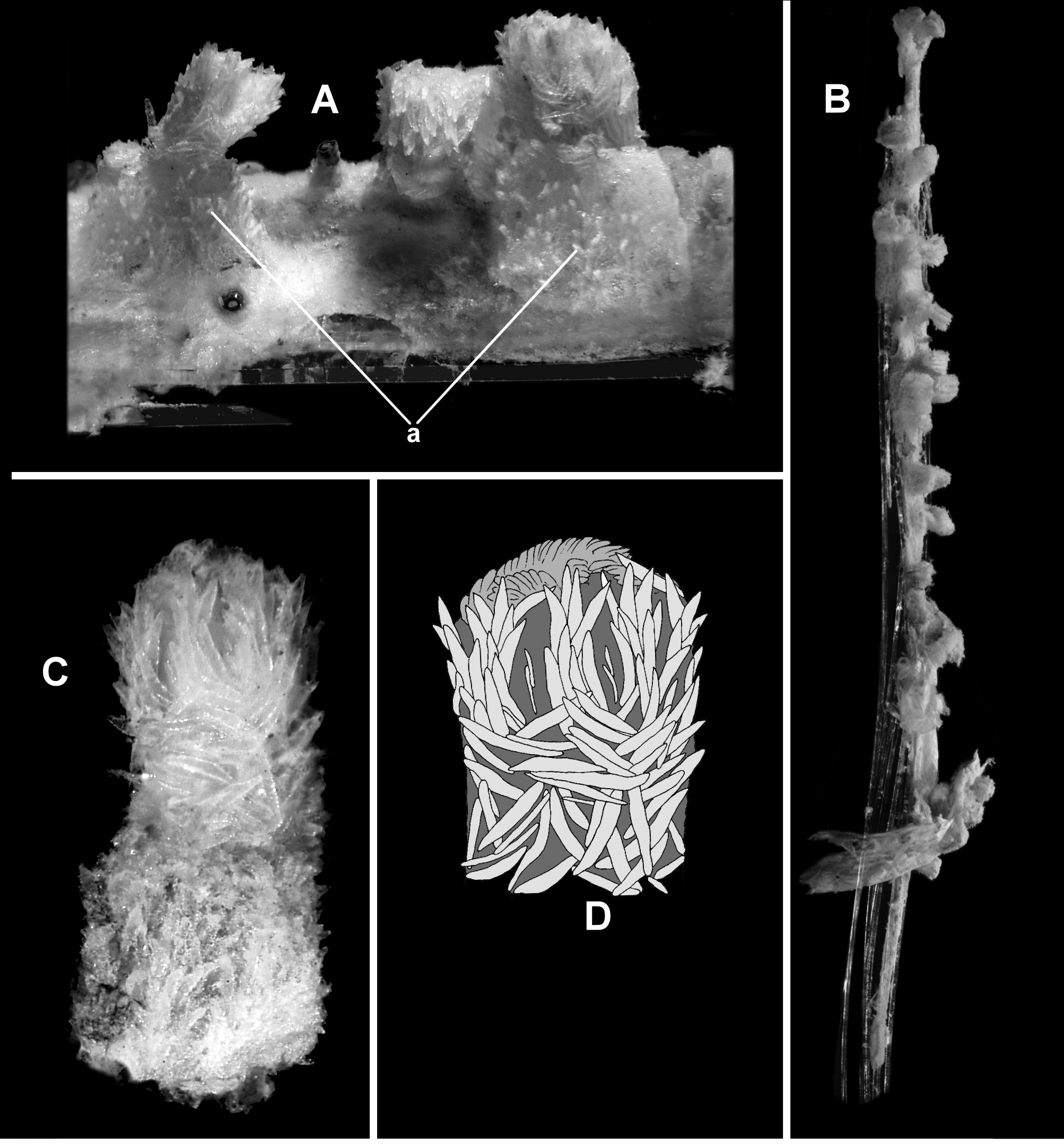
Colony form: The holotype is broken into 6 small, irregular pieces of branches, all with calyces and polyps ( Fig. 42 View FIGURE 42 A). It is not possible to confidently reconstruct the shape or size of the colony, however the slightly twisted nature of the branches and the many bifurcation points indicate the colony form was probably tangled with irregular branching. The pieces of colony range in length from 10.6 mm to 24.6 mm and all are narrow (1.2–2.2 mm) and relatively delicate. The branches are usually circular in cross-section although they tend to flatten or distort at bifurcation points and where calyces arise. One piece has some evidence of anastomoses. All the colony pieces are in good condition with many intact polyps and undamaged surfaces.
On three of the colony pieces, calyces are crowded into clavate terminal bunches with no space between the bases ( Fig. 42 View FIGURE 42 B). Proximal to the terminal bunches and on the remainder of the colony pieces, calyces occur sparsely, on all sides of the branches and project at right angles. There are sections of the branches which have no calyces; the largest of these spaces is 8.8 mm long.
Colour: There is no record of live colour for this specimen; it is now light beige in alcohol.
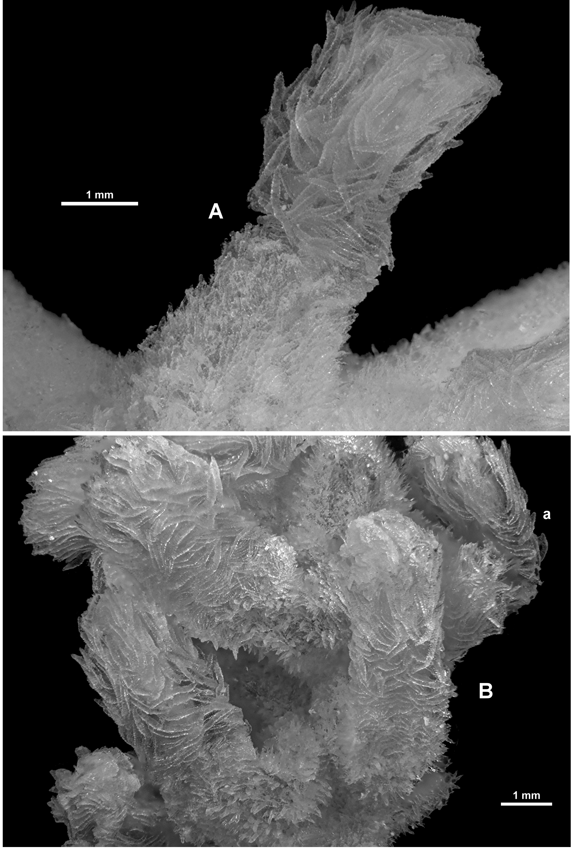
Polyps and Calyces: Calyces are large relative to the branch diameter, and range from 1.5–2.5 mm in height and 2–2.5 mm in width. They tend to be conical, and are clearly differentiated from the polyp neck and head by the arrangement and alignment of the sclerites ( Fig. 43 View FIGURE 43 A), which are small, crowded, arranged longitudinally and project out from the surface of the calyx giving it a prickly appearance. In contrast, immediately above the calyx lip, on the polyp neck and head much larger sclerites are arranged transversely, covering the polyp neck with no obvious thinning of the dense arrangement as is usually the case in other Anthothela species. These large sclerites continue obliquely to longitudinally up eight well-defined and quite spectacular points. All polyps are exsert with little or no invagination of the neck region and often with the polyp head bent over ( Fig. 43 View FIGURE 43 Ba), protruding 2.2–3.2 mm from the lip of the calyx and having a diameter of approximately 1.2–2.2 mm. The eight tentacles fold over the mouth of the polyp creating eight rounded ridges on the top of the polyp head. There are approximately 10 pinnules arranged in a single row along each side of the tentacles.
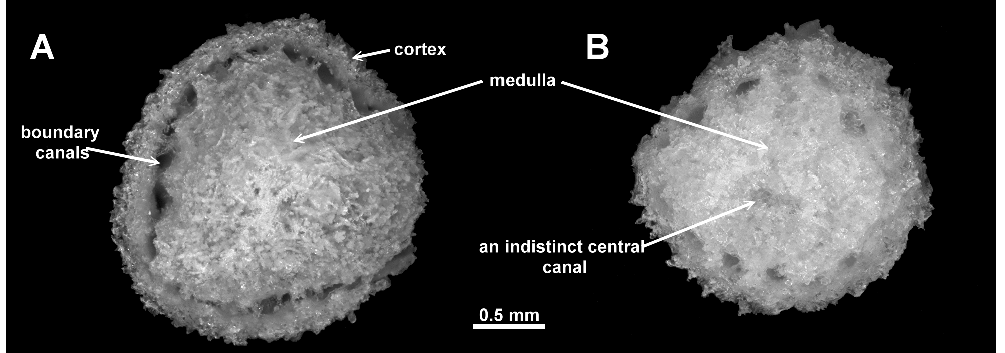
Medulla and Cortex: The branches of the colony are composed of a central medulla, made up of tightly packed longitudinally arranged sclerites, that is surrounded by a cortex that is approximately 0.1–0.2 mm thick. The cortex and medulla are separated by a crowded series of adjacent longitudinal canals which encircle the medulla, allowing it to be easily separated from the cortex. A cross-section taken at the widest available part of the branches clearly shows the boundary canals, with no obvious, internal coelenteric canals ( Fig. 44 View FIGURE 44 A). In a narrower part of the colony, another cross-section demonstrates the same clear boundary canals with perhaps some indistinct canals in the central medulla ( Fig. 44 View FIGURE 44 B) which are more likely a thinning of the sclerites rather than defined canals. The body cavities of the polyps along the branches terminate at the medulla while the gastric canals of the polyps that are arranged in bunches at the tips of some branches tend to extend internally down the branch a short distance.
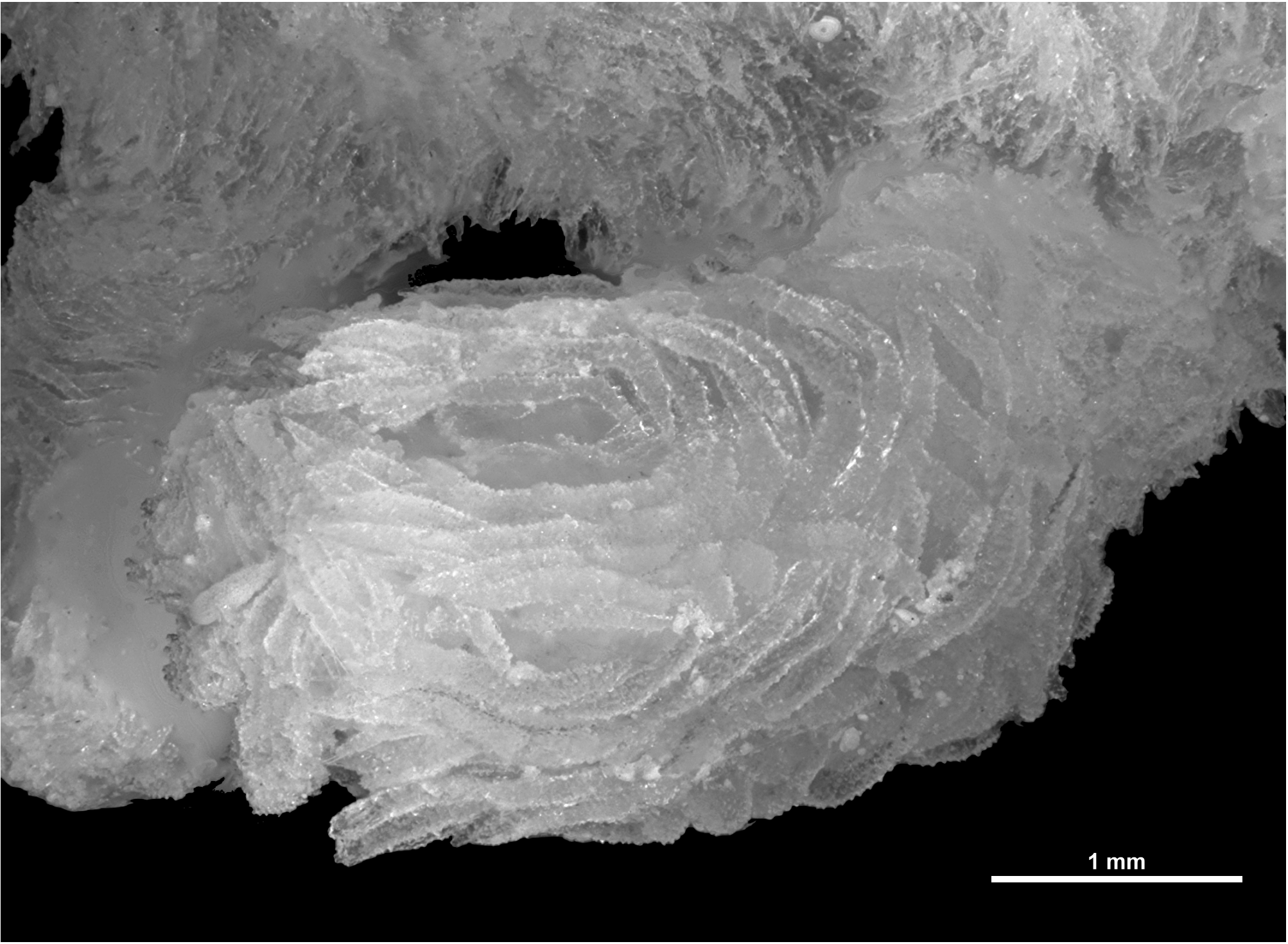
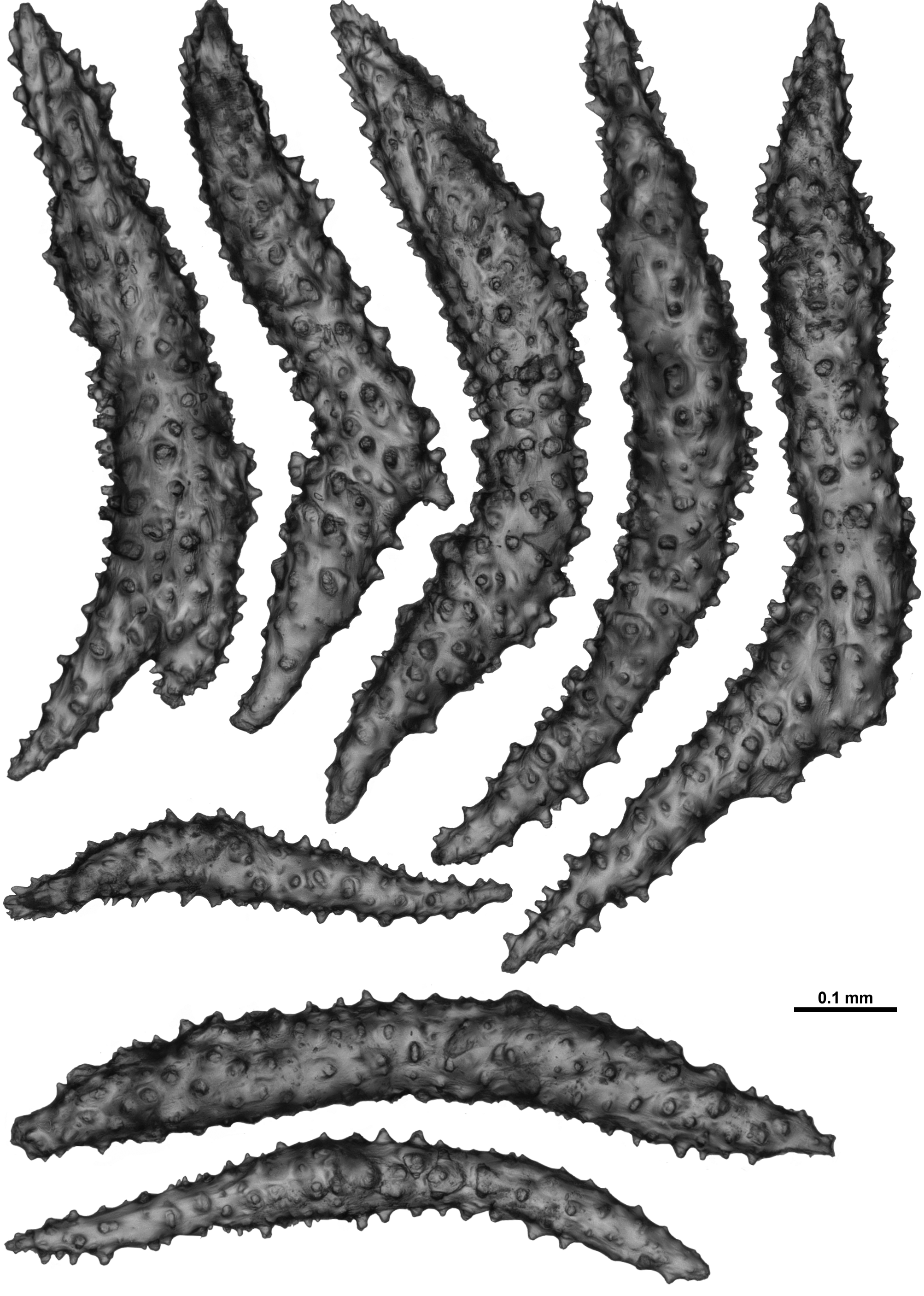
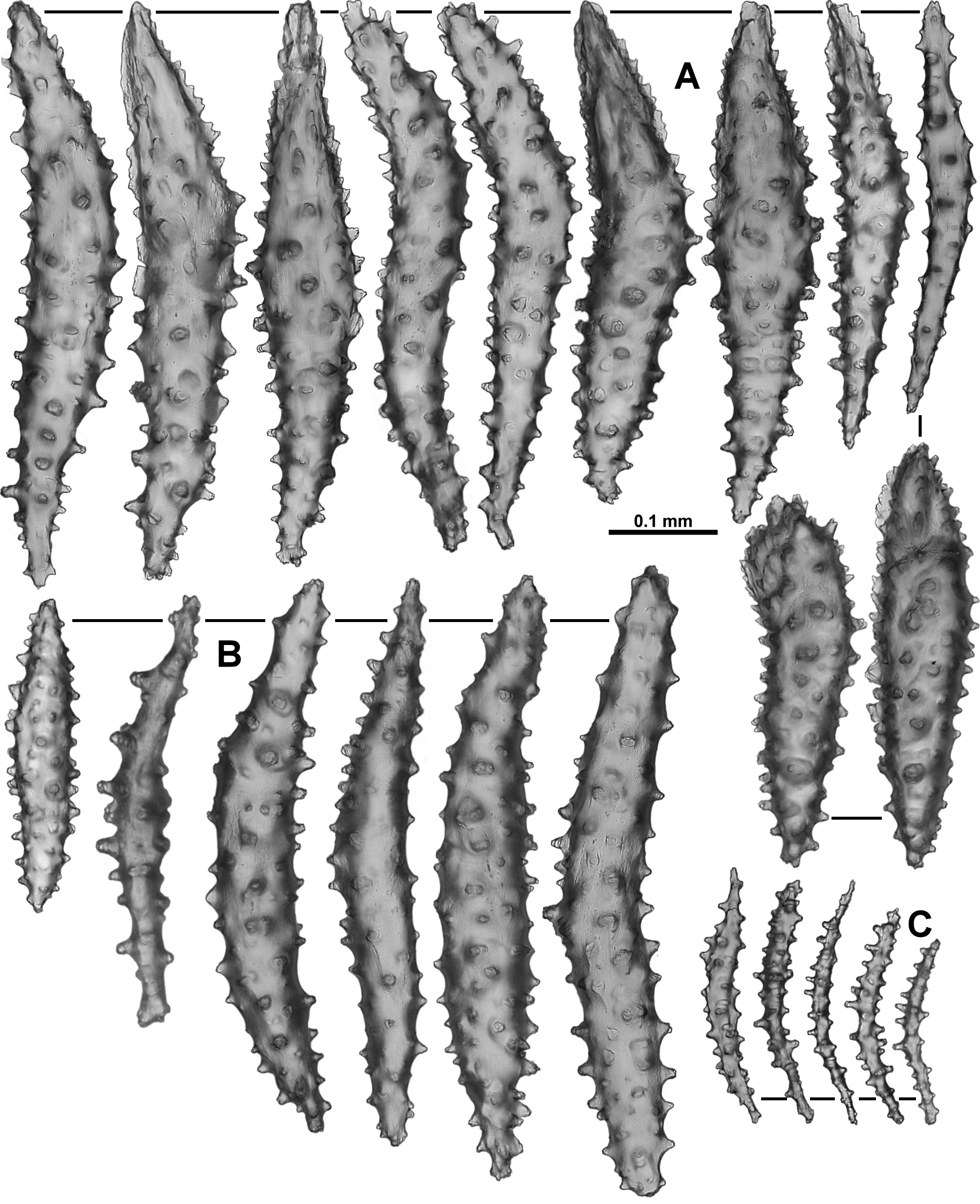
Sclerites: The polyps and calyces are covered with a dense layer of crowded sclerites which are mostly tuberculate sticks and spindles on the polyp head and spiky thorn clubs on the calyx and colony surface. On the polyp head sclerites are very large, relative to the polyp, and are not as crowded as elsewhere on the colony. The largest sclerites are crescents or bent tuberculate hockey-sticks, with the straight, longest part of the sclerite arranged longitudinally in the points and the proximal portion curving to be transverse at the base of the points ( Figs. 43 View FIGURE 43 A; 45). Some have roots (or small branches) at the base and many have a serrated, thorny tip ( Fig. 46 View FIGURE 46 ) that can project out from the polyp head and above the back of the folded tentacles ( Fig. 45 View FIGURE 45 ). There is no true collaret, rather curved spindles are arranged transversely and obliquely down the polyp neck with no diminution of the sclerite cover at the neck area. Sclerites in the points range in size from 0.40–0.90 mm approximately, while those from the neck are slightly smaller (0.26–0.77 mm).
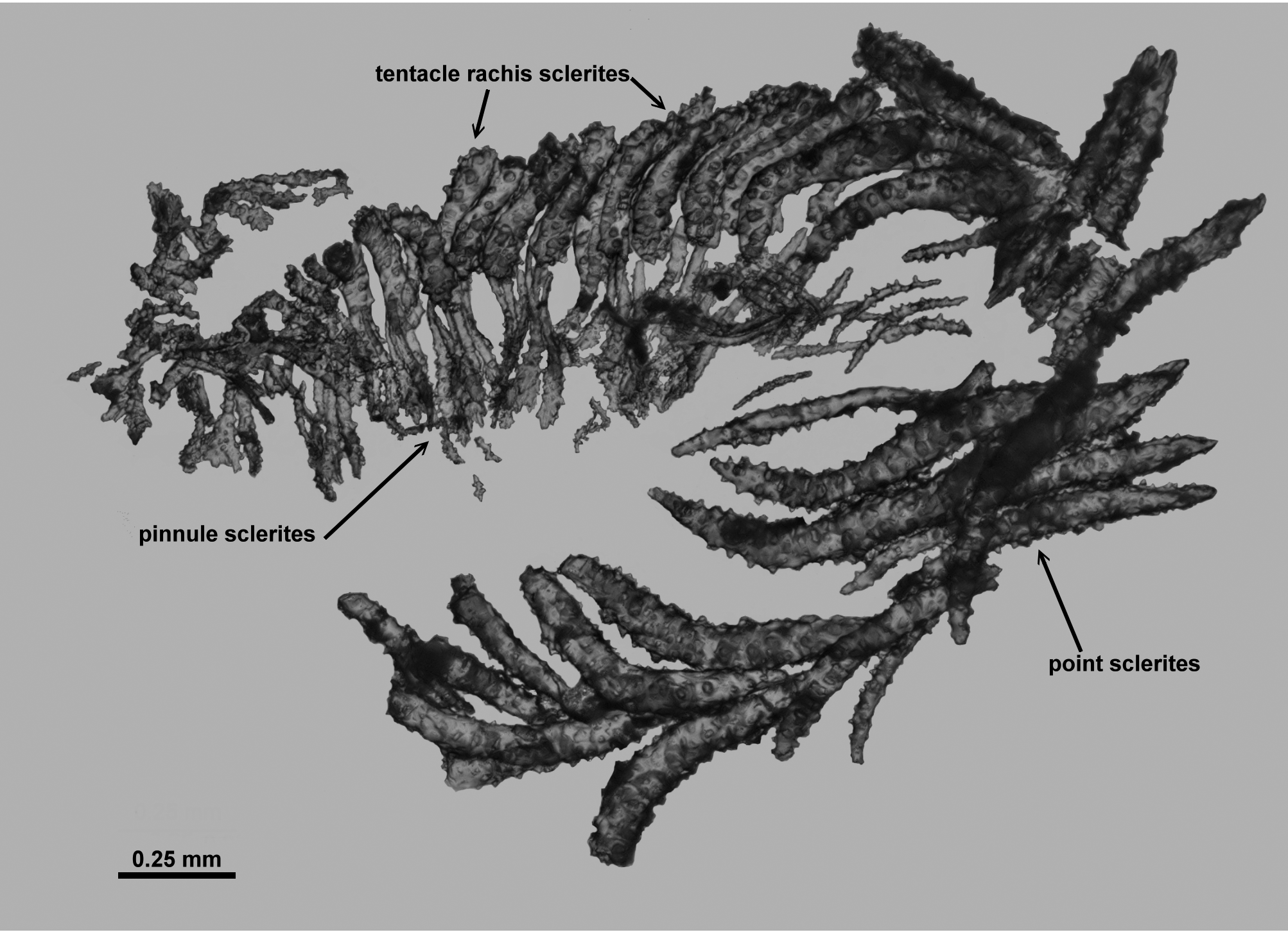
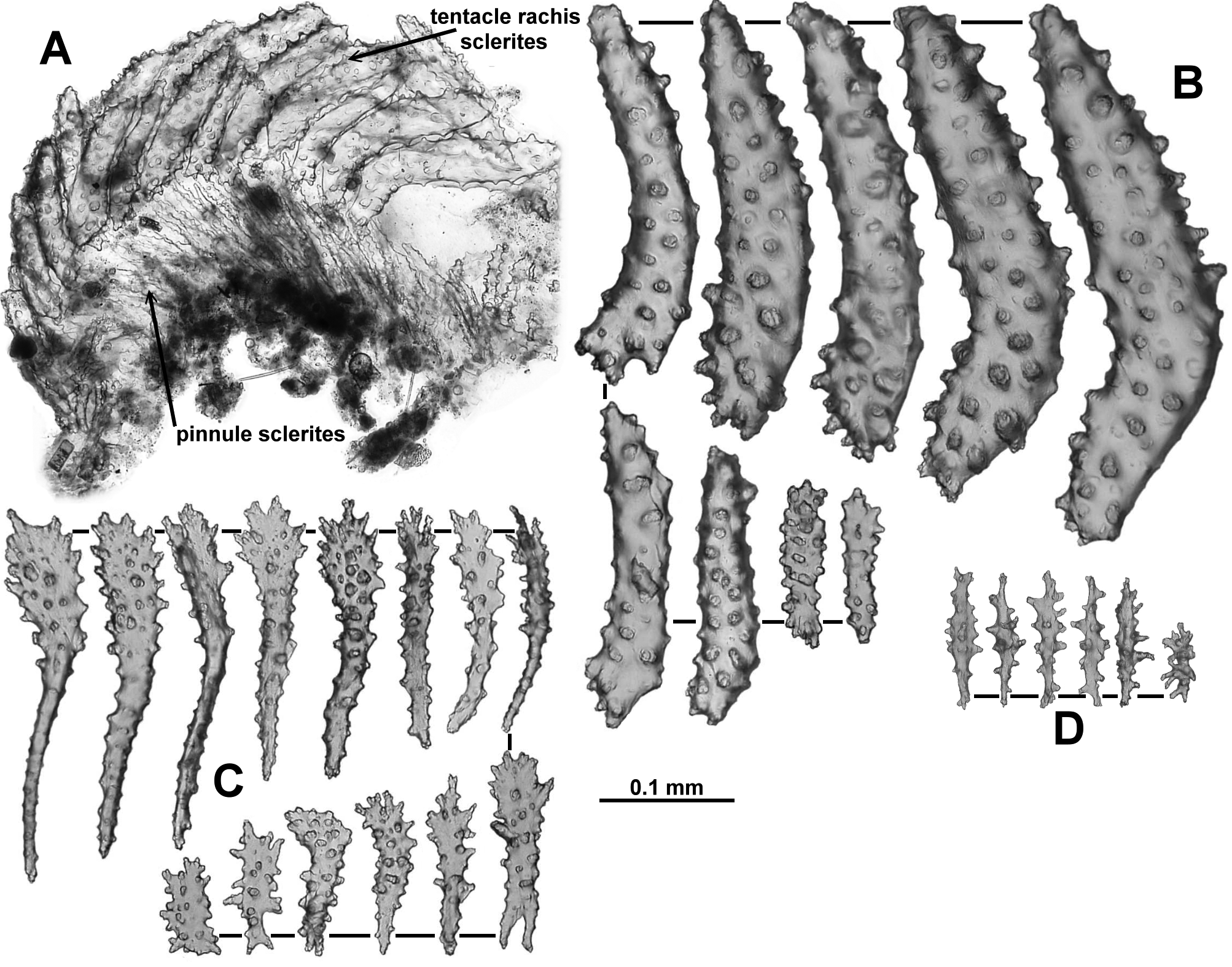
From the tip of the points, sclerites continue obliquely along the tentacle rachis ( Fig. 47 View FIGURE 47 ). These sclerites are bent or straight tuberculate rods and sticks and spindles often with the shorter, curved end ( Fig. 48 View FIGURE 48 Aa) extending onto the tentacle flanks towards the pinnules. Straight sclerites are more commonly along the middle ridge of the tentacle rachis. The sclerites diminish in size along the tentacle; on the proximal end, the largest sclerites are approximately 0.58 mm long grading to the distal end of the tentacle where the smallest sclerites are approximately 0.20 mm long.
In the pinnules, sclerites are crowded longitudinally and are delicate and easily broken ( Fig. 47 View FIGURE 47 ). Spatulate clubs are common, with a tapered handle and a broad, spatulate, almost leaf-like end, that is oriented distad in the pinnules ( Fig. 48 View FIGURE 48 B). These sclerites vary in length from 0.12–0.32 mm and the handle can be narrow and cylindrical or wide and flat. The smaller sclerites grade from spatulate clubs to simple tuberculate rods. There are also short flat rods (0.08–0.1 mm long) with sparse tubercles and narrow curved sticks and spindles (0.14–0.21 mm long) inter-dispersed with the spatulate clubs ( Fig. 48 View FIGURE 48 C, D).
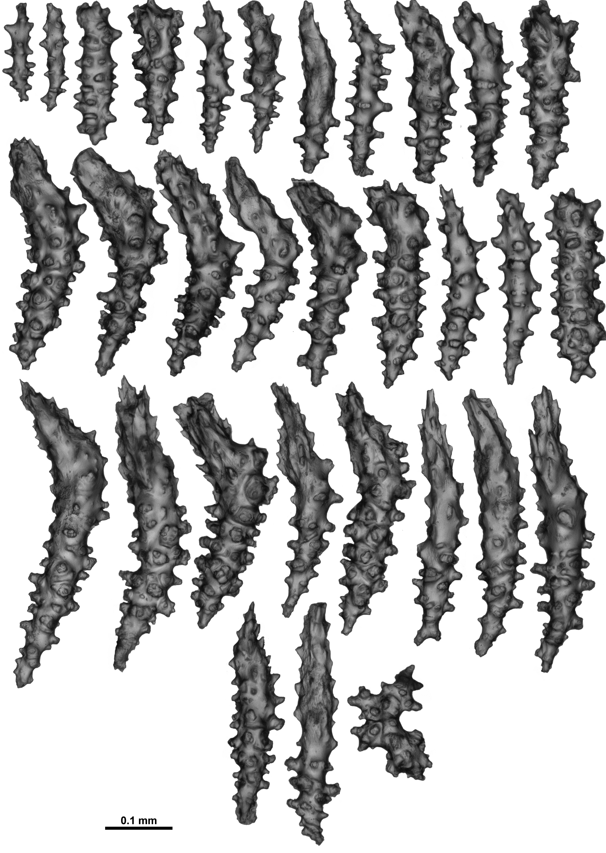
Calyces are covered in a dense and prickly layer of sclerites, almost all of which are small, bent, tuberculate thorn clubs ( Fig. 49 View FIGURE 49 ), orientated with the foliaceous, thorny tips distal on the calyx and angled out from the surface giving the calyx its prickly appearance. For the smaller sclerites there is some gradation between thorn clubs and those which are less developed at the tip and could be termed a wart club. Most calyx sclerites range from 0.22– 0.52 mm, however there are some smaller, straight, tuberculate sticks and spindles mingled with the thorn clubs, which only reach approximately 0.17 mm in length.
Very small sclerites with conical spines occur in the pharynx ( Fig. 50 View FIGURE 50 A). They are quite numerous and tend to occur in bunches. The size ranges from 0.05–0.12 mm long.
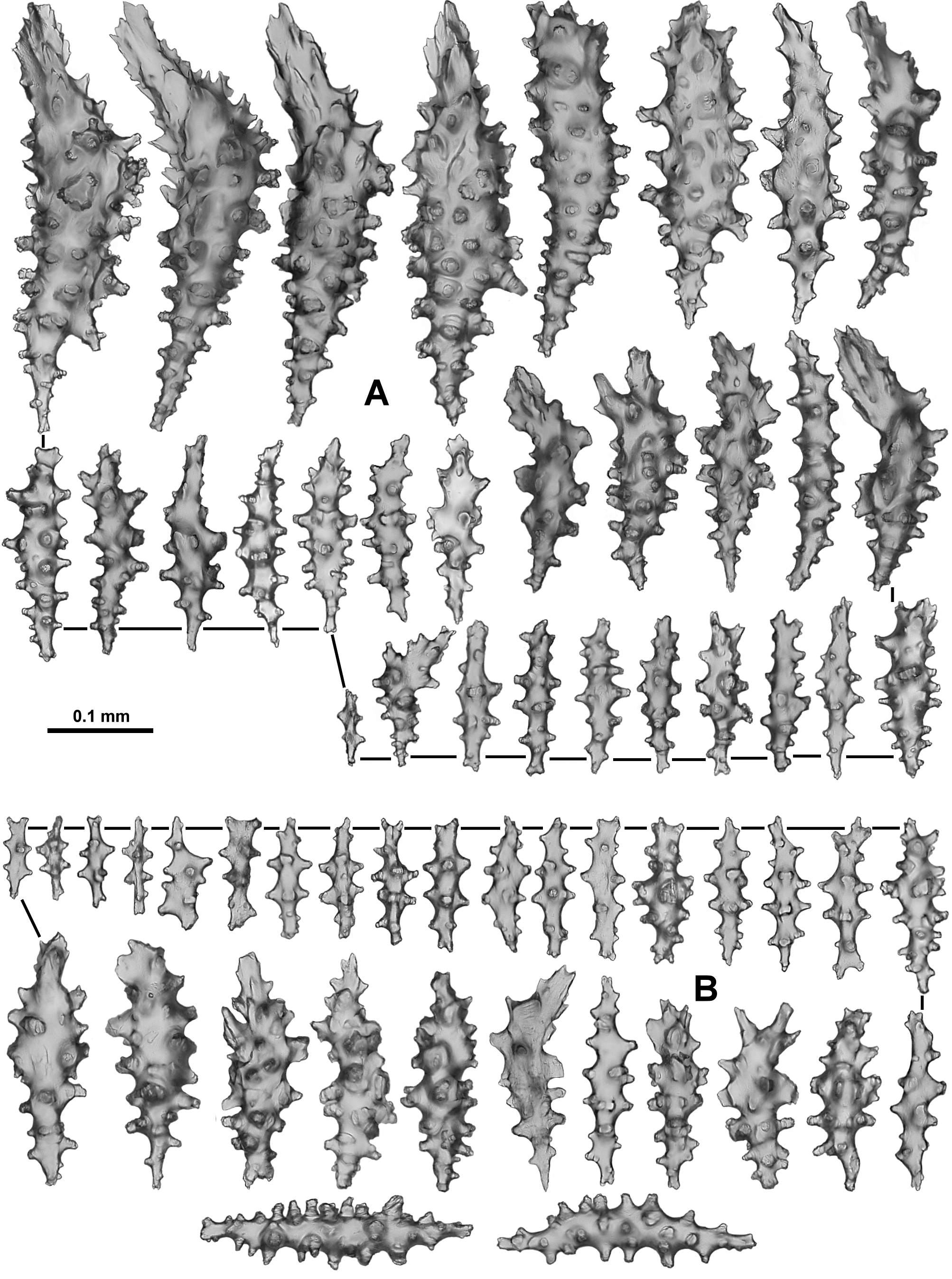
The cortex contains sclerites very similar to those in the calyx—small, bent, warty thorn clubs with quite complex, at times foliose, spear-tips ( Fig. 50 View FIGURE 50 B). They are tightly packed with the tips projecting out from the surface giving the branches a very prickly appearance. Size does not vary much with most of the sclerites being from 0.19–0.43 mm long but occasionally there are some up to 0.53 mm long and as small as 0.12 mm. Amongst these short thorn clubs are some simple, tuberculate sticks and spindles (of a similar length) but the thorn clubs are far more common.
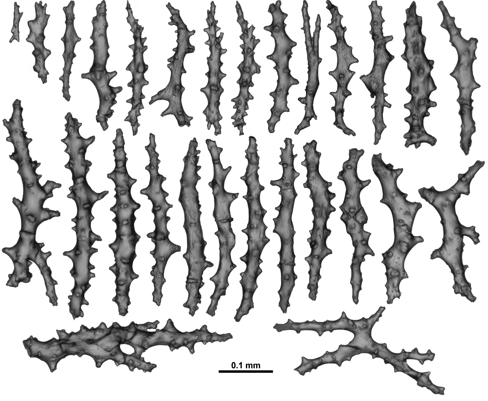
The medulla is composed of tightly packed, longitudinally arranged sclerites—mostly sparsely tuberculate sticks and spindles ( Fig. 51 View FIGURE 51 ). Occasionally there are larger sticks and spindles with only sparse tubercles, often with branches, forks and fused areas. Most sclerites are from 0.20–0.53 mm long although many of those sampled showed evidence of breakage. It was difficult to ensure these long sclerites remained undamaged during sampling so the prevalence of these cannot be estimated. Occasionally there are small spindles only 0.1 mm in length.
Sclerites are uniformly transparent under transmitted light.
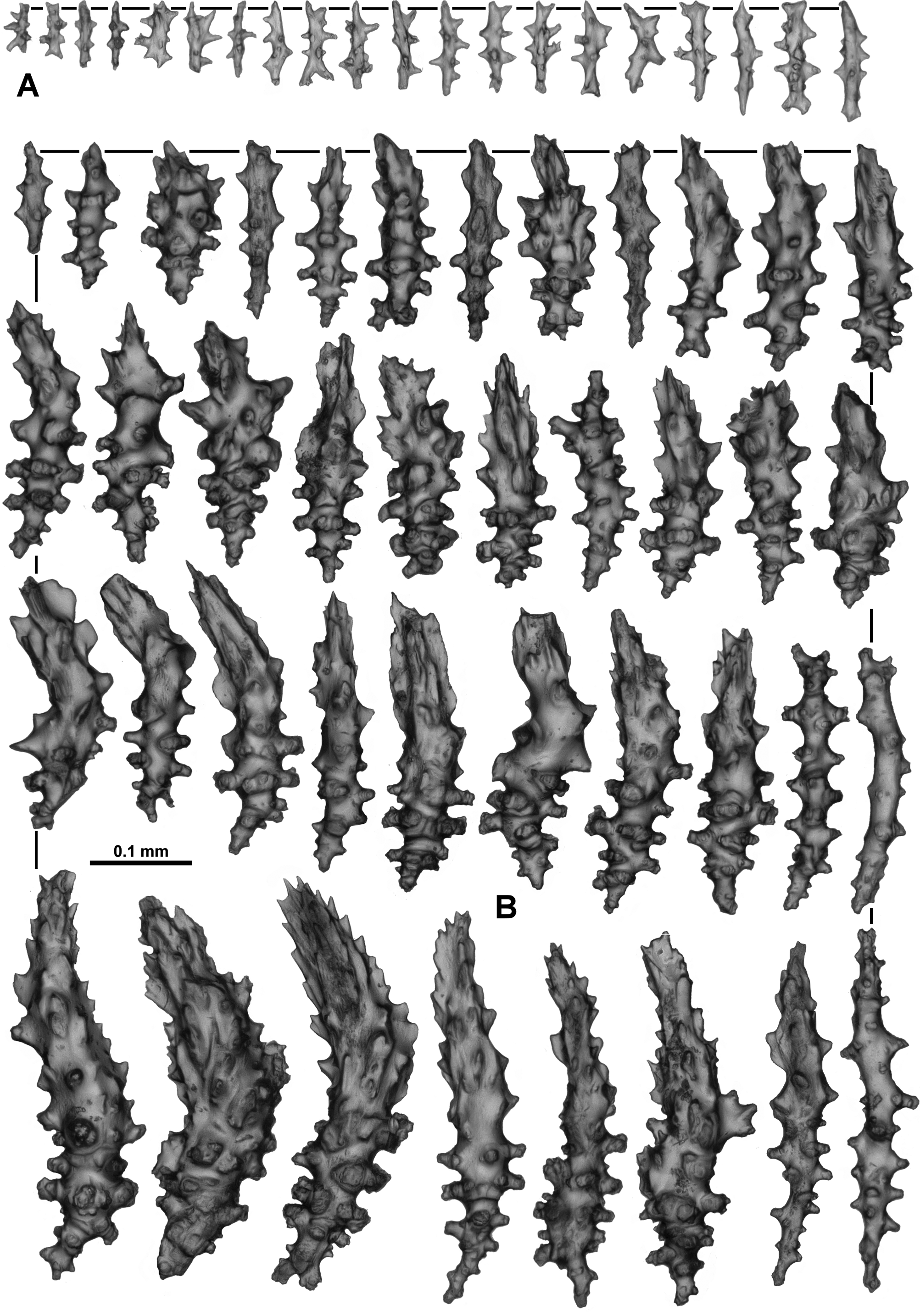
Variation: The paratype, WAM Z13059 View Materials , is membranous only, thinly encrusting large, straight sponge spicules ( Fig. 52 View FIGURE 52 A, B). It is from a site close to where the holotype was collected and was found at a similar depth. There is a similarly obvious delineation between the calyx and the polyp body ( Fig. 52 View FIGURE 52 A, C) and no retracted polyps were noted. Straightened out, the largest polyp is 5 mm long with the head 1.7 mm long and 1.4 mm wide. The sclerites on the points and neck region are smaller than those of the holotype (the largest measured at 0.58 mm) and slightly more crowded ( Fig. 52 View FIGURE 52 C, D) but the serrated ridges and thorny tubercles on the distal tips of the point sclerites resemble those of the holotype ( Fig. 53 View FIGURE 53 A) as do those sclerites from the neck ( Fig. 53 View FIGURE 53 B). Between each group of point sclerites there are two small intermediate sclerites which are narrow, curved spindles ( Fig. 53 View FIGURE 53 C). The arrangement of sclerites in the tentacles is similar to that in the holotype ( Fig. 54 View FIGURE 54 A) with bent tuberculate rods and bars ( Fig. 54 View FIGURE 54 B) arranged obliquely along the aboral side of the tentacle rachis and down onto the flanks, diminishing in size towards the distal tip, and the pinnules packed with spatulate clubs arranged longitudinally ( Fig. 54 View FIGURE 54 C). Some small rodlets with sparse, simple tubercles were found in the pharynx ( Fig. 54 View FIGURE 54 D). The calyces have well-developed, foliose thorn clubs similar to those in the holotype ( Fig. 55 View FIGURE 55 A), which project out from the surface giving it a prickly appearance ( Fig. 52 View FIGURE 52 Aa). The basal membrane of the colony contains predominantly rodlets, up to 0.2 mm long ( Fig. 55 View FIGURE 55 B). The larger spindles and small foliose, thorn clubs shown in the lower part of this figure are not common.
The other paratype colony is also membranous only, growing on straight sponge spicules, but otherwise corresponds well with the holotype. Interestingly, it is geographically separated from the other two specimens, occurring in the Perth Canyon off southern Western Australia.
Distribution: Western Australian coast
Depth: 570–600 metres.
Remarks: This species is different to other species in the genus Anthothela in having such large sclerites on the neck and in the points and predominately small thorn clubs in the calyces and cortex.
The paratype is membranous only. Colony form has been such a large part of historical determinations in octocorals that linking this colony with scleraxonians which are predominantly branched is not immediately intuitive. In the absence of a medulla, the presence of spatulate clubs in the pinnules, well-developed calyces and clavate sclerites can provide a trigger to assess specimens with regards to Anthothela .
It was only possible to obtain successful sequences of the two mitochondrial gene regions mtMutS and igr1– cox1 from the holotype. Across the length of the two gene regions combined there was only a single nucleotide difference from a clade consisting of A. grandiflora and A. vickersi n. comb. specimens. In the phylogenetic analysis this was sufficient for the A. aldersladei n. sp. specimen to be positioned outside the A. grandiflora / A. vickersi clade but with low support. A single nucleotide in a single specimen may be no more than sequencing error so further attempts to sequence other specimens are necessary for a more robust result.
Etymology: This species is named in honour of Dr Philip Alderslade, the first author’s PhD supervisor, who originally recognised the paratype as a possible Anthothela species and used the specimen as the catalyst for this revision.

No known copyright restrictions apply. See Agosti, D., Egloff, W., 2009. Taxonomic information exchange and copyright: the Plazi approach. BMC Research Notes 2009, 2:53 for further explanation.
|
Kingdom |
|
|
Phylum |
|
|
Class |
|
|
Order |
|
|
Family |
|
|
Genus |