
Rhynchelmis (Rhynchelmis) tetratheca Michaelsen, 1921
|
publication ID |
https://doi.org/ 10.5281/zenodo.194252 |
|
DOI |
https://doi.org/10.5281/zenodo.6201843 |
|
persistent identifier |
https://treatment.plazi.org/id/03C5878E-FFE6-D40C-B2CF-FC117DE36C14 |
|
treatment provided by |
Plazi |
|
scientific name |
Rhynchelmis (Rhynchelmis) tetratheca Michaelsen, 1921 |
| status |
|
Rhynchelmis (Rhynchelmis) tetratheca Michaelsen, 1921 View in CoL
( Figs. 1–2 View FIGURE 1. R View FIGURE 2. R )
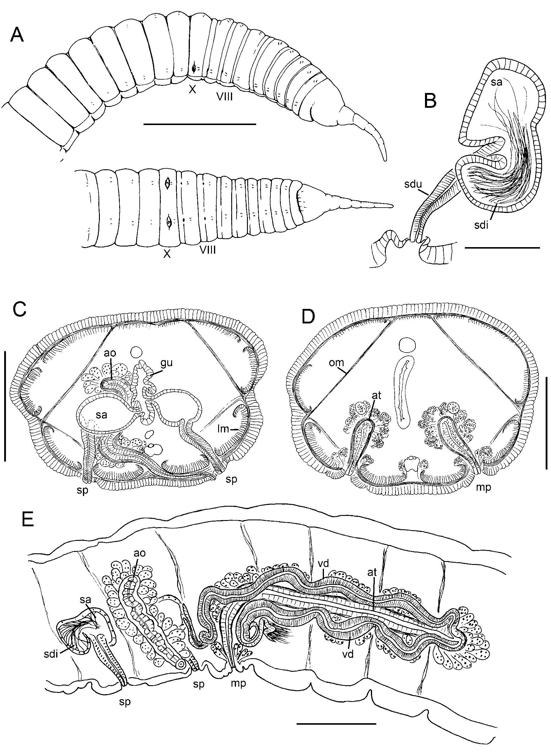
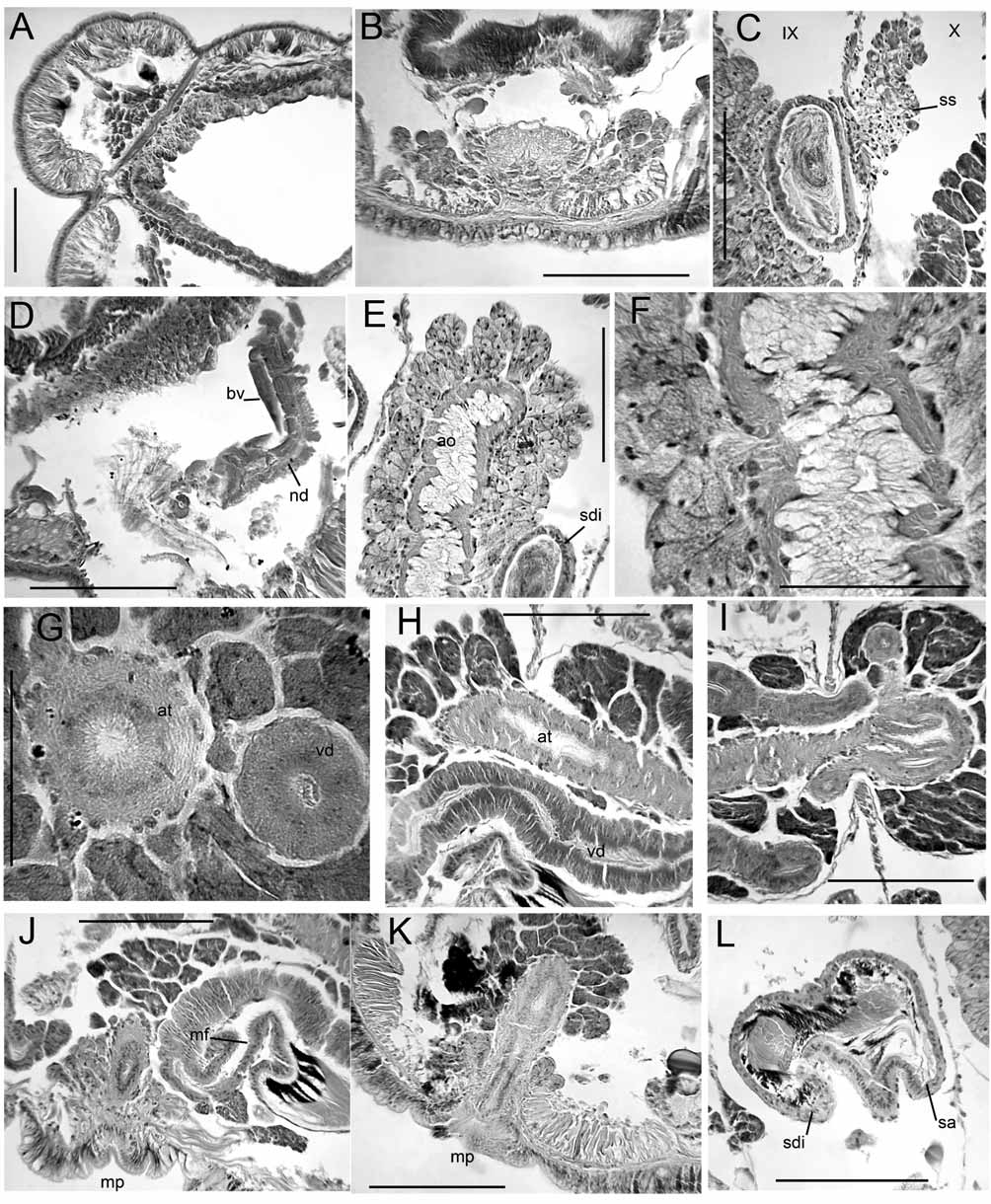
Supplementary description. Body usually tetragonal in transverse section, with distinct longitudinal ridges in anterior segments, associated with muscle bands. Annulations relatively strong, even in clitellar region ( Fig. 1 View FIGURE 1. R A). Longitudinal muscle bands strongly tapered at lateral margins; the thinner margins curl into the coelom ( Figs. 1 View FIGURE 1. R C, 2A). Longitudinal sheets of oblique, dorsolateral muscles insert dorsally and at the lateral line in segments posterior to about VI, but most prominent behind the clitellum ( Fig. 2 View FIGURE 2. R A). Clitellum from IX to XX or XXII. Small, petiolate clumps of granular cells on either side of ventral nerve cord in segments anterior to VIII; cells appear to have indistinct ducts to epidermis on either side of nerve cord, but without a distinct ectal pore ( Fig. 2 View FIGURE 2. R B). Pharynx from about II to IV, walls about 50–60 μm thick, at most only slightly thicker dorsally than ventrally, without a distinct dorsal pad.
Ventral blood vessel divides in VII; the two branches join anteriorly in front of the brain at the dorsal vessel. One pair lateral blood vessels in posterior part of each segment; they are unbranched and convoluted in XII and anterior, and branched in posterior segments. Anteriorly to about XXXV lateral vessels (or a main branch) terminate in the ventral vessel. In more posterior segments (to at least XLV) a thick branch joins the perivisceral sinus, and a thin branch joins the ventral vessel. Nephridia paired, occurring regularly in most segments posterior to XII. A small, bulbous postseptal expansion is followed by a tubule loop that follows commissural blood vessel to near the dorsal vessel ( Fig. 2 View FIGURE 2. R D). Nephropores just anterior to ventral chaetae; ectal ducts of nephridia without vesicles.
An anterior outpocketing of the spermathecal ampulla forms an indistinct, roughly tubular diverticulum, which may have sperm heads lined up in it [ Figs. 1 View FIGURE 1. R B, 2L; see also Fig. 4 View FIGURE 4. R in Timm (1970)]; remainder of spermathecal ampulla with loose sperm cells; ampulla ovate, entirely in originating segment, with a very short, simple duct to the gut ( Fig. 1 View FIGURE 1. R C). Small clumps of cells (possibly vestigial sacs) on septum above anterior funnels in X ( Fig. 2 View FIGURE 2. R C).
Accessory organ in IX consists of a muscular tube lined with a single layer of epithelial cells; outer layer of densely-packed, pyriform clumps of cells, similar to prostates, except that the cells are very granular and do not stain darkly ( Figs. 2 View FIGURE 2. R E–F). As in prostate glands, extensions of the outer cell clusters penetrate the muscle layer and are continuous with the cells of the inner layer ( Fig. 2 View FIGURE 2. R F). Inner cell layer thick, appearing vacuolate. Pore posterior to chaetae, slightly to one side after duct passes beneath the nerve cord ( Fig. 1 View FIGURE 1. R C).
Posterior sperm funnels larger than anterior, folded into the sperm sacs; anterior male funnels without sperm ( Figs. 1 View FIGURE 1. R E, 2J). Vasa deferentia thick, diameter 60–80 Μm but thinner at funnels and at atrium; tube formed of thick uniserial layer of elongate, glandular cells; no distinct muscle layer ( Figs. 2 View FIGURE 2. R G–H). Vasa deferentia enter atrium near ental end ( Figs. 1 View FIGURE 1. R E, 2I), but do not penetrate 10/11. Prostates very darkly staining, covering atrium almost to male pore ( Figs. 1 View FIGURE 1. R E, 2J–K). Atria tubular, diameter 80–100 μm, muscle layer to 10– 14 μm; extending to XIV–XVI. The single pair of male and both pairs of spermathecal pores just anterior to posterior septum of respective segment, on ventral chaetal line between ventral and ventrolateral muscle bands ( Figs. 1 View FIGURE 1. R A, 1D, 2K). No distinct penes or penial sac; male pore on a minute and weakly defined protrusion of duct and epidermal cells within a slight concavity ( Fig. 2 View FIGURE 2. R K). Male pores associated with cluster of glands, similar to prostates but not as darkly staining.
Rhynchelmis (Rhynchelmis) granuensis Hrab ĕ, 1961
Supplementary description. Epidermis strongly annulated, even in clitellum. Clitellum to XVIII. Very large septal cell masses (possibly vestigial sacs) in IX–X. Nephridia with elongate postseptal expansion; tubules loop dorsally in some segments; ectal ducts simple without vesicle.
Spermathecal pores on short papillae; ectal duct cylindrical, with columnar epithelial cells. Spermathecal ampulla forms an indistinct diverticulum, projecting anteriad as described for R. tetratheca (see above).
Posterior male funnel large, convoluted, with sperm; anterior funnel smaller, conical, without sperm. Atria to XV; covered with with granular, petiolate prostate bundles beginning near the ectal end, continuing to ental end. Anterior vas deferens narrow (25 Μm) near funnel, widening to nearly 60 Μm; posterior vas diameter 50– 60 Μm. Both vasa deferentia appear to have glandular epithelium; both join atrial muscle slightly before ental end, entering lumen apically. Male pore a slight papilla in a shallow sac, without a distinct penis, surrounded by a thick mass of granular, prostate-like cells.

No known copyright restrictions apply. See Agosti, D., Egloff, W., 2009. Taxonomic information exchange and copyright: the Plazi approach. BMC Research Notes 2009, 2:53 for further explanation.