
Rhynchelmis shamanensis, Martin & Kaygorodova, 1998, Martin & Kaygorodova, 1998
|
publication ID |
https://doi.org/ 10.5281/zenodo.194252 |
|
DOI |
https://doi.org/10.5281/zenodo.6201859 |
|
persistent identifier |
https://treatment.plazi.org/id/03C5878E-FFF6-D419-B2CF-FE4C7CA06DFF |
|
treatment provided by |
Plazi |
|
scientific name |
Rhynchelmis shamanensis |
| status |
|
Pseudorhynchelmis shamanensis View in CoL ( Martin & Kaygorodova, 1998)
( Figs. 9 View FIGURE 9. P A–L)
(= Pseudorhynchelmis olchonensis sensu Hrabė, 1982)
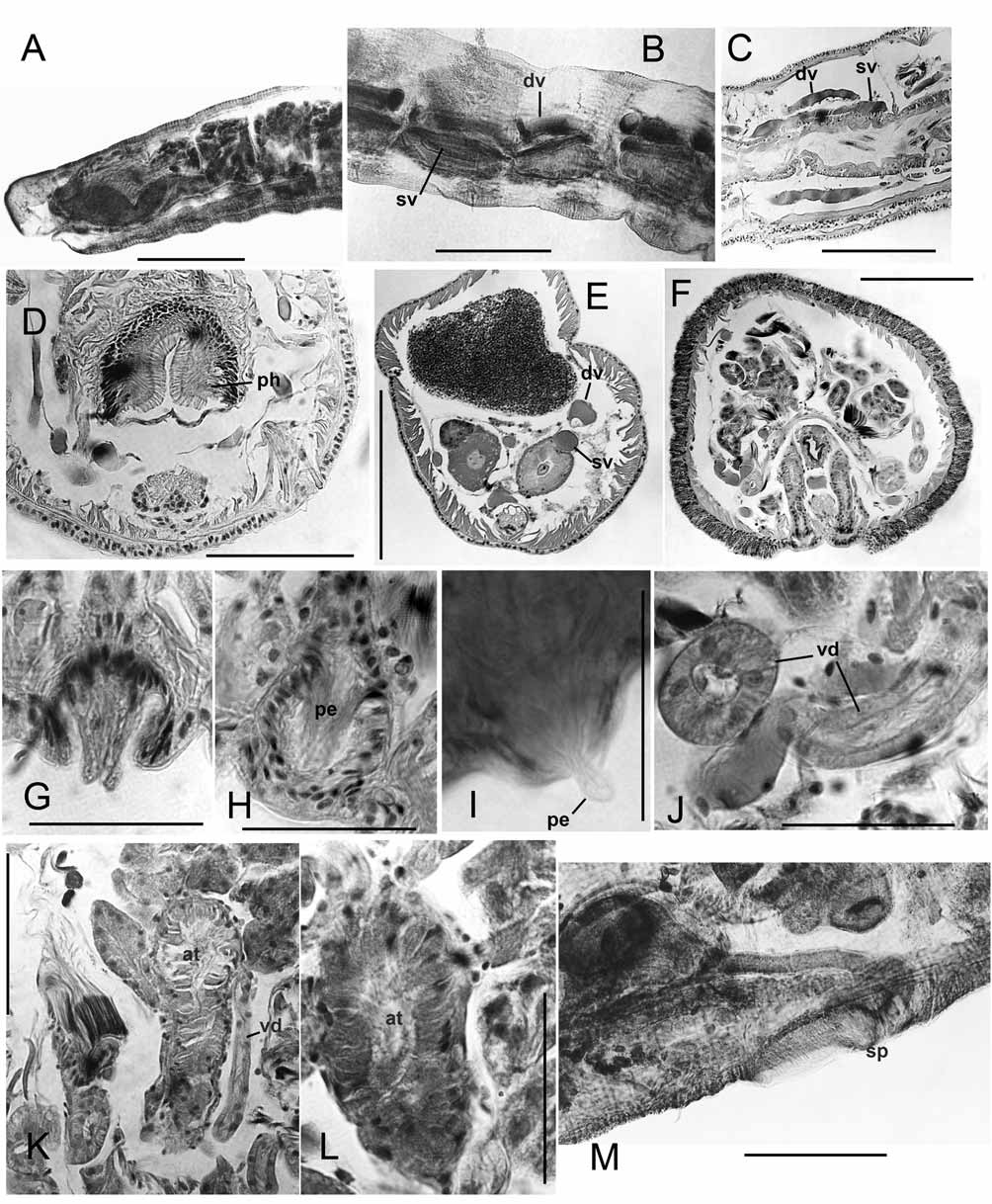
Supplementry description. Martin collection: Pharyngeal pad strongly developed ( Fig. 9 View FIGURE 9. P A). The first lateral blood vessel is visible as a loop in II, but appears to join dorsal and ventral vessels at mid-segment in III. Commissures in IV–V as in II; those in VI–X also unbranched and convoluted, with forward loops, but apparently stay in one segment. No lateral vessels were visible beyond X, but there may be loops into sperm and egg sacs from anterior commissures; dorsal and ventral vessels very thick. Starting about XV, posterior segments with a normal, thick dorsal vessel having cardiac cells, and a supra-intestinal vessel mostly in contact with the perivisceral sinus ( Fig. 9 View FIGURE 9. P B).
Spermathecal duct narrow and elongate (length about 5 times width), spermathecal ampulla sac-like, filled with loosely-bundled, unordered sperm. Major, ental portion of atrium tubular, with thin muscle and granular lining cells to over 30 Μm thick; lumen very narrow, about 5 Μm. Ectal end of the atrial duct widens slightly to about 40 μm, and protrudes slightly as a low papilla ( Fig. 9 View FIGURE 9. P I). Duct lining cells oblique and elongate, protruding slightly within the widened opening of the papilla, suggesting that the penis is formed by extruded atrial lining cells; some cells of atrial lining extruded beyond the papilla ( Fig. 9 View FIGURE 9. P I).
Sectioned Hrabĕ specimens: Pharyngeal pad and supra-intestinal blood vessel as described above ( Figs. 9 View FIGURE 9. P C–E). Small nephridia occur irregularly in posterior segments; postseptal mass not strongly developed, with unmodified duct to ectal pore, and convoluted tubules adjacent to ventral blood vessel. Male pores distinctly inside chaetal line, near 10/11 ( Fig. 9 View FIGURE 9. P F). Distinct, conical penes are extensions of terminal lining cells, within a shallow sac formed by an ectal expansion of the duct ( Figs. 9 View FIGURE 9. P G–H). Vasa deferentia very wide, to 30–40 Μm at ental end; narrowing to about 15–20 Μm in XI along atria ( Figs. 9 View FIGURE 9. P J–K). Atrial histology as described above; irregular prostate bundles sparsely cover atria ( Figs. 9 View FIGURE 9. P K–L). Pseudorhynchelmis shamanensis differs from the description of P. olchonensis primarily by the lack of glandular cells associated with the chaetae of X. The absence of these glands in the material used by Hrabĕ (1982) in his redescription of P. olchonensis indicate that these specimens should be attributed to P. shamanensis ( Martin et al. 1998).
No known copyright restrictions apply. See Agosti, D., Egloff, W., 2009. Taxonomic information exchange and copyright: the Plazi approach. BMC Research Notes 2009, 2:53 for further explanation.