
Pseudopolydora antennata ( Claparède, 1868 )
|
publication ID |
https://doi.org/ 10.5852/ejt.2021.773.1519 |
|
publication LSID |
lsid:zoobank.org:pub:A018A660-F0D8-4411-AC25-77C089A75A57 |
|
DOI |
https://doi.org/10.5281/zenodo.8338150 |
|
persistent identifier |
https://treatment.plazi.org/id/03CF9F22-FF9A-AE77-6D59-36C8FEBDF92F |
|
treatment provided by |
Felipe |
|
scientific name |
Pseudopolydora antennata ( Claparède, 1868 ) |
| status |
|
Pseudopolydora antennata ( Claparède, 1868) View in CoL View at ENA
Figs 1A View Fig , 4 View Fig , 5A–C View Fig , 6–9 View Fig View Fig View Fig View Fig
Polydora antennata Claparède, 1868: 320−321 , pl. XXI fig. 3, 3a−c.
Polydora antennata – Claparède 1869: 60–61, pl. XXI fig. 3, 3a−c; 1870: 60–61, pl. XXI fig. 3, 3a−c. — Carus 1885: 257. — Carazzi 1893: 25−26, pl. 2 figs 11−12. — Lo Bianco 1893: 30. — Bhaud 1972: 125.
Pseudopolydora antennata View in CoL – Czerniavsky 1881: 362. — Guille & Laubier 1966: 271. — Mohammad 1971: 296. — Lardicci 1989: 137−138. — Simboura & Nicolaidou 2001: 78. — García-Arberas & Rallo 2002: 50. — Dauvin et al. 2003: 84. — Castelli et al. 2008: 356. — Mikac 2015: 121. — Radashevsky 2021: 2–7, figs 1–2.
Pseudopolydora sp. A – Al-Kandari et al. 2019: 9. — Radashevsky et al. 2020: table 1, fig. 1.
Description
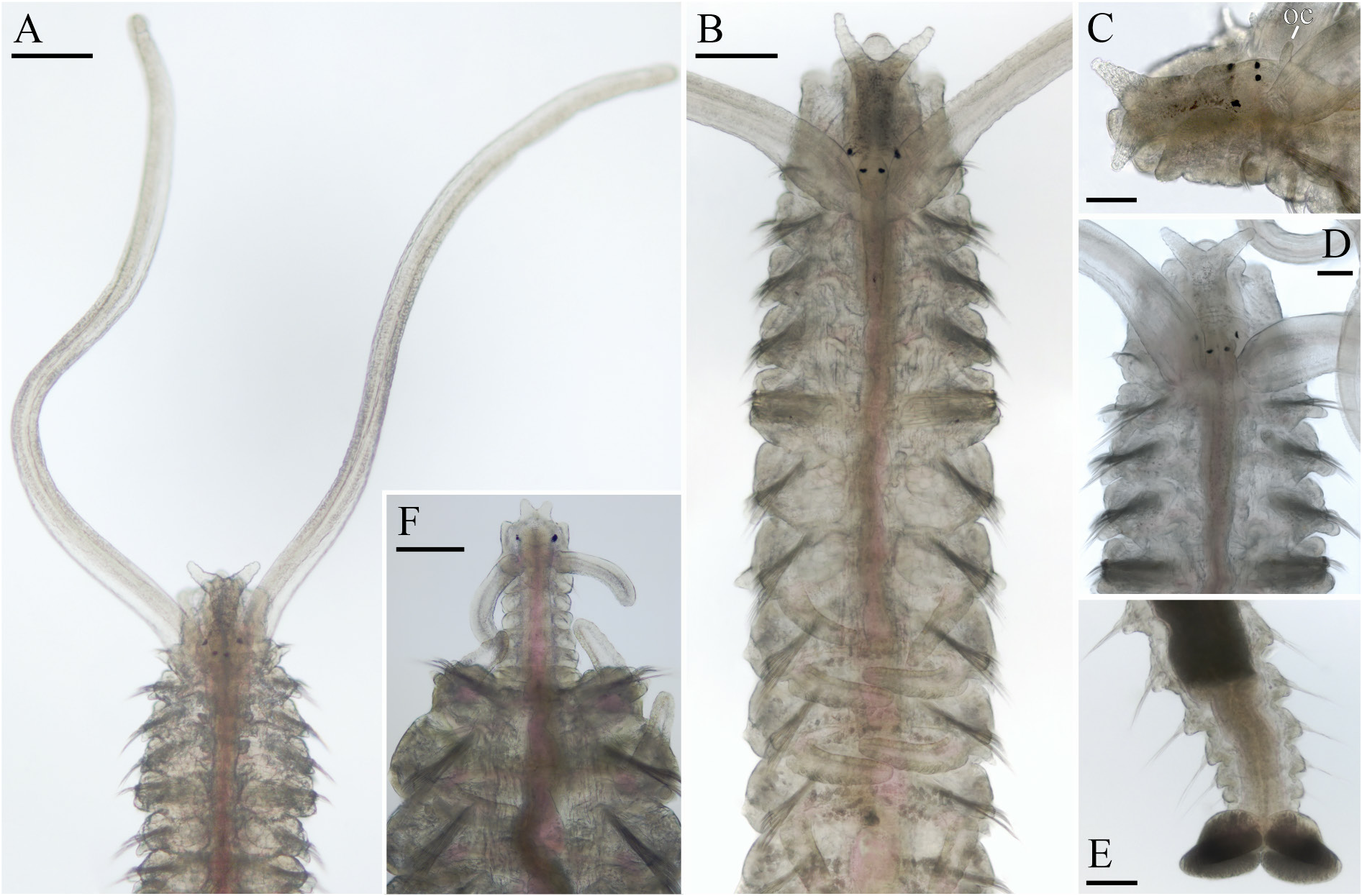
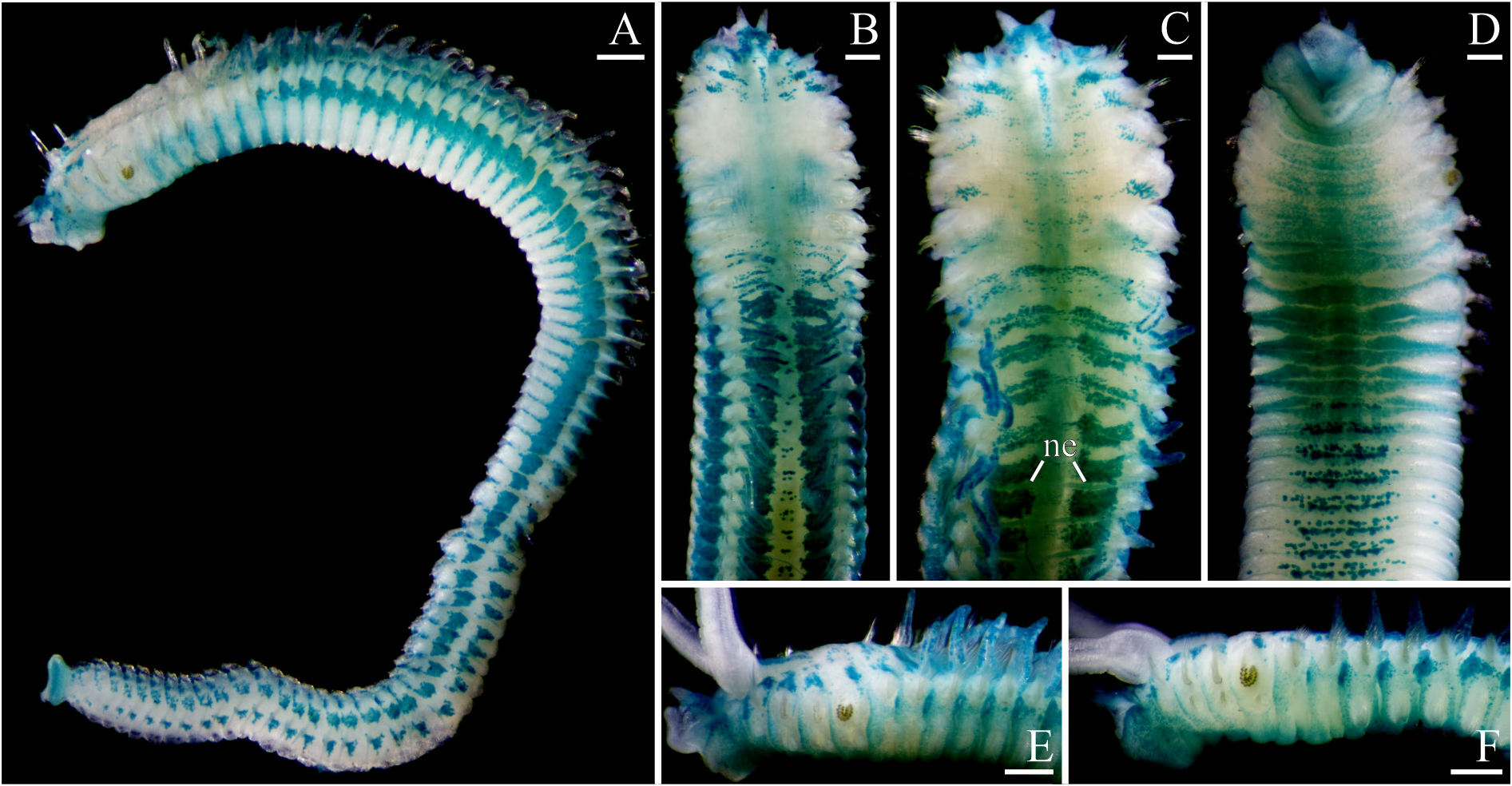
Adults up to 16 mm long, 1 mm wide with 86 chaetigers. No pigmentation on body and palps; fine black pigment scattered on dorsal side of anterior chaetigers without particular pattern in some individuals. Prostomium anteriorly bifurcated, with two long, distally narrowing lobes ( Fig. 4A–D View Fig ), posteriorly extending to end of chaetiger 4 as a low caruncle, shorter in small individuals. Long cirriform occipital antenna present on caruncle between palps ( Fig. 4C View Fig ). Two pairs of black eyes arranged trapezoidally, comprising one pair of median eyes, and one pair of lateral eyes situated anteriorly and set wider apart. Palps as long as 15–25 chaetigers, with frontal longitudinal groove lined with fine cilia, and short compound non-motile cilia arising directly from palp surface sparsely arranged in line on sides of groove and sparsely scattered on lateral and abfrontal palp surfaces.
Chaetiger 1 with short capillaries in neuropodia, short notopodial lamellae and well-developed neuropodial postchaetal lamellae; notochaetae absent. Chaetiger 2 notochaetae all slender capillaries. Anteriorrow notopodial capillaries on chaetiger 3 with slightly enlarged wing; capillaries on chaetiger 4 with wing slightly larger than on chaetigers 3. Anterior-row capillaries on chaetigers 3, 4, 6 and 7 arranged in J-shaped series; posterior-row capillaries on these chaetigers arranged in vertical series ( Fig. 6A View Fig ). Posterior notopodia with a few long alimbate capillary chaetae arising from elongated fleshy notopodial lobes. Posterior neuropodia with elongated fleshy lobes, with hooks arranged in line on top of lobes.
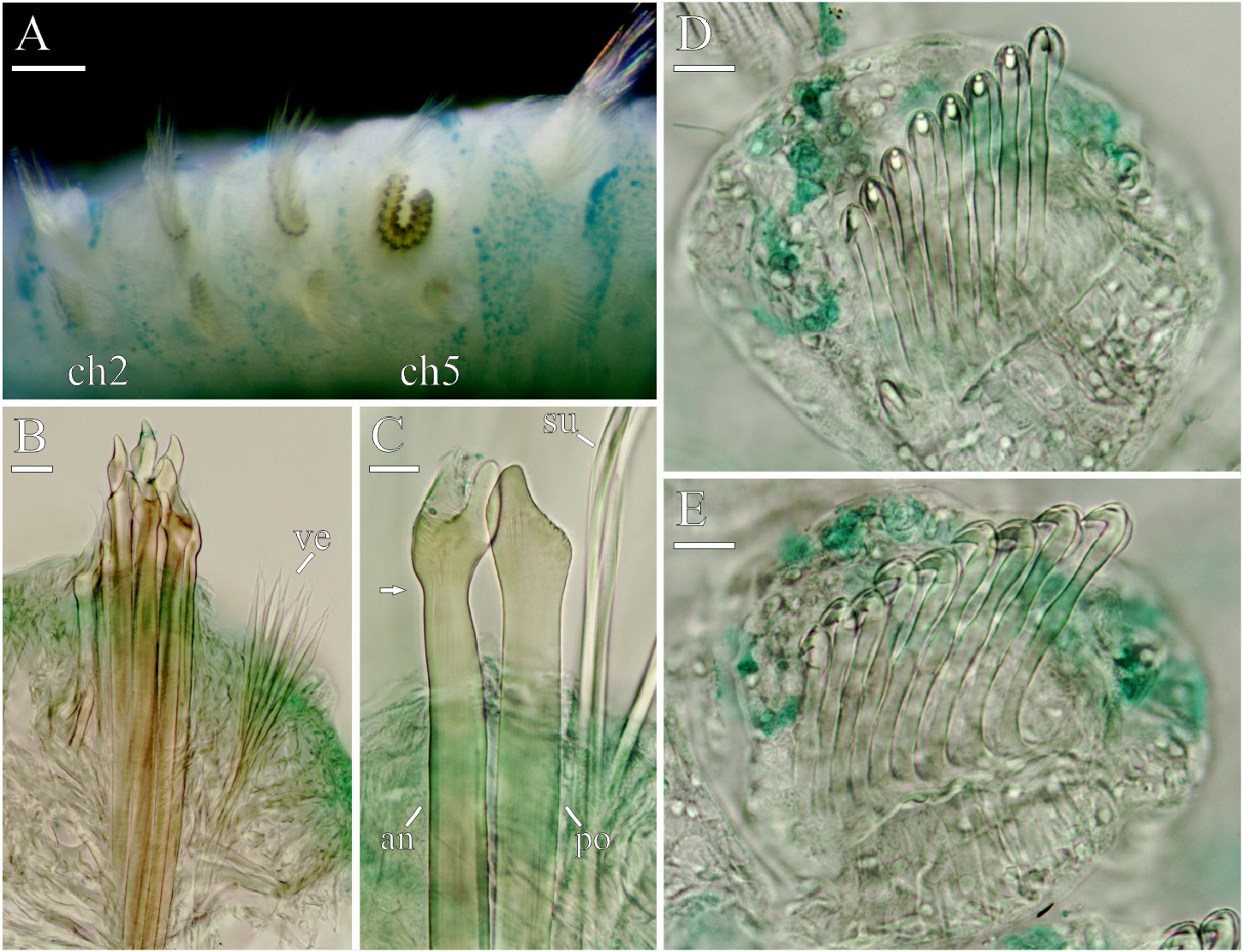
Chaetiger 5 same in size as chaetiger 4 or 6, with dorsal superior capillaries, two kinds of notopodial spines arranged in a double U-shaped row, and ventral capillaries; notopodial postchaetal lamellae reduced, but neuropodial lamellae same as on chaetigers 4 and 6 ( Fig. 6B View Fig ). Dorsal superior capillaries shorter and fewer than those capillaries on chaetigers 4 and 6. Ventral capillaries same in size, number and arrangement (in three groups) as those on chaetiger 4 ( Fig. 6B View Fig ). Upper posterior part of double U-shaped row of spines equal to or slightly lower than upper anterior part. Newly developed spines in posterior upper part of U-shaped row slightly larger than older spines in anterior upper part of row. Outer (anterior-row) notopodial spines up to 11 in a series, with distal part of stem enlarged, with concavity on top and large triangular tooth on its side directed upwards and facing towards the inside of the U-shaped row of spines; fine bristles arising from concavity forming long flag-like pointed transparent tip which usually broken in worn old spines in anterior upper part of row ( Fig. 6B–C View Fig ). Inner (posterior-row) notopodial spines up to eight in a series, falcate, with short rounded distal part geniculate, with subdistal bulbous swelling bearing very short fine bristles and facing towards the inside of the U-shaped row of spines ( Fig. 6B–C View Fig ).
Hooks in neuropodia from chaetiger 8, up to 30 in a series, not accompanied by capillaries. Hooks bidentate, with upper tooth closely applied to main fang; upper part of shaft with constriction; lower part of shaft bent at right angle in hooks in posterior neuropodia ( Fig. 6D–E View Fig ).
Branchiae up to 42 pairs from chaetiger 7 to chaetiger 48, fewer in small individuals ( Fig. 5A View Fig ), present beyond midbody in individuals with more than 30 chaetigers ( Fig. 5C View Fig ). Branchiae full-sized from chaetigers 10–11, free from notopodial postchaetal lamellae, flattened, with surfaces oriented perpendicular to body axis, with longitudinal ciliation (extension of nototroch) running on inner anterior edge.
Nototrochs usually from chaetiger 7 onwards, occasionally from chaetiger 6 or 8, composed of single rows of cilia in both sexes. On branchiate chaetigers, nototroch cilia long, arranged in transverse lines and extending onto branchiae; on posterior abranchiate chaetigers, cilia arranged in U-shaped bands, with arms directed posteriorly. Additional ciliation on chaetigers absent.
Pygidium bilobed, with two semi-oval lateral lobes ( Fig. 4E View Fig ), white due to great number of spindleshaped glandular cells with striated content.
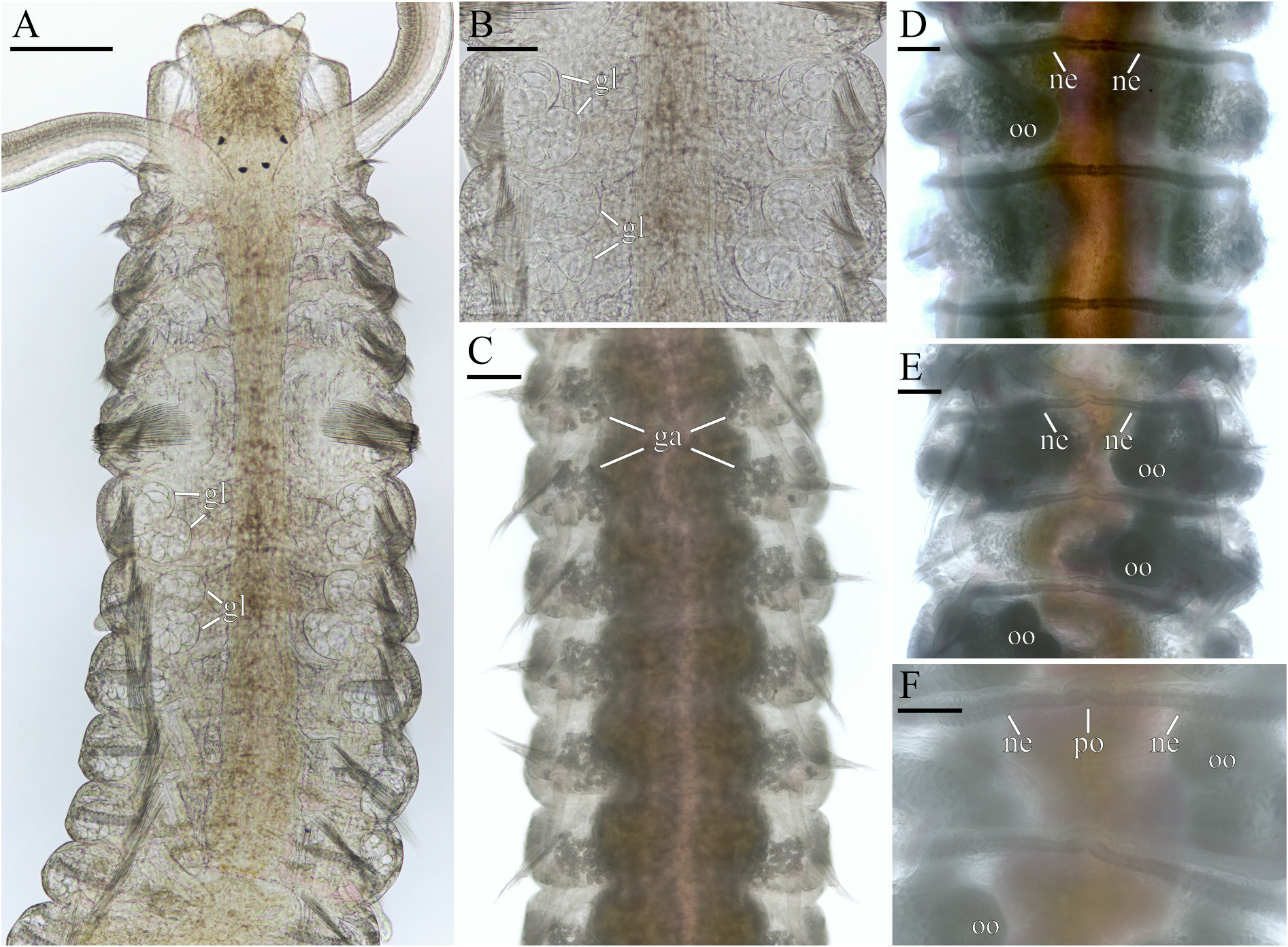
Subspherical and of irregular shape glandular cells with striated content present on dorsal side of chaetigers. Cells few on anterior and posterior chaetigers, forming distinct paired gatherings from chaetigers 8–11 through two thirds of body, making this part of dorsum whitish in life ( Figs 4B View Fig , 7C View Fig ).
Glandular pouches in neuropodia from chaetiger 1, largest and paired in each neuropodium in chaetigers 6 and 7, single in other neuropodia ( Fig. 7A–B View Fig ).
Digestive tract without ventral buccal bulb and gizzard-like structure, not pigmented.
Main dorsal blood vessel transformed into gut sinus in anterior part of midgut. Heart body absent. Blood red, without globules or other elements.
Nephridia from chaetiger 4 onwards, very narrow in chaetigers 4–6, well-developed from chaetiger 7. In female fertile chaetigers, paired nephridia on each chaetiger opening to exterior via common middorsal nephridiopore situated anterior to nototroch; walls of distal parts of nephridia containing glandular cells marking nephridia in live individuals ( Fig. 7D–F View Fig ) and also in fixed specimens stained with MG ( Fig. 8C View Fig ).
MG staining
Intensely stained dorsal sides of the prostomium and peristomium; most intensely stained blotches on the dorsal and lateral sides from chaetigers 7–9 onwards; wide transverse bands on the ventral side from chaetigers 7–8 to chaetigers 10–11 ( Fig. 8A–F View Fig ).
Regeneration
One broken individual regenerated 7 anterior chaetigers ( Fig. 4F View Fig ).
Habitat
Adult P. antennata inhabit silty tubes in muddy sand intertidally and in shallow waters. The population density in some local places can reach a maximum of 50 000 individuals per 1 m 2.
Reproduction
Pseudopolydora antennata is gonochoristic. Both females and males become mature after growing to about 10 mm long with 70 chaetigers. Of 21 examined mature individuals, 14 were females and seven were males. The gametes developed from chaetiger 12 to chaetigers 40−45 ( Fig. 5B View Fig ). Paired testes or ovaries were attached to segmental blood vessels in fertile chaetigers.
Oogenesis was partly intraovarian. Developed coelomic oocytes were spherical, about 130 µm in diameter, with smooth envelope less than 1 µm thick ( Fig. 7E–F View Fig ).
Spermatogonia proliferated in testes; spermatogenesis occurred in the coelomic cavity. Spermatids, each about 4 µm in diameter, were joined in tetrads. Spermatozoa were introsperm with pointed acrosome about 1 µm long, straight elongated nucleus about 1.5 µm in diameter and 6 µm long, midpiece 33 µm (head + midpiece about 40 µm long), and a flagellum about 40 µm long (the precision of measurement of spermatids and parts of head of spermatozoa were at maximum to the nearest 0.5 µm and flagellum to the nearest 2 µm).
Larval development in the plankton
The spermatophores and egg capsules, typically produced by Pseudopolydora , were not observed in P. antennata from the Arabian Gulf. The 13−15-chaetiger larvae of the species were collected in plankton in March 2016. The larvae were identified by the morphology of the heavy spines in the notopodia of chaetiger 5.Although similar spines are also present in worms of two other species described below, those species were quite rare and not found in the area where numerous larvae were collected; P. antennata worms were common in that area and the larvae are therefore referred to this species. The larvae were maintained in Petri dishes in the laboratory but did not settle during the study.
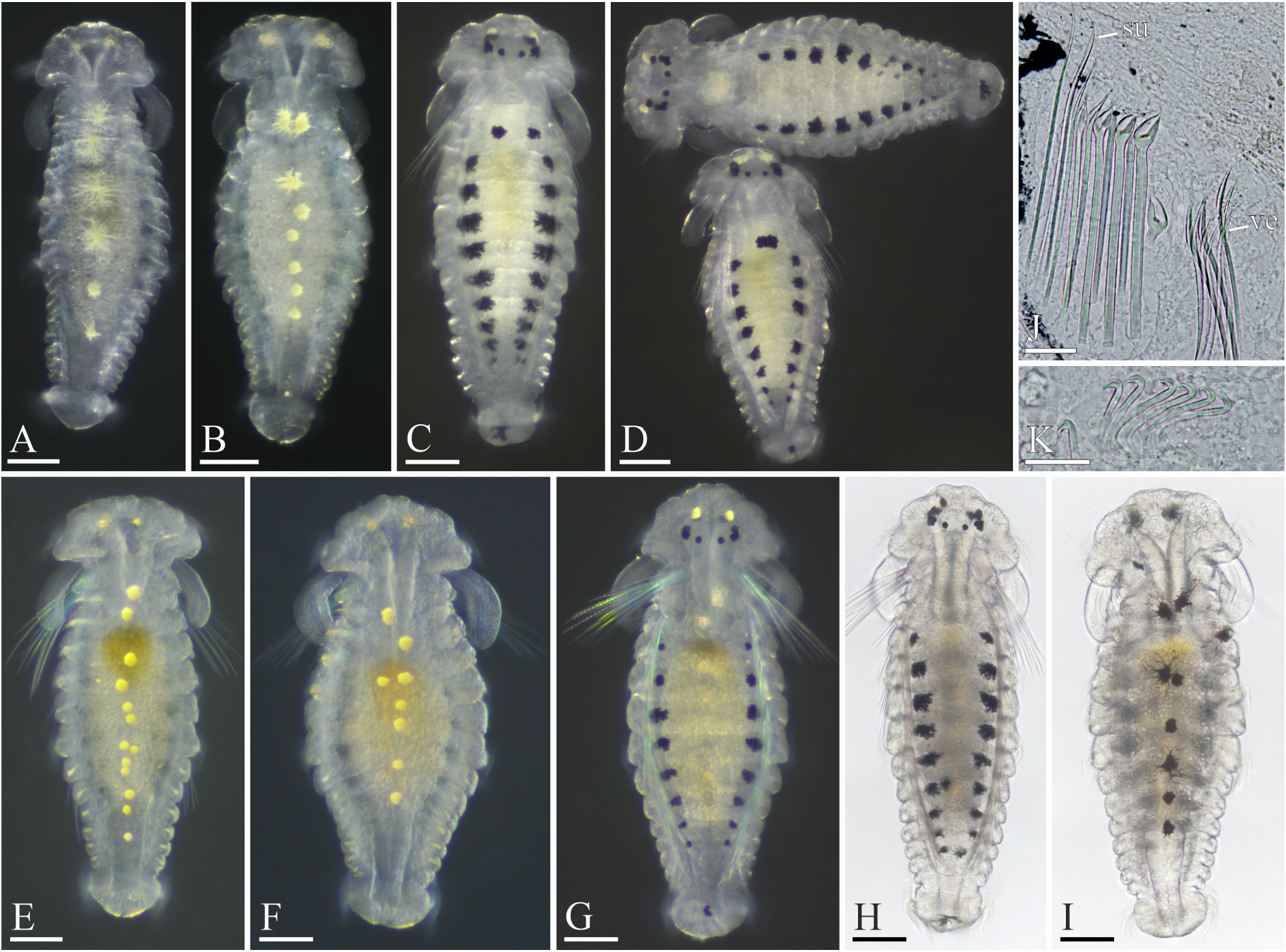
13–14-chaetiger larvae
Larvae750–850 µm long and up to300 µm wide in middle part, intensely pigmented with large melanophores and chromatophores ( Fig. 9A–I View Fig ). Distinct black spots present between median and lateral eyes, and small ramified melanophores of variable shape situated anterior to each pair of lateral eyes. Of 29 examined larvae, 13 (45%) larvae with one pair of large ramified dorsal melanophores on chaetiger 3 ( Fig. 9C–D View Fig ); 3 (10%) larvae with only one of these melanophores (either left or right) present; and 13 (45%) larvae without dorsal melanophores on chaetiger 3 ( Fig. 9D, G–H View Fig ). Large ramified dorso-lateral melanophores invariably present from chaetiger 4 onwards. Small to large ramified middorsal melanophore present on pygidium, situated posterior to telotroch. Melanophore in lateral peristomial lips, middorsal and lateral melanophores on chaetigers absent. One, rarely two pairs of subspherical yellow chromatophores present in front of lateral eyes, near melanophores in anterior part of prostomium. Larger subspherical yellow midventral chromatophores arranged from chaetiger 2 to chaetigers 7–12; shape and arrangement of these chromatophores greatly variable ( Fig. 9A–B, E–F, I View Fig ). They are usually single, occasionally double per chaetiger, rarely missing on chaetiger 6 or 7 or both. Chromatophore of chaetiger 3 often expanded onto posterior part of chaetiger 2. Yellow pigment diffused in anterior part of prostomium and in pygidium. Chromatophores absent in lateral peristomial lips and on ventral side of pygidium. Both melanophores and chromatophores changing their shape and size according to light intensity, thus appearing small and compact ( Fig. 9E–F View Fig ) or greatly expanded and ramified ( Fig. 9A, C View Fig ).
Prostomium wide and rounded anteriorly, posteriorly narrow, extending to end of chaetiger 1 as a low caruncle.Occipital antenna absent.Three pairs of black eyes arranged almost in a transverse line, comprising one pair of small rounded median eyes and two pairs of lateral eyes; lateral eyes on each side situated close to each other and appearing as one comma-shaped eye. Black pigment spot situated between lateral and median eyes on each side. One pair of spherical unpigmented ocelli, each about 20 µm in diameter, situated in front of pigmented eyes. Palps arising from posterior dorso-lateral edges of peristomium, posterior to prototroch; each palps as long as about three chaetigers. Ciliation on palps not yet developed.
Prototroch and telotroch well developed. Nototrochs and grasping cilia from chaetiger 3 onwards. Triangular neurotroch extending from ventral peristomial lip to end of chaetiger 1. Two small ciliated cells situated on each side of neurotroch. Ventral ciliary pit absent. Gastrotroch on chaetiger 3 composed of two small cells with short cilia; those on chaetigers 5 and 7 each composed of 8–10 large cells with long numerous cilia; additional gastrotroch usually present on chaetiger 9, and in some larvae also on chaetiger 10 and/or chaetiger 11, each composed of one to two pairs of small cells with short cilia.
Larval serrated bristles present in all notopodia including chaetiger 5; those in chaetiger 1 longest, up to 250 µm long; bristles in chaetigers 2 and 3 175–200 µm long, then gradually becoming shorted in succeeding chaetiger. Few short adult capillaries present in notopodia from chaetiger 2, and in neuropodia in chaetigers 1–7.
Chaetiger 5 with two kinds of heavy spines in notopodia in addition to short adult capillaries and long provisional serrated bristles. Spines comprising 2–4 anterior-row spines with enlarged distal end of stem and pointed tip, and 1–2 posterior-row simple falcate spines ( Fig. 9J View Fig ).
Hooks in neuropodia from chaetiger 8 onwards, up to seven in a series, not accompanied by capillaries. Hooks bidentate, with upper tooth closely applied to main fang, with outer hood; lower part of shaft bent almost at right angle ( Fig. 9K View Fig ).
Branchiae short, on chaetigers 7–9. Pygidium semispherical.
Lateral sensory organs each 5–6 µm in diameter with thin non-motile cilia up to 20 µm long situated between noto- and neuropodia from chaetiger 1 onwards, including chaetigers 3–5.
Glandular pouches in neuropodia from chaetigers 2–3, very small in first chaetigers, large in chaetigers 6 and 7.
Large lateral lips and small ventral lip of peristomium forming voluminous vestibulum. Narrow oesophagus extending through 2–3 chaetigers. Gizzard-like structure absent in digestive tract. Wall of voluminous midgut with numerous lipid globules, each up to 15 µm in diameter.
Circulatory system developed and functional; blood transparent, without pigment.
Two pairs of protonephridia in chaetigers 1 and 2. Metanephridia not yet developed.
14–15-chaetiger larvae
Larvae 850–900 µm long, with glandular pouches in neuropodia from chaetiger 1, protonephridia in chaetigers 1 and 2, and metanephridia from chaetiger 4 onwards.
Remarks
Adult Pseudopolydora from the Arabian Gulf described above appear identical to P. antennata from Italy recently re-described by Radashevsky (2021), and are consequently referred to this species. They have the prostomia anteriorly bifurcated, with two long, distally narrowing lobes, caruncles extending to the end of chaetiger 4, comparatively short occipital antennae, chaetiger 5 spines of the same morphology, bilobed pygidia with two fleshy semi-oval lateral lobes, and gametes developing from chaetiger 12.
The exact dimensions of the gametes of P. antennata from the Mediterranean remain unknown but worms from the Arabian Gulf are unusual among Pseudopolydora in having spermatids joined in tetrads instead of octads, and the spermatozoa with an extremely long midpiece, about 33 µm instead of 3.2−4.6 µm long as in other examined species (see review by Blake & Arnofsky 1999: table 2).
The morphology of genuine P. antennata larvae from the Mediterranean has never been described or illustrated (see review by Radashevsky 2021). Hannerz (1956) described and illustrated pelagic, ready to metamorphose 11- and 13−14-chaetiger Pseudopolydora larvae, and a 19-chaetiger juvenile from Gullmar Fjord, Sweden. He referred them to P. antennata and reported that the larvae occurred in the Fjord from July through November but were relatively scarce. The Arabic larvae herein referred to P. antennata are similar to those described by Hannerz (1956) only in having dorso-lateral ramified melanophores from chaetiger 4 onwards, but differ significantly by having 1−2 pairs of subspherical yellow chromatophores in front of lateral eyes, large yellow melanophores on the ventral side of chaetigers, main gastrotrochs on chaetigers 3, 5, 7, and small additional gastrotrochs on chaetigers 9, 10 and/or chaetiger 11, instead of gastrotrochs on chaetigers 5 and 7 only. Moreover, the Arabic larvae are unique among Pseudopolydora larvae in the absence of large ramified middorsal melanophore on chaetiger 1. Rasmussen (1973: 113) described and perfectly illustrated egg capsules laid by Pseudopolydora in Isefjord, Denmark, and noted that “The hatched larvae from the Isefjord agreed closely with the description [of P. antennata ] given by Hannerz (1956, pp 126−130)”. Following Hannerz (1956), Rasmussen (1973) referred Danish worms to P. antennata . Obviously, larvae from the Arabian Gulf referred to P. antennata in the present study are not conspecific with those from Gullmar Fjord described by Hannerz (1956). Which of them belong to P. antennata remains to be clarified.
The larvae from the Arabian Gulf referred to P. antennata are characterised by the following features: one pair of large ramified dorsal melanophores on chaetiger 3 (one or both of them can be absent), large ramified dorso-lateral melanophores from chaetiger 4 onwards, middorsal melanophore on the pygidium, paired yellow chromatophores in the anterior part of prostomium, and large median, usually unpaired, yellow chromatophores on the ventral side from chaetiger 2 onwards. Melanophores in lateral peristomial lips, middorsal and lateral melanophores on chaetigers, and chromatophores on the ventral side of pygidium, characteristic for larvae of other species of Pseudopolydora , are absent.
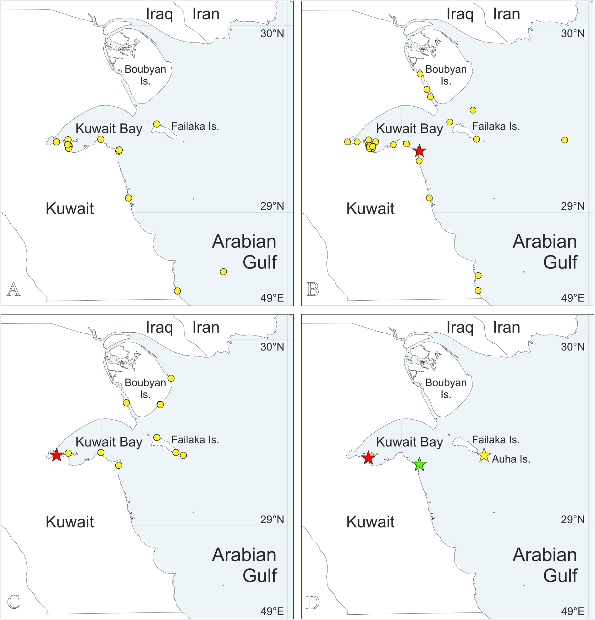
Distribution
Mediterranean Sea; Arabian Gulf ( Fig. 1A View Fig ). See comments by Radashevsky (2021) about other records of this species.

No known copyright restrictions apply. See Agosti, D., Egloff, W., 2009. Taxonomic information exchange and copyright: the Plazi approach. BMC Research Notes 2009, 2:53 for further explanation.
|
Kingdom |
|
|
Phylum |
|
|
Class |
|
|
Order |
|
|
Family |
|
|
Genus |
Pseudopolydora antennata ( Claparède, 1868 )
| Radashevsky, Vasily I., Al-Kandari, Manal, Malyar, Vasily V. & Pankova, Victoria V. 2021 |
Pseudopolydora sp. A
| Al-Kandari M. & Sattari Z. & Hussain S. & Radashevsky V. I. & Zhadan A. 2019: 9 |
Pseudopolydora antennata
| Mikac B. 2015: 121 |
| Castelli A. & Bianchi C. N. & Cantone G. & Cinar M. E. & Gambi M. C. & Giangrande A. & Sareri D. I. & Lanera P. & Licciano M. & Musco L. & Sanfilippo R. & Simonini R. 2008: 356 |
| Dauvin J. - C. & Dewarumez J. - M. & Gentil F. 2003: 84 |
| Garcia-Arberas L. & Rallo A. 2002: 50 |
| Simboura N. & Nicolaidou A. 2001: 78 |
| Lardicci C. 1989: 137 |
| Mohammad M. - B. M. 1971: 296 |
| Guille A. & Laubier L. 1966: 271 |
| Czerniavsky V. 1881: 362 |
Polydora antennata
| Bhaud M. 1972: 125 |
| Carazzi D. 1893: 25 |
| Lo Bianco S. 1893: 30 |
| Carus J. V. 1885: 257 |
| Claparede E. 1869: 60 |
Polydora antennata Claparède, 1868: 320−321
| Claparede E. 1868: 321 |