
Eremidrilus gilita, Fend & Rodriguez, 2020
|
publication ID |
https://doi.org/ 10.11646/zootaxa.4809.1.6 |
|
publication LSID |
lsid:zoobank.org:pub:6E4829CF-1476-4FDB-941D-9EAFCA29D011 |
|
DOI |
https://doi.org/10.5281/zenodo.10499419 |
|
persistent identifier |
https://treatment.plazi.org/id/C090F3C9-BBF0-4FCF-A8BD-517C92FDC4F5 |
|
taxon LSID |
lsid:zoobank.org:act:C090F3C9-BBF0-4FCF-A8BD-517C92FDC4F5 |
|
treatment provided by |
Plazi |
|
scientific name |
Eremidrilus gilita |
| status |
sp. nov. |
Eremidrilus gilita View in CoL n. sp.
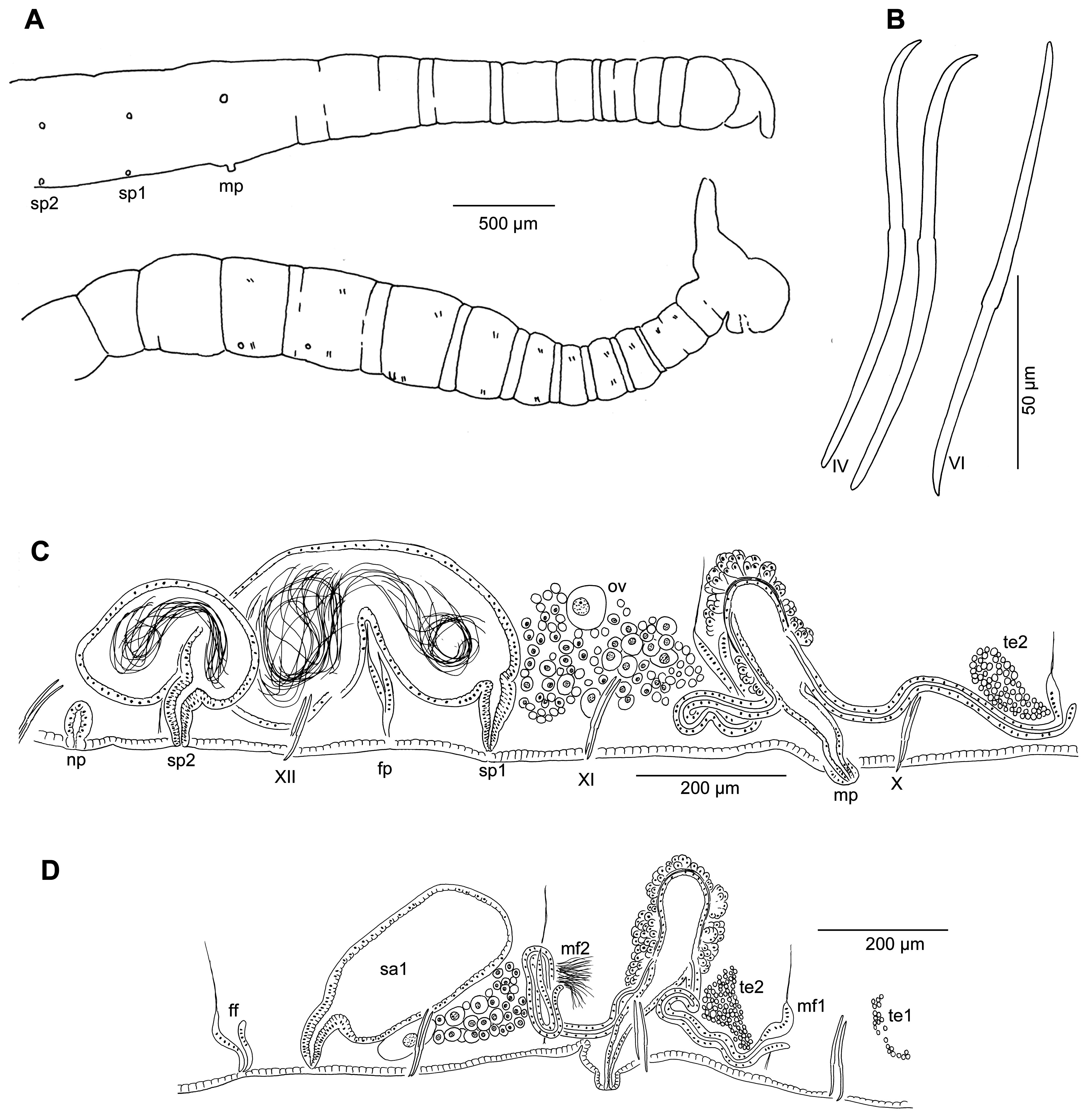
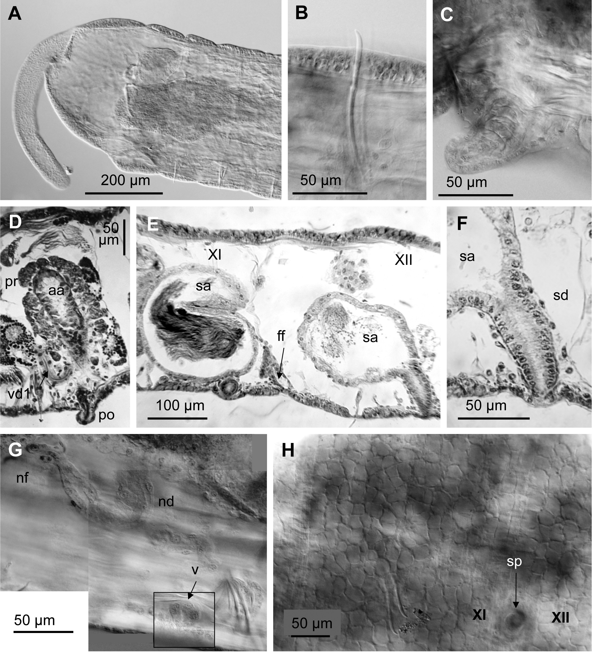
( Figures 7 View FIGURE 7 , 8 View FIGURE 8 , 11E View FIGURE 11 )
Holotype. USNM 1618768 View Materials . Dissected worm, incomplete but with sperm in spermathecae and mature egg; slidemounted in Canada balsam, stained with carmine.
Type Locality. USA, New Mexico, Catron Co., Gilita Creek at Willow Creek (Site 14, Table 1), in riffle, 22 Apr 1996, collected by S. Fend.
Paratypes. From the type locality (Site 14), same collection data; all mature: USNM 1618770 View Materials , 1 View Materials sagittally sectioned worm on 2 slides, stained with hematoxylin and eosin; USNM 1618769 View Materials , 1 View Materials dissected, incomplete. MNCN 16.3 View Materials /3115, 1 whole-mount, complete. All slide-mounted in Canada balsam .
Etymology. Named for the type locality, Gilita Creek. Noun in apposition.
Description. Length of one complete, preserved worm 17 mm, 58 segments. Diameter at clitellum 0.35–0.5 mm. Proboscis 260–480 µm long, about 85–95 µm diameter at middle ( Fig. 7A View FIGURE 7 , 8A View FIGURE 8 ). Secondary annulation from IV to clitellum ( Fig. 7A View FIGURE 7 ). Chaetae with nodulus 0.4 to 0.5 from distal end in mid-body, but may be more distal posteriorly; length 112–133 µm, shorter in II ( Figs. 7B View FIGURE 7 , 8B View FIGURE 8 ). Male pores on the ventral chaetal line, midway between chaetae and posterior septum, on narrow porophores ( Figs. 7A,C View FIGURE 7 , 8C,D View FIGURE 8 ). Simple spermathecal pores ( Fig. 8E,F View FIGURE 8 ), behind ventral chaetae, on the ventral chaetal line, the first at 1/3 the distance from posterior septum to ventral chaetae, second close to intersegment 12/13 ( Figs. 7C View FIGURE 7 , 8E,H View FIGURE 8 ). Female pores intersegmental at 11/12.
Epidermis 5–10 µm thick, up to 12 µm in clitellum. Clitellum X to XIII. Pharynx in II–IV, dorsal pad not prominent; pharyngeal glands in IV–VI (VII). Sperm sacs extend anteriorly to VII, posteriorly to XIII or XIV. Egg sacs to XV. Nephridia in several postclitellar segments, terminating in a round vesicle (30 µm wide), anterior to ventral chaetae ( Figs. 7C View FIGURE 7 , 8G View FIGURE 8 ).
Atria 240–340 µm long, entirely in X, club-shaped, maximum diameter 52–77 µm; muscle layer thin, 2–4 µm; epithelial cells cuboidal, to 6–7 µm high. Atrial ampulla covered with a prostate layer, 20–30 µm thick, prostate cells in densely packed clusters ( Figs. 7C,D View FIGURE 7 , 8D View FIGURE 8 ). Atrium narrows to 20 µm before porophore, and to about 10 µm near the male pore, which opens on a narrowly cylindrical porophore (26–50 µm long, 30–33 µm wide). Atrium length about 9–10 times porophore width, and 0.6–0.7 times body width. Two vasa deferentia per atrium, diameter 19–24 µm, length about 300 µm; posterior vas loops in XI, and both vasa join the atrial muscle layer near the middle, entering the lumen at about one third the distance from tip. ( Fig. 7C,D View FIGURE 7 )
Spermathecae in XI and XII, ampullae may extend into next segment ( Fig. 7C View FIGURE 7 ). Ampullae irregular sac-like, epithelial layer about 8 µm thick ( Fig. 8E View FIGURE 8 ). Ectal ducts of spermathecae 60–90 µm long and up to 50 µm wide, usually tapered to about 20 µm near the pore; muscle layer thin (ca. 1 µm), lining cells somewhat columnar, about 12 µm high; lumen very narrow ( Fig. 8F View FIGURE 8 ). Spermathecal pores without glands or distinct epidermal modifications.
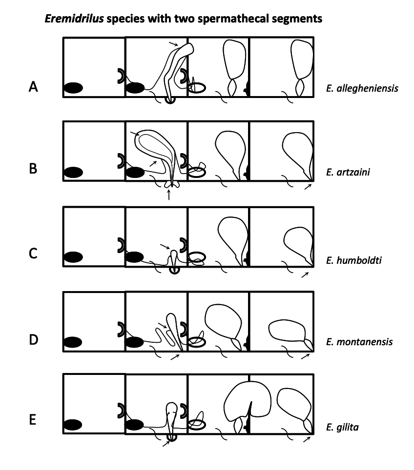
Remarks. The position of the spermathecal pores in line with the ventral chaetae, with the second pair close to intersegment 12/13, is similar to that of other Eremidrilus species having two spermathecal segments. Male pores open on a short porophore, narrower than that of E. humboldti n. sp. and E. allegheniensis (see below), and markedly different in shape from the concave, annular porophore of E. artzaini (a porophore is absent in E. montanensis n. sp.). The small size of the atrium and the thin atrial musculature most closely resemble those of E. humboldti , but in that species the atrium is shorter and the porophore is relatively large (see Table 2). The nephridial vesicles, observed in middle segments, appear more distinctive than those of E. montanensis .
No known copyright restrictions apply. See Agosti, D., Egloff, W., 2009. Taxonomic information exchange and copyright: the Plazi approach. BMC Research Notes 2009, 2:53 for further explanation.
|
Kingdom |
|
|
Phylum |
|
|
Class |
|
|
Order |
|
|
Family |
|
|
Genus |