
Eremidrilus artzaini, Fend & Rodriguez, 2020
|
publication ID |
https://doi.org/ 10.11646/zootaxa.4809.1.6 |
|
publication LSID |
lsid:zoobank.org:pub:6E4829CF-1476-4FDB-941D-9EAFCA29D011 |
|
DOI |
https://doi.org/10.5281/zenodo.4329191 |
|
persistent identifier |
https://treatment.plazi.org/id/2CA46EB4-8AAB-4D9E-9316-B643743CB534 |
|
taxon LSID |
lsid:zoobank.org:act:2CA46EB4-8AAB-4D9E-9316-B643743CB534 |
|
treatment provided by |
Plazi |
|
scientific name |
Eremidrilus artzaini |
| status |
sp. nov. |
Eremidrilus artzaini View in CoL n. sp.
( Figures 1 View FIGURE 1 , 2 View FIGURE 2 , 11B View FIGURE 11 )
Holotype. USNM 1618756 View Materials . Whole worm, incomplete (tail segments missing), mated with sperm in anterior spermathecae, slide-mounted in Canada balsam.
Type Locality: USA, Idaho, Custer Co., East Fork Big Lost River above Willow Creek (Site 4 in Table 1), 1 Jul 2000, DG .
Paratypes. USNM 1618757 View Materials , from type locality ( Site 4), 1 Jul 2000, DG, 1 dissected . USNM 1618758 View Materials , Site 4, 1 Jul 2000, transverse histological sections . USNM 1618759 View Materials , Site 6, 27 Sep 2019, SF, 1 whole-mounted anterior end, DNA voucher (C. Erséus pers. com.) . MNCN 16.03 View Materials /3108, Site 1, 30 Jun 2000, DG, sagittal sections, and MNCN 16.03 View Materials /3109, Site 1, 30 Jun 2000, DG, one dissected . MNCN 16.03 View Materials /3110, Site 4, 1 Jul 2000, DG, 1 whole-mount. All slide-mounted in Canada balsam .
Other material. several sites in the Big Lost River Basin, Idaho ( Table 1): Site 1, 30 Jun 2000, DG, 1 wholemount and 6 dissected. Site 2, 5 Sep 2004, DG, 1 whole-mount. Site 3, 1 Jul 2000, DG, 3 dissected and 3 wholemounts; 27 Sep 2019, SF, 6 whole-mounted DNA vouchers. Site 4, 1 Jul 2000, DG, 1 sagittally sectioned, 6 dissected and 2 whole mounts. Site 5, 14 Sep 2003, DG, 3 dissected and 7 whole-mounts. Site 6, 13 Sep 03, DG, 4 dissected; 27 Sep 2019, SF, 6 whole-mounted DNA vouchers. Site 7, 5 Jun 1999, DG, 1 dissected, 1 whole-mount; 1 Jun 2008, SF, 1 dissected. Site 8, 14 Sep 2003, DG, 1 dissected. Site 9, 30 Apr 2000, DG, 1 dissected. All slidemounted in Canada balsam. Collectors: DG = Daniel L. Gustafson, SF = Steven Fend.
Etymology. From “artzain”, shepherd in the Basque language. The species is named for the many Basques who migrated to Idaho to work as shepherds in remote valleys.
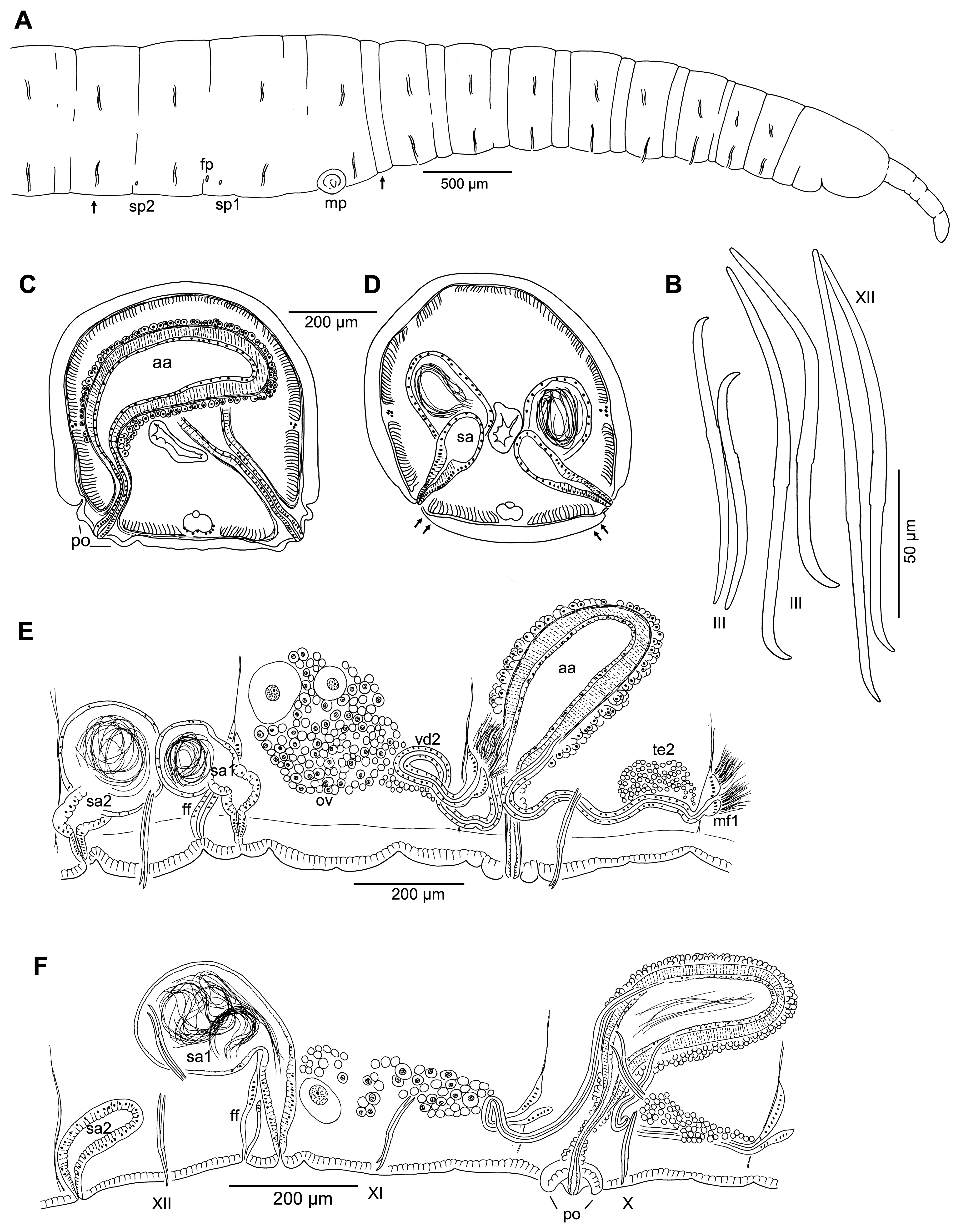
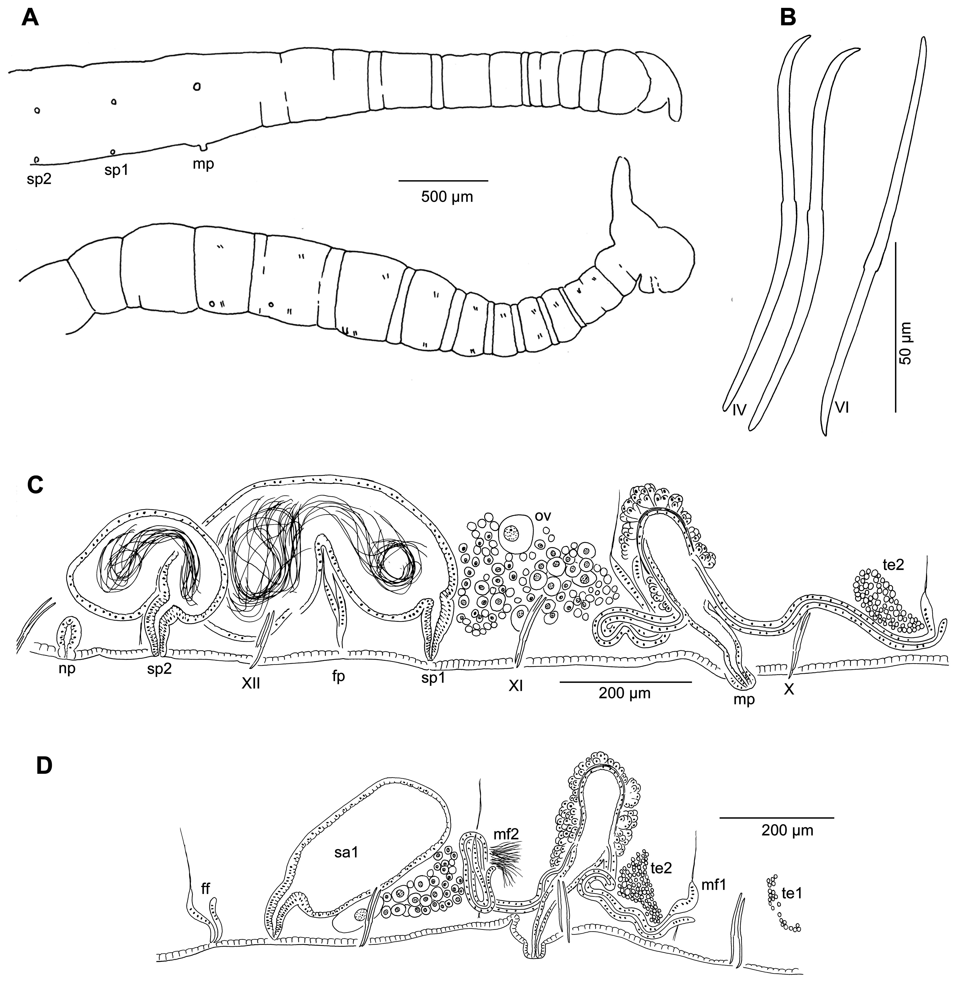
Description (based on populations from the Big Lost River Basin, Idaho). Length of preserved worms 11–19 mm; 56–75 segments; diameter in X 0.4–0.7 mm. Prostomium with proboscis, the latter 240–530 µm long, about 65 µm diameter at middle ( Fig. 1A View FIGURE 1 ). Segmentation distinct in anterior segments; anterior secondary annulus about ¼ segment width, beginning segment IV and extending throughout body length except for clitellum. Chaetae ( Figs. 1B View FIGURE 1 , 2A View FIGURE 2 ) with nodulus median to slightly distal (about 0.4–0.5 from distal end), similar in anterior and posterior segments; within each pair, the inner chaeta is slightly longer, with a more proximal nodulus; ventral chaetae in preclitellar segments 122–192 µm, shorter in II (102–115 µm); in middle and posterior segments 127–192 µm long; dorsal chaetae 122–175 µm in anterior segments, and 120–175 µm in postclitellar segments. One pair of male pores in X, midway between chaetae and posterior septum ( Fig. 1A,E,F View FIGURE 1 ), on short papillae (ca. 30–40 μm long) within broad, concave, ring-shaped porophores ( Figs. 1C,F View FIGURE 1 , 2G View FIGURE 2 ); porophore width 94–134 µm, height 30–72 µm; in some unmated specimens the atrial duct opens in a simple pore without a porophore. Two pairs of simple spermathecal pores, well behind ventral chaetae, on or slightly lateral to the ventral chaetal lines in XI and XII ( Fig. 1A,D View FIGURE 1 ); those in XII very close to the posterior septum (12/13). Female pores on 11/12.
Epidermis 16–28 µm thick in anterior segments, longitudinal muscles 26–44 µm thick. Clitellum (IX) X to XII, up to 36 µm thick. Pharynx mostly in II–IV, without a prominent dorsal pad; pharyngeal glands in IV–VI (VII). First nephridium on 12/13, nephridial duct without vesicle at pore.
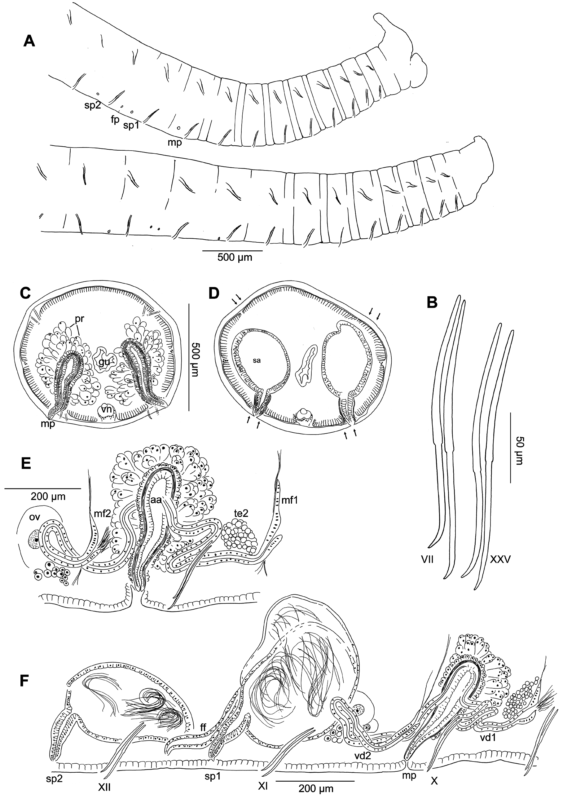
Paired testes in IX and X, ovaries in XI. Sperm sacs extend anteriorly to VIII, posteriorly to XIII or XIV; egg sacs to XV. Male funnels on 9/10 and 10/11, of similar size. Free portion of both anterior and posterior vasa deferentia 14–25 µm wide and about 320–600 µm long. Posterior vas forms a short loop in XI before entering X; both join basal part of atrial ampulla, run along outer surface a short distance, then under the muscle layer to enter atrial lumen near midpoint of the ampulla ( Figs. 1E,F View FIGURE 1 , 2E View FIGURE 2 ). Atrium petiolate, entirely in X, total length 440–720 μm; atrial duct narrow (180–300 µm long, diameter 18–30 µm), gradually widening into the elongate ampulla, and widening slightly near the ectal pore ( Figs. 1C,E,F View FIGURE 1 , 2B View FIGURE 2 ). Atrial ampulla covered by a layer of prostate cells, 10–24 µm high; layer may appear continuous ( Fig. 2D View FIGURE 2 ), or as small, tightly-packed multicellular clusters ( Fig. 2C View FIGURE 2 ); maximum atrium diameter in ental part of ampulla 127–205 µm; epithelial cells cuboidal, not glandular, forming a narrow layer, 3–10 µm high; atrial musculature very thick in ampulla (14–42 µm); muscle fibers arranged in an irregular, somewhat diagonal direction in the median part of the ampulla ( Fig. 2 View FIGURE 2 D–F). Atrium length 4–7 times porophore width, and 0.7–1.5 times body diameter in X.
Spermathecae paired in XI and XII ( Fig. 1E,F View FIGURE 1 ), either both with sperm, or only the anterior pair well-developed and filled with sperm. Spermathecal ampullae may extend into adjacent segments; ducts usually in segments XI and XII, but the second pair may extend into XIII, due to the pore location near the posterior intersegment. Ampullae irregular, sac-like, with uniform epithelial layer not glandular, about 8 µm thick; sperm appears unordered within ampulla ( Fig. 2H View FIGURE 2 ). Spermathecal ducts short and strongly tapered, 40–150 µm long, with gradual transition to ampulla; usually with columnar cells and a narrow lumen; muscle layer thin (ca. 1 µm) ( Fig. 2I,J View FIGURE 2 ). Ectal pores of spermathecae without glands or distinct epidermal modification.
Specimens from other drainages: Two specimens from sites in other drainages (1 each from Muldoon and Hayden Creeks, both within the Snake River catchment) are similar to typical specimens in diagnostic characters, with elongate (but somewhat shorter, 320–595 µm), petiolate atria, with thick muscle layer and thin prostate layer. Male porophores are broad and slightly concave.
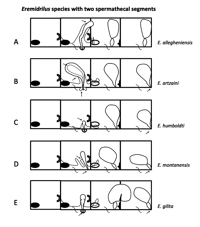
Remarks. Externally, E. artzaini n. sp. is distinguished from other Eremidrilus species by the low, concave (annular) porophore, which surrounds a small papilla terminating in the male pore. Atrial morphology differs from that of all other species in the following combination of characters: large dimensions ( Table 2), petiolate form (with elongate, narrow duct), thick and complex muscular layer, very thin epithelium, and thin layer of prostate glands (see the descriptions and Fig. 11 View FIGURE 11 below; see also Fig. 5 View FIGURE 5 in Fend & Rodriguez 2003). Spermathecal pores are close to the posterior septa, and approximately on the ventral chaetal lines, as in other species described here; in contrast, they tend to be more laterally positioned in species having one spermathecal segment (cf. Fig. 7 View FIGURE 7 in Fend & Rodriguez 2003). The posterior spermathecae (in XII) are usually smaller than the anterior pair (in XI), and may not contain sperm.

No known copyright restrictions apply. See Agosti, D., Egloff, W., 2009. Taxonomic information exchange and copyright: the Plazi approach. BMC Research Notes 2009, 2:53 for further explanation.
|
Kingdom |
|
|
Phylum |
|
|
Class |
|
|
Order |
|
|
Family |
|
|
Genus |