
Eremidrilus montanensis, Fend & Rodriguez, 2020
|
publication ID |
https://doi.org/ 10.11646/zootaxa.4809.1.6 |
|
publication LSID |
lsid:zoobank.org:pub:6E4829CF-1476-4FDB-941D-9EAFCA29D011 |
|
DOI |
https://doi.org/10.5281/zenodo.4329193 |
|
persistent identifier |
https://treatment.plazi.org/id/03D287B7-A57F-FFC7-FF7D-6A20F70DF80E |
|
treatment provided by |
Plazi |
|
scientific name |
Eremidrilus montanensis |
| status |
sp. nov. |
Eremidrilus montanensis View in CoL n. sp.
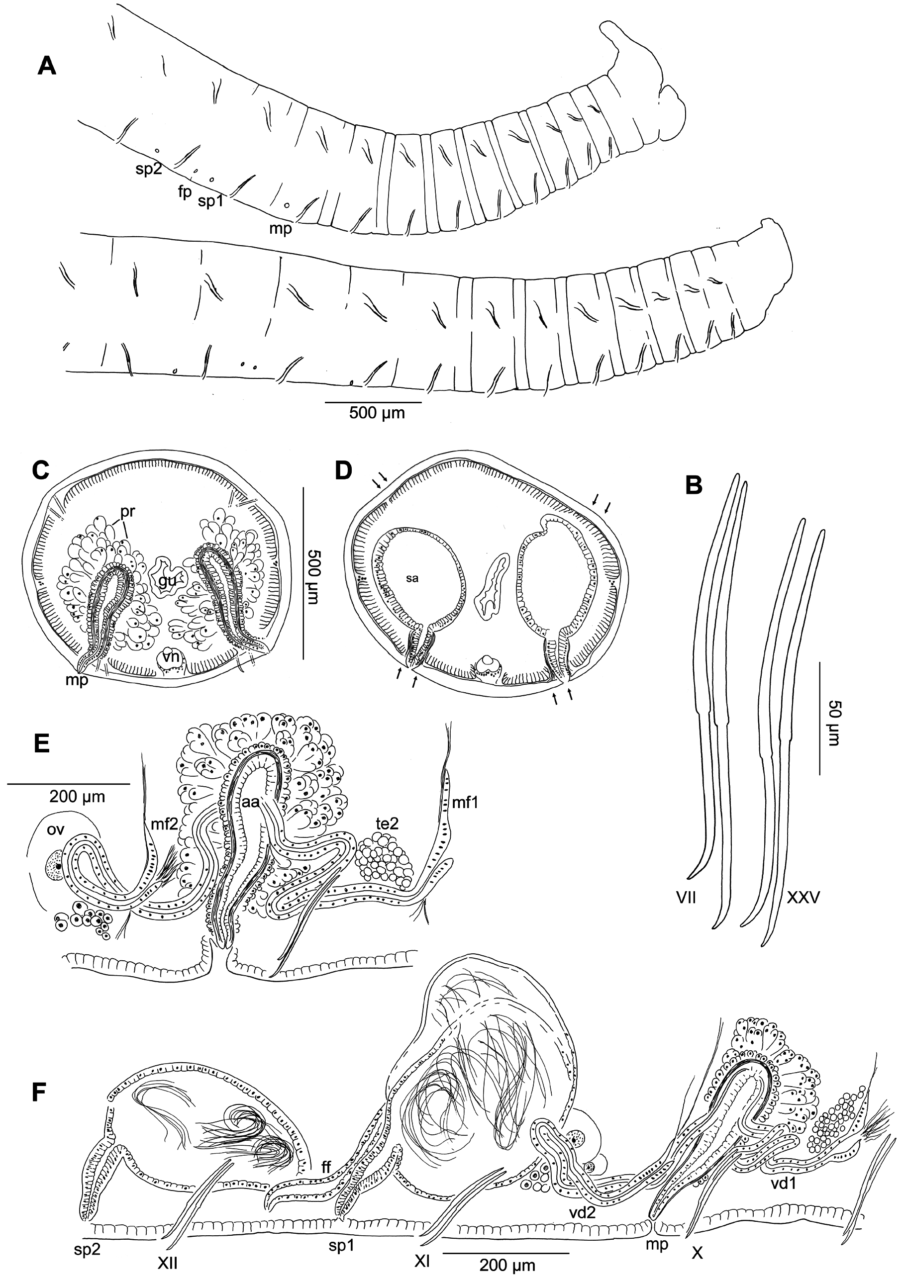
( Figures 5 View FIGURE 5 , 6 View FIGURE 6 , 11D View FIGURE 11 )
Holotype. USNM 1618764 View Materials . One dissected worm, tail broken, mated (all spermathecae with sperm), with mature egg and well-developed clitellum, slide-mounted in Canada balsam.
Type locality. USA, Montana, Broadwater Co., Eureka Creek at Crow Creek , 14 Nov 1999, coll. D.L. Gustafson (Site 16, Table 1) .
Paratypes. All from the type locality, same collection data (Site 16) . USNM 1618767 View Materials , transverse histological sections, stained in hematoxylin and eosin . USNM 1618765–1618766 View Materials , 1 View Materials dissected and 1 whole-mount . MNCN 16.03 View Materials /3113 and 16.03/3114, 1 dissected and 1 sagittally sectioned. All slide-mounted in Canada balsam .
Other material. From type locality (Site 16), 14 Nov 1999, 2 whole mounts, 3 dissected, and 1 transversely sectioned. Site 15, 21 Nov 1997, 1 whole mount. Site 17, 14 Nov 1999, 1 dissected. All collected by D.L. Gustafson.
Etymology. Named for the State of Montana, location of the type locality.
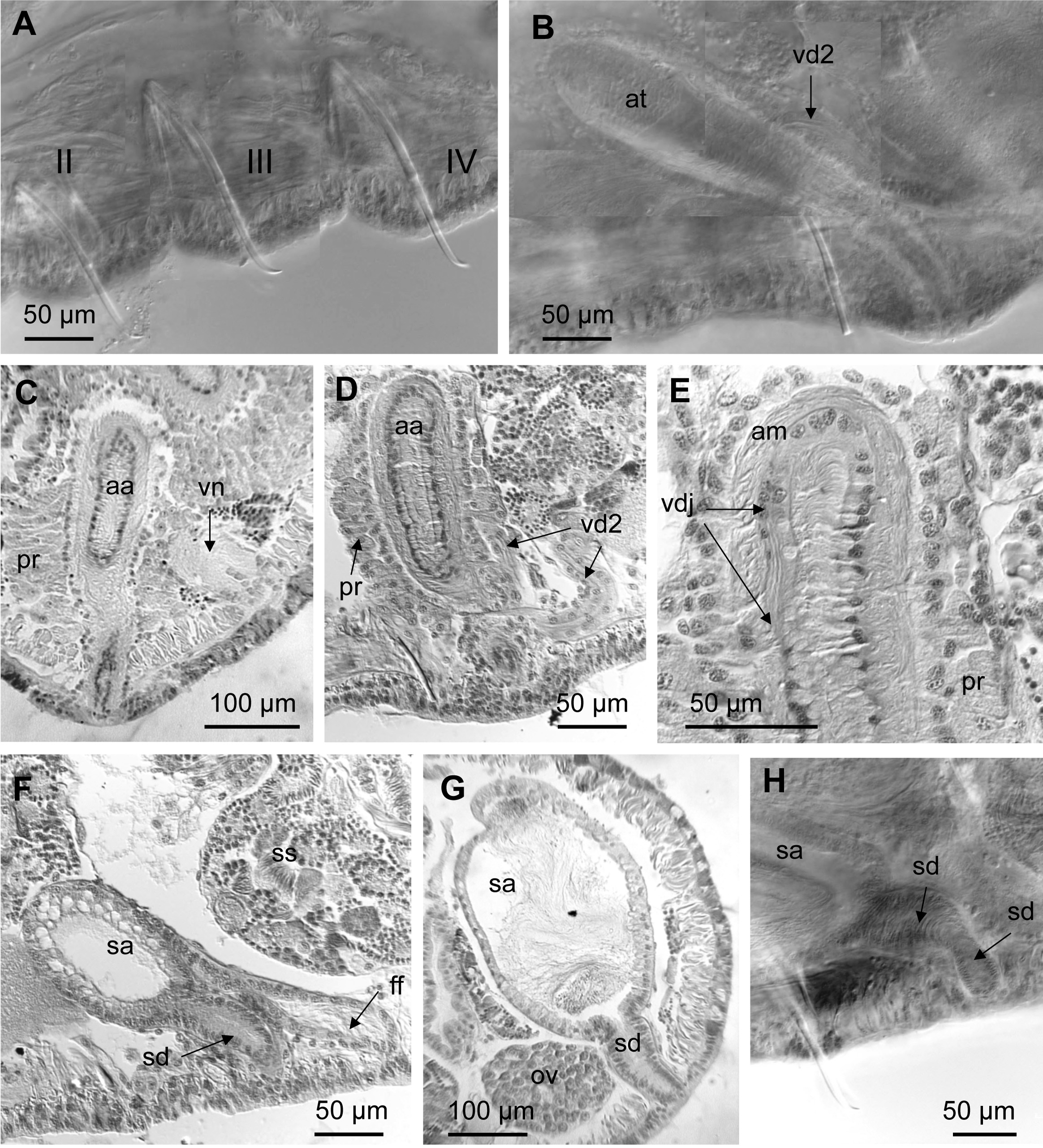
Description (based on specimens from the type locality). Length of preserved worms 17–26 mm; 61–91 segments; diameter in X 0.5–0.8 mm. Prostomium with proboscis, the latter 250 µm long in the single individual where complete, diameter 30–80 µm. Secondary segmentation from IV, weak in posterior segments ( Fig. 5A View FIGURE 5 ). Chaetae two per bundle, with nodulus slightly distal (0.4–0.5 distance from tip) ( Figs. 5B View FIGURE 5 , 6A View FIGURE 6 ); in preclitellar ventral bundles length 142–218 µm, shorter in II (115–146 µm); in middle segments 162–226 µm, in posterior segments 142–232 µm; length of dorsal chaetae similar to ventrals. Male pores open on or slightly lateral to ventral chaetal lines ( Figs. 5A,C View FIGURE 5 , 6B View FIGURE 6 ), between chaetae and posterior septum ( Fig. 5E View FIGURE 5 ); porophores absent or inconspicuous ( Figs. 5C View FIGURE 5 , 6B,C View FIGURE 6 ). Two pairs simple spermathecal pores in XI and XII, behind ventral chaetae, on the ventral chaetal line ( Figs. 5D View FIGURE 5 , 6F,G View FIGURE 6 ), the anterior pair about 2/3 distance from chaetae to posterior septum, the posterior pair very close to 12/13. Female pores on 11/12.
Epidermis 12–26 µm thick in preclitellar segments, thinner (8-15 µm) in post-clitellar segments. Clitellum in X to XIV, clitellar epidermis somewhat thickened (20-35 µm), with glandular cells in specimens with mature eggs Pharyngeal glands in segments IV or V to VI (VII). First visible nephridium at 12/ 13 in most specimens, pore anterior to ventral chaetae; in posterior segments the duct may be slightly expanded (to about 30 µm) at the nephridiopore, forming a small vesicle.
Paired testes in IX and X, ovaries in XI; sperm sacs extend anteriorly to VIII or more, posteriorly to XIII–XVII; egg sacs extend 1–2 segments beyond. Male funnels on 9/10 and 10/11, 110– 165 µm high. Two vasa deferentia per atrium, both 20–30 µm in diameter, and about 400–600 µm long. Posterior vasa deferentia loop back into XI; both anterior and posterior vasa join atrium at about the ental third of ampulla, running within atrial musculature and opening to atrial lumen near the apex ( Figs. 5E,F View FIGURE 5 , 6E View FIGURE 6 ). Atria entirely in X, 280–408 µm long, or 0.5–0.7 times body width at segment X; club-shaped, ampulla not clearly separated from the duct; duct narrows gradually towards pore. Maximum atrium diameter 73–89 µm; epithelial cells somewhat columnar, 7–19 µm high in ampulla ( Fig. 6E View FIGURE 6 ); atrial lumen variable, to 25 µm. Atrial muscle layer 12–16 µm thick; all but a short ectal portion of atrium covered with a dense layer of short cells (11–16 µm high) beneath a thick layer (60–150 µm high) of densely packed, multicellular prostate glands that appear highly granular ( Fig. 6 View FIGURE 6 C–E). No obvious glands at the male pore ( Fig. 6C View FIGURE 6 ).
Spermathecal ampullae oval or sac-like, the first pair 360–530 µm long, the second pair about 2/3 the size of the first pair, each restricted to one segment, sometimes filling most of the segment. Ampullar epithelial layer not obviously glandular, mostly 9–15 µm thick, cells may be somewhat vacuolized near duct, up to 30 µm thick ( Fig. 6G View FIGURE 6 ). Ectal ducts of spermathecae 80–160 long, sharply differentiated from ampulla, diameter 40–68 µm near ampulla, tapered to the pore ( Figs. 5D,F View FIGURE 5 , 6G,H View FIGURE 6 ); the ducts of the second pair as long as or slightly shorter than those of the first pair.
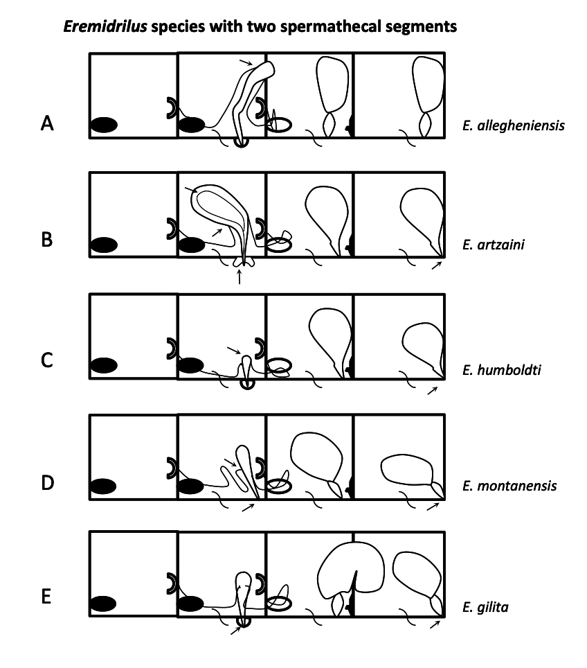
Remarks. The species is distinguished from all other Eremidrilus species by the absence of a distinct male porophore. The club-shaped atria, with very large, dense prostate glands, and the thick epithelium and muscle layer are also distinctive. As in other congeners with two spermathecal segments, spermathecae open on the ventral chaetal lines, with the second pair posteriorly placed, close to intersegment 12/13. Unlike E. artzaini and E. humboldti n. spp., spermathecal ducts are sharply distinguished from the ampulla. Although this is the only Eremidrilus species without a distinct male porophore, other characters such as the proboscis, the club-shaped, semiprosoporous atrium and the postatrial spermathecae support its attribution to the genus. A continuous layer of cells covering the atrial ampulla, basal to the prostate gland layer has been also observed in E. elegans and E. coyote Fend & Rodriguez, 2003 ( Fend & Rodriguez 2003).
No known copyright restrictions apply. See Agosti, D., Egloff, W., 2009. Taxonomic information exchange and copyright: the Plazi approach. BMC Research Notes 2009, 2:53 for further explanation.
|
Kingdom |
|
|
Phylum |
|
|
Class |
|
|
Order |
|
|
Family |
|
|
Genus |