
Ctenoneura simulans Bey-Bienko, 1969
|
publication ID |
https://doi.org/ 10.11646/zootaxa.4237.2.3 |
|
publication LSID |
lsid:zoobank.org:pub:30330D9E-BC76-449B-9C99-2B5EEDA0F8F5 |
|
DOI |
https://doi.org/10.5281/zenodo.6053098 |
|
persistent identifier |
https://treatment.plazi.org/id/03D2F661-FFC5-FF80-658E-FA3EFF381949 |
|
treatment provided by |
Plazi |
|
scientific name |
Ctenoneura simulans Bey-Bienko, 1969 |
| status |
|
Ctenoneura simulans Bey-Bienko, 1969 View in CoL
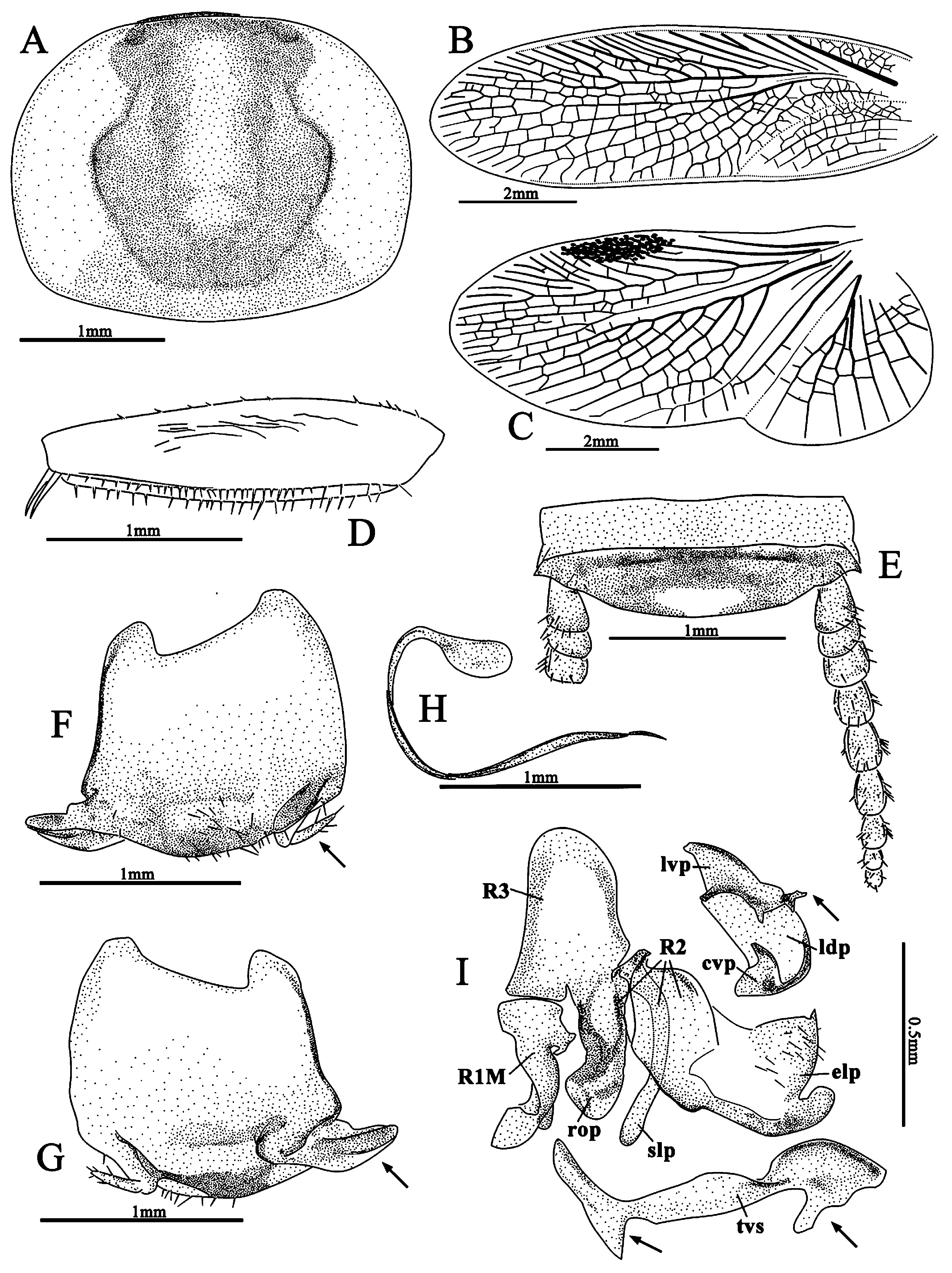
( Figs. 9 View FIGURE 9 , 13 View FIGURE 13 B, 16 A–D, 21)
Ctenoneura simulans Bey-Bienko, 1969: 833 View in CoL , figs. 2–3, ♂ holotype (In Russian); Bey-Bienko, 1970: 528, figs 2–3, ♂ holotype, type locality: “S. China, Yunnan: Hsiaomonyang (vicinity) [around Xiaomengyang Town, Jinghong City], 900–1100m ” (English version of Bey-Bienko, 1969); Roth 1993: 107, figs. 21 E–F (redrawn from Bey-Bienko, 1969); Feng, Guo & Woo 1997: 34.
Material examined. CHINA: Yunnan: 1 male ( IZCAS), Damenglong († AEAE), Jinghong City , Xishuangbanna Prefecture , 650 m, 19.IV.1958, Xu-Wu Meng leg.
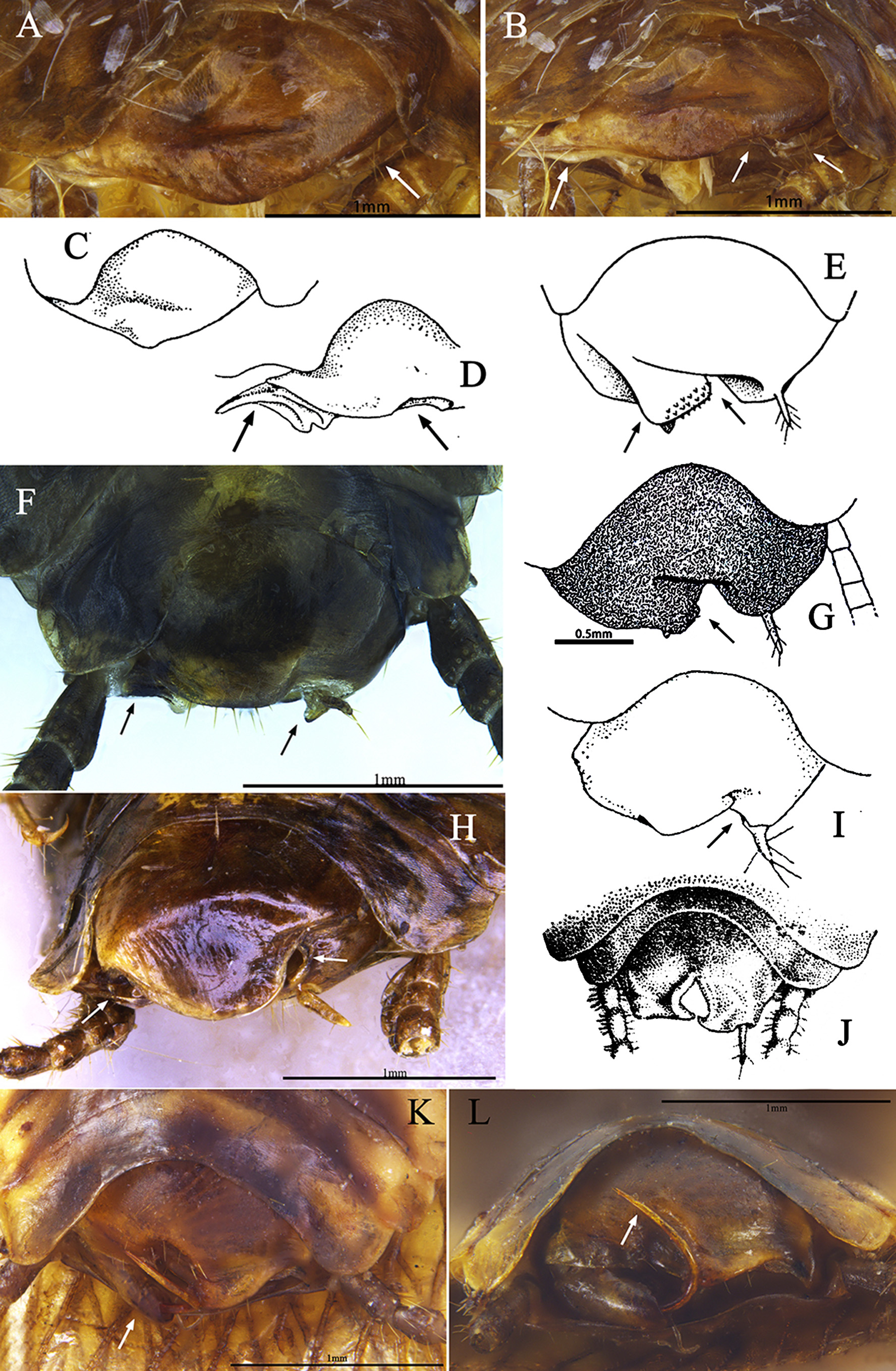
Diagnosis. C. simulans differs from any other species in this genus by the simple shape of subgenital plate in ventral view ( Figs. 16 View FIGURE 16 A, C) and the quite elongated eds in subgenital plate ( Fig. 9 F View FIGURE 9 , 16 View FIGURE 16 D).
Redescription. Male. Body length 7.1 mm; overall length including tegmen 11.1 mm; pronotum length × width 2.2 × 2.8 mm.
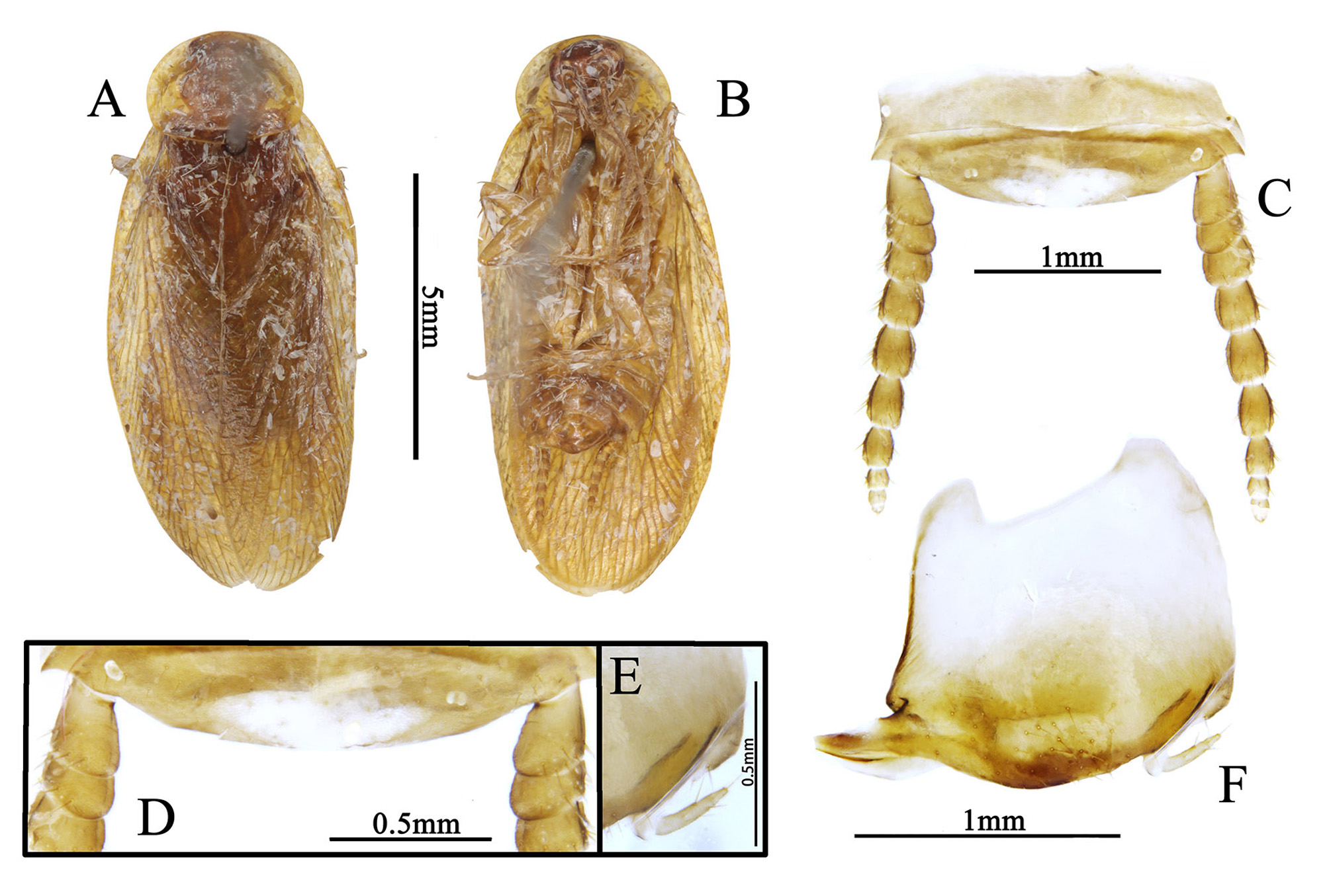
Coloration. Body brownish yellow. Head and eyes brown, antennae light brown. Pronotal disk brown, lateral areas yellow, subtransparent, posterior margin subtransparent narrowly. Tegmina brownish yellow, wings transparent, RP area brownish yellow. Legs, abdomen, cerci brownish yellow ( Figs. 9 A–B View FIGURE 9 ).
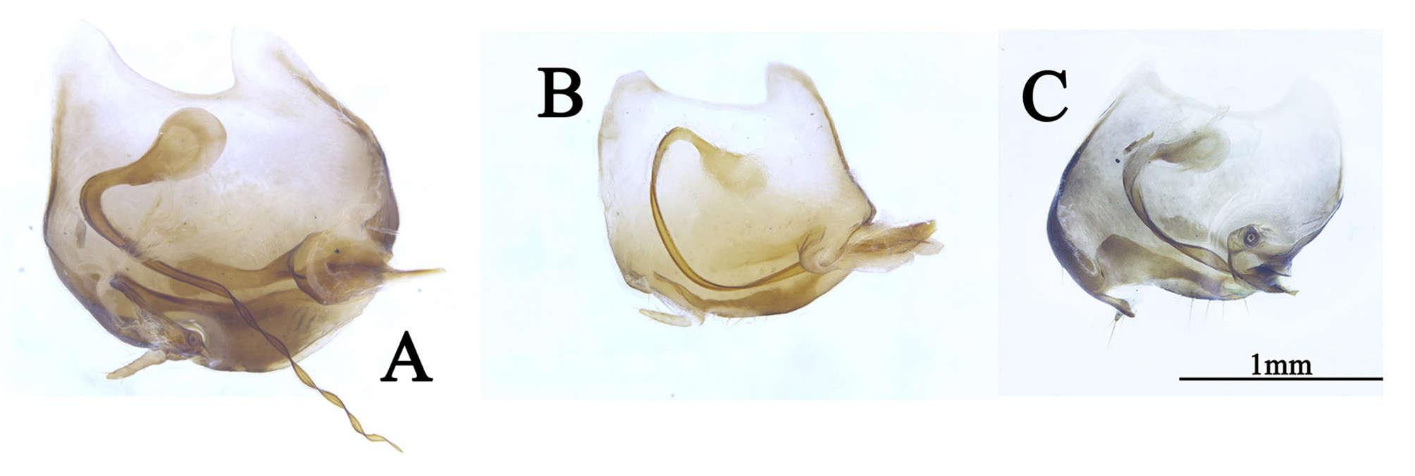
Head: almost hidden ( Fig. 21 View FIGURE 21 A), eyes wide apart, interocular space greater than the distance between antennal sockets, ocelli small, antennae from the second subsegment of flagellum with much small pubescence. Pronotum: suborbicular, lateral corners of posterior slightly quadrate ( Fig. 21 View FIGURE 21 A). Tegmina and wings: fully developed extending well beyond end of the abdomen; tegmen with a thick Sc, M with 5–7 branches, between R and M presents an intercalary vein, CuA short, not bifurcate ( Fig. 21 View FIGURE 21 B); hind wing with intercalary vein present, M bifurcate, CuA with 6–7 branches, CuP long and thin ( Fig. 21 View FIGURE 21 C). Legs: front femur type C1, anterior margin with several spinules ( Fig. 21 View FIGURE 21 D). Pulvilli absent, tarsal claws symmetrical, simple, arolia minute. Abdomen: supra-anal plate in dorsal view transverse, simple, median with a small transparent area; cerci long, slightly pubescent, segments round ( Figs. 9 C–D View FIGURE 9 , 21 View FIGURE 21 E). Subgenital plate asymmetrical, when observing it in the specimen, simple ( Figs. 16 View FIGURE 16 A, C), when observing it separately, apex with several setae, right with a very long eds, forming a groove towards right ( Figs. 9 F View FIGURE 9 , 21 View FIGURE 21 F–G); in rear view, left with an incision ( Figs. 16 View FIGURE 16 B, D), forming a small process, with one stylus on it ( Fig. 9 E View FIGURE 9 ); in dorsal view, the sgs above the subgenital plate slender and curved, but with one side expanded, flat and round, another side sharpened, the sharpened side nested in the eds ( Figs. 13 View FIGURE 13 B, 21 H). Genitalia: left phallomere: well sclerotized, lvp with some small irregular processes posteriorly; right margin straight, and anterior apex slightly curved; ldp with thickened cvp. Right phallomere: R1M tortuose, R2 with strong rop, thick slp, the elp with setose membrane, R3 large, with round anterior apex. Transverse sclerite (tvs): not regular, with two processes posteriorly, one in the right part, another near left end; left apex flat, right portion bent near the process ( Fig. 21 View FIGURE 21 I).
Female. Unknown.
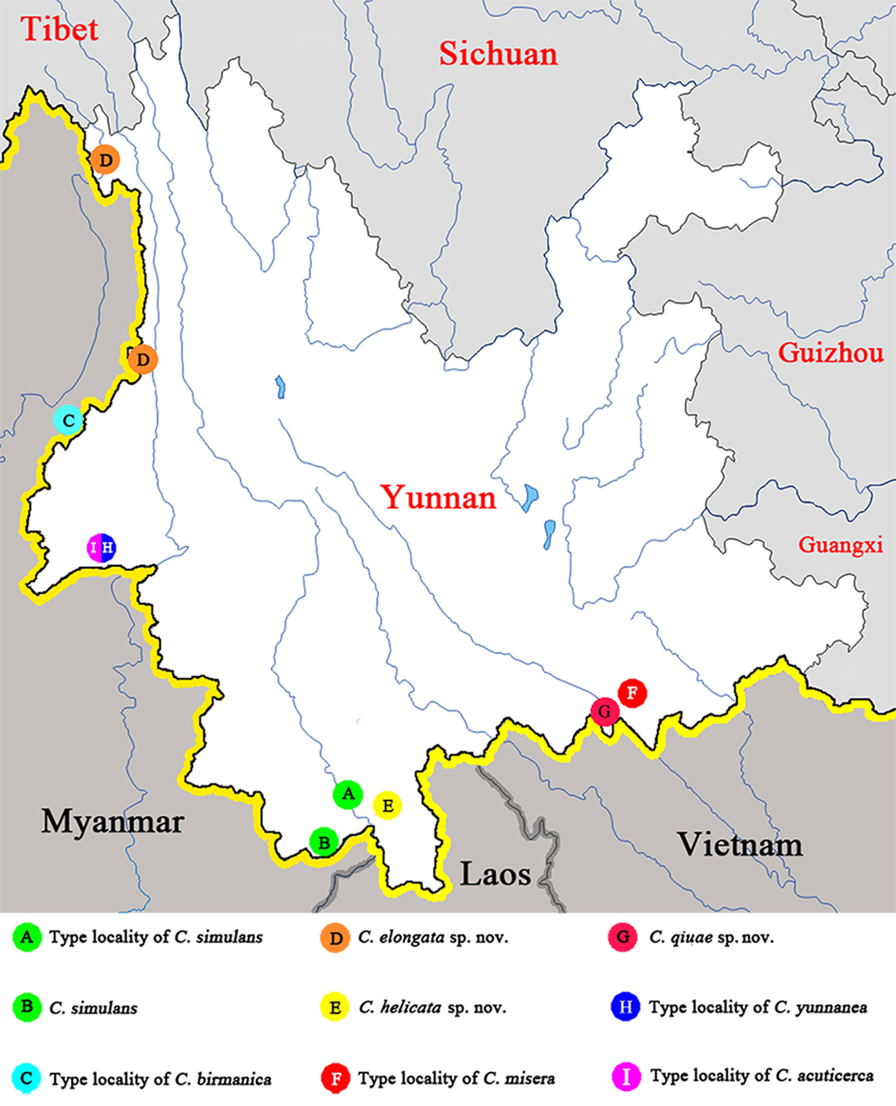
Distribution. China: South Yunnan ( Fig. 3 View FIGURE 3 ).
Remarks. The senior author has been to IZCAS for examining the type specimens of C. acuticerca and C. yunnanea , but only found this none type specimen of C. simulans from Yunnan; the surface of the specimen was blotted with many Lepidoptera scales. We recognized it as C. simulans . Bey-Bienko (1969) didn’t mention whether there is a stylus; Roth (1993) thought the stylus could be small and invisible. We found one small stylus hidden in the incision ( Fig. 16 View FIGURE 16 B); after dissection the stylus appears clearly ( Fig. 9 E–F View FIGURE 9 ), thus this species actually possesses a stylus.
| IZCAS |
Institute of Zoology, Chinese Academy of Sciences |
No known copyright restrictions apply. See Agosti, D., Egloff, W., 2009. Taxonomic information exchange and copyright: the Plazi approach. BMC Research Notes 2009, 2:53 for further explanation.
|
Kingdom |
|
|
Phylum |
|
|
Class |
|
|
Order |
|
|
SuperFamily |
Corydioidea |
|
Family |
|
|
Genus |
Ctenoneura simulans Bey-Bienko, 1969
| Qiu, Lu, Che, Yan-Li & Wang, Zong-Qing 2017 |
Ctenoneura simulans
| Feng 1997: 34 |
| Roth 1993: 107 |
| Bey-Bienko 1970: 528 |
| Bey-Bienko 1969: 833 |