
Ctenoneura elongata, Qiu, Lu, Che, Yan-Li & Wang, Zong-Qing, 2017
|
publication ID |
https://doi.org/ 10.11646/zootaxa.4237.2.3 |
|
publication LSID |
lsid:zoobank.org:pub:30330D9E-BC76-449B-9C99-2B5EEDA0F8F5 |
|
DOI |
https://doi.org/10.5281/zenodo.6053102 |
|
persistent identifier |
https://treatment.plazi.org/id/78D6856C-846D-46E4-A5E8-FF7EAE9483B9 |
|
taxon LSID |
lsid:zoobank.org:act:78D6856C-846D-46E4-A5E8-FF7EAE9483B9 |
|
treatment provided by |
Plazi |
|
scientific name |
Ctenoneura elongata |
| status |
sp. nov. |
Ctenoneura elongata sp. nov.
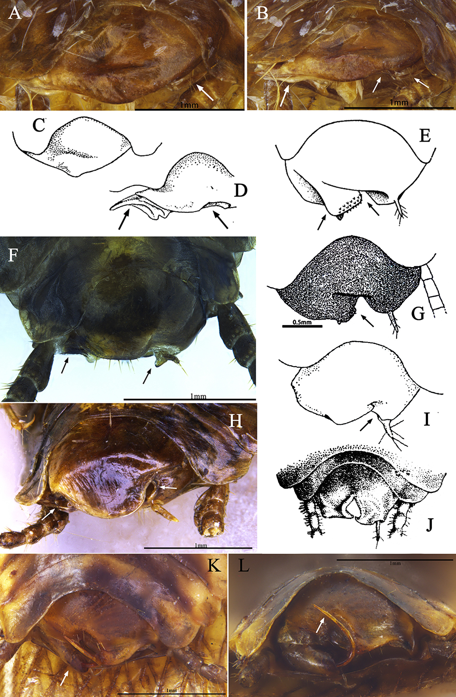
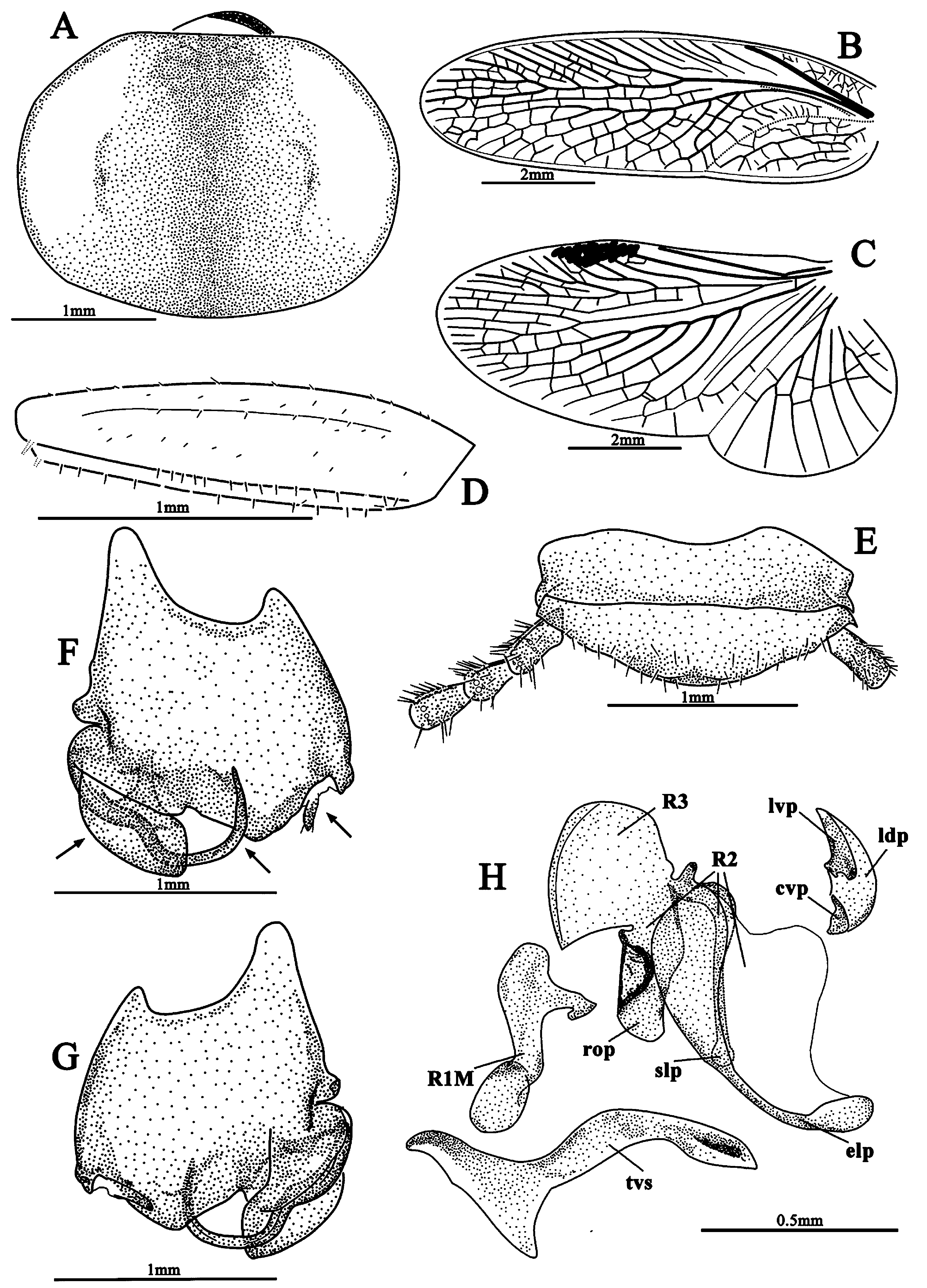
( Figs. 10 View FIGURE 10 , 16 View FIGURE 16 K–L, 22)
Type material. Holotype: CHINA: Yunnan: male ( MHBU), Pianma Town (ÉŞffi), Lushui County, Nujiang Prefecture, 11.V.2004, Xiu-Juan Yang & Yu-Shuang Liu leg . Paratype: CHINA: Yunnan: 1 male ( SWU), Dulongjiang Township (ÊAEỮş), Gongshan County, Nujiang Prefecture, VI.2016, by Malaise trap, Zhi-Wei Dong leg.
Diagnosis. This species superficially resembles C. qiuae but can be easily distinguished from the latter by the exposed and unseparated sgs and enlarged eds ( Figs. 10 F View FIGURE 10 , 16 View FIGURE 16 K–L, 22 F–G).
Description. Male. Body length 7.1–7.3 mm; overall length including tegmen 10.5–10.8 mm; pronotum length × width 1.6–2.0× 2.3–2.6 mm.
Coloration. Body brown. Head and maxillary palpi dark brown. Pronotum with lateral parts transparent widely, disc, anterior and hind areas dark brown, the hind area with wider brown portion that the anterior area. Tegmina brownish yellow with lighter colored basal margins, especially the Sc areas, wings transparent, venation of both tegmina and wings brown. Abdomen brown, each segment with lateral parts brownish yellow, cerci brown ( Figs. 10 A–B View FIGURE 10 ).
Head: eyes wide apart, interocular space greater than the distance between antennal sockets, ocelli minute, antennae missing, frons convex ( Fig. 22 View FIGURE 22 A). Pronotum: transversely oval, anterior margin truncated, slightly convex above head ( Fig. 22 View FIGURE 22 A). Tegmina and wings: fully developed extending well beyond the end of the abdomen; tegmen with major veins well defined, branches of R with some intercalary branches distally, M with 6 branches, an intercalary vein present between R and M, CuA slightly curved, not bifurcate ( Fig. 22 View FIGURE 22 B); wing with intercalary vein present, M bifurcate, CuA with 5 branches, CuP very slender ( Fig. 22 View FIGURE 22 C). Legs: most legs of the holotype missing, front femur type C1, surface slightly with fine setae ( Fig. 22 View FIGURE 22 D). Abdomen: supra-anal plate in dorsal view transverse, simple, median without a transparent area; cerci long, slightly pubescent, segments normal ( Figs. 10 C–D View FIGURE 10 , 22 View FIGURE 22 E). Subgenital plate asymmetrical, when observing it in the specimen, right with an cylinder shaped eds, the sgs protruding out from apex of the structure, slender and curved, formed like a long needle, the apex of subgenital plate deeply sunken on the right, forming a pit with the eds, the stylus situated in a notch on the left side of subgenital plate, but hidden inside which makes it invisible ( Figs. 16 View FIGURE 16 K–L); when observing it separately, the eds unfolded (perhaps the result of NaOH), which can also be observed originate from the inner surface of subgenital plate, the curved sgs originating from the right corner, the basal part robust, then gradually necked, apex acute, the base partly covered by the cylindrical eds, the stylus is clearly visible from the left side of subgenital plate, originating from a notch ( Figs. 10 E–F View FIGURE 10 , 22 View FIGURE 22 F–G); in dorsal view, the roots of the cylindrical eds and sgs are better observed, the unfolded eds formed with two lobes, the dorsal lobe small, while the ventral lobe much larger, these two lobes curved forming a cylindrical process which can be distinctly seen in specimen, the curved sgs partly resting inside it, the root of the stylus with hyaline membrane ( Fig. 10 E View FIGURE 10 , 22 View FIGURE 22 F–G). Genitalia: left phallomere: more reduced than any other species described in this article, lvp narrow, reduced, ldp narrow, cvp short. Right phallomere: R1M short, tortuose, base with an irregular process towards left, R2 with short rop, median with a large depression, slp with a round apex, elp slender, membraned, apex becoming larger. Transverse sclerite (tvs): curved, left portion reduced in holotype, while paratype slightly expanded, right portion bent, with one process towards anterior ( Fig. 22 View FIGURE 22 H).
Female. Unknown.
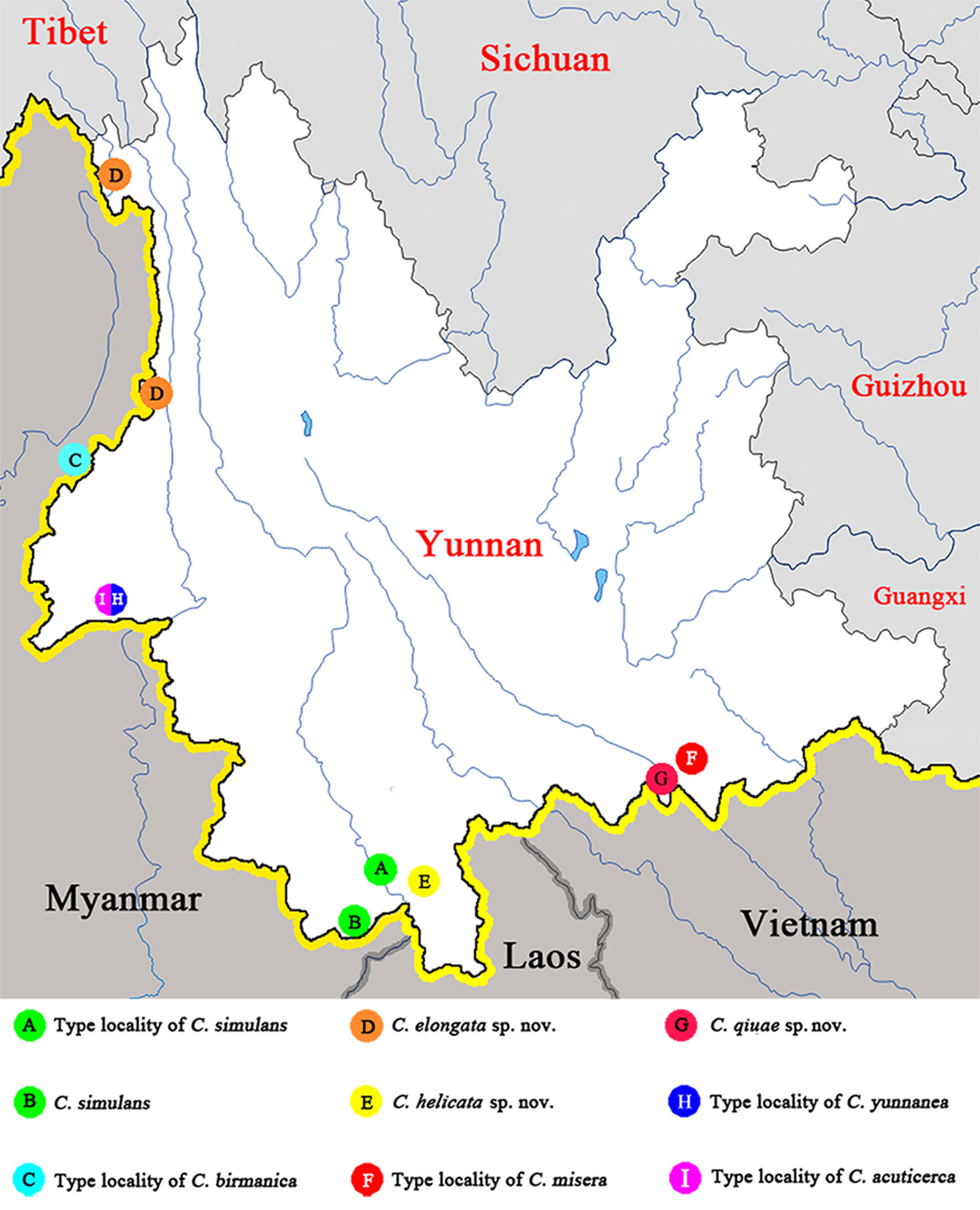
Distribution. China: West Yunnan ( Fig. 3 View FIGURE 3 ).
Remarks. This species is remarkable with an unseparated sgs which originates from the right lateral of subgenital plate, this sclerite is distinctly visible even before dissection, and the eds is well developed, large and protruding, functioning as a groove to hold the sgs. The supra-anal plate is generally the simulans -group type, but without transparent median part and rounded segments of cerci, the left phallomere is more reduced than in any other dissected species. We still regard it as a member of the simulans -group, the peculiar aberrant structure of subgenital plate makes it a transition between the simulans -group and other species groups which remain unknown to us.

| SWU |
Sungshin Women's University |
No known copyright restrictions apply. See Agosti, D., Egloff, W., 2009. Taxonomic information exchange and copyright: the Plazi approach. BMC Research Notes 2009, 2:53 for further explanation.
|
Kingdom |
|
|
Phylum |
|
|
Class |
|
|
Order |
|
|
SuperFamily |
Corydioidea |
|
Family |
|
|
Genus |