
Polydora websteri Hartman
|
publication ID |
https://doi.org/ 10.11646/zootaxa.4969.2.2 |
|
publication LSID |
lsid:zoobank.org:pub:14358F21-D335-44BA-9F8E-866B62E3881D |
|
DOI |
https://doi.org/10.5281/zenodo.5915226 |
|
persistent identifier |
https://treatment.plazi.org/id/03DA8792-FF88-0920-9DC5-FE6DFDE00ACA |
|
treatment provided by |
Plazi |
|
scientific name |
Polydora websteri Hartman |
| status |
|
Polydora websteri Hartman View in CoL , in Loosanoff and Engle 1943
( Figs 4–7 View FIGURE 4 View FIGURE 5 View FIGURE 6 View FIGURE 7 )
Polydora websteri: Hartman View in CoL , in Loosanoff & Engle 1943: 70–72, Fig. 1 View FIGURE 1
Polydora websteri: Blake 1969: 814–815 View in CoL , Fig. 2 View FIGURE 2 ; Blake 1971: 6–8, Fig. 3 View FIGURE 3 ; Foster 1971: 26; Blake & Kudenov 1978: 258–259, Figs 43k–n; Handley & Bergquist 1997: 191–205; Radashevsky & Williams 1998: 212–216; Radashevsky 1999; 107–113, Fig. 1 View FIGURE 1 ; Sato-Okoshi 1999: 832–834, Fig. 2B View FIGURE 2 ; Surugiu 2005: 67; Bonifácio 2009; Read 2010: 9–11, Figs 1H–J View FIGURE 1 , 2B, 2D, 2F View FIGURE 2 , 4D–G View FIGURE 4 ; Surugiu 2012: 50–53 View Cited Treatment , Fig. 3 View FIGURE 3 ; Sato-Okoshi & Abe 2013: 1280–1281, Fig. 2 View FIGURE 2 ; Sato-Okoshi et al. 2013, 153–159, Fig. 5 View FIGURE 5 ; Ye et al. 2017; Rice et al. 2018
Polydora cf. ciliata: Simon 2011 View in CoL
Polydora haswelli: Sato-Okoshi et al. 2008: 495 View in CoL , Fig. 4F, G View FIGURE 4
Polydora cf. websteri: Williams 2015 View in CoL ; Simon 2015; Simon & Sato-Okoshi 2015; Williams et al. 2017
Material examined. South Africa: Eastern Cape, Nelson Mandela Bay ; 33°50′0″ S, 25°50′0″ E; SAMC-A089084 - SAMC-A089088 (1 specimen each); SAMC-A089094 (16 complete and 2 incomplete specimens), SAMC-A089181 (1 specimen); August 2017; C. A. Simon; from cultured Crassostrea gigas . Type material examined GoogleMaps . United States of America: Connecticut, Long Island Sound; Lectotype ( LACM-AHF POLY 1628 ) ; 4 January 1943; J. B. Engle; mouth of Milford River ; from vesicles on empty oyster shells. Additional material examined . South Africa: Eastern Cape, Nelson Mandela Bay ; 33°50′0″ S, 25°50′0″ E; 26 specimens sacrificed for genetic analysis; August 2017; C. A. Simon; from cultured C. gigas GoogleMaps . South Africa: Northern Cape, Kleinzee ; 29°39′59″ S, 17°04′60″ E; 7 specimens sacrificed for genetic analysis; November 2012; C. A. Simon; from cultured C. gigas GoogleMaps .
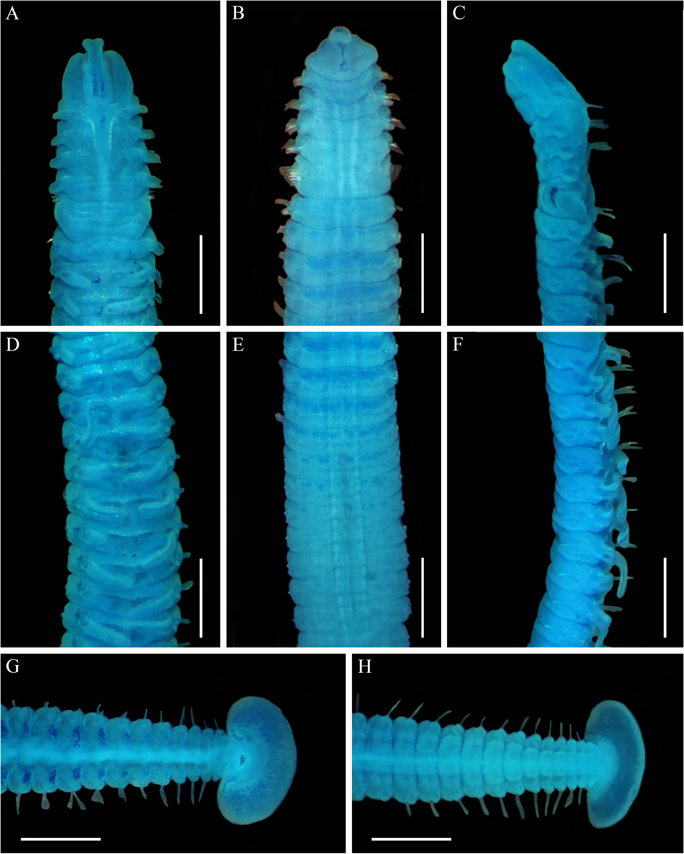
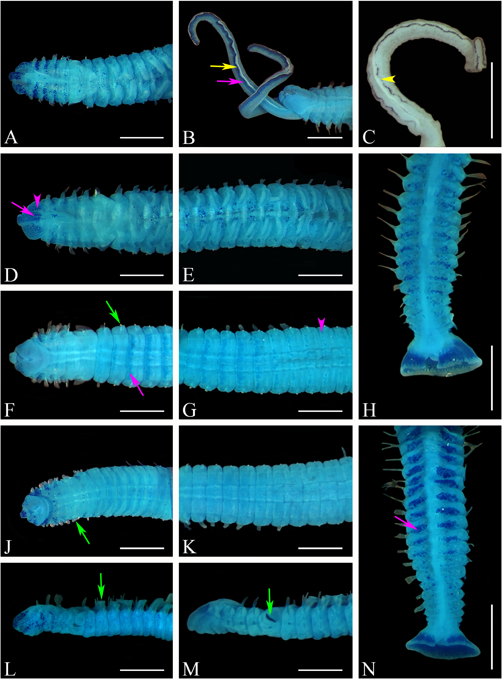
Description of P. websteri from South Africa. Complete specimens 4.7 to 22 mm long and 0.32 to 0.95 mm wide at chaetiger 5 (n = 22), for up to 39 to 123 chaetigers (n = 20). Prostomium anteriorly bilobed or weakly bilobed; caruncle extending to mid chaetiger 2 or up to end chaetiger 3 ( Figs 4A, C View FIGURE 4 , 5A, D, L, M View FIGURE 5 ); eyes usually absent, but up to 4 arranged in trapezoid when present; occipital antenna absent ( Figs 4C View FIGURE 4 , 5L, M View FIGURE 5 ). Body pigmentation absent; palps with distinct continuous black pigmentation lines adjacent to the food groove ( Fig. 5B, C View FIGURE 5 ).
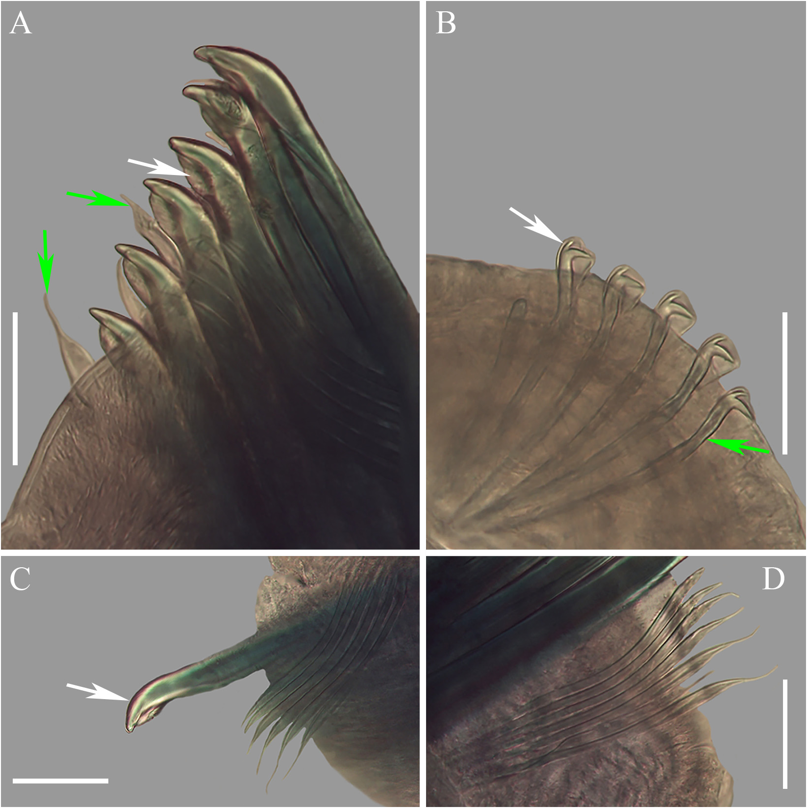
Notochaetae absent on chaetiger 1, notopodial lobe small. Winged capillary notochaetae with postchaetal lamellae on chaetigers 2 to 4 and 6. Capillary notochaetae with postchaetal lamellae on posterior chaetigers reducing in size posteriorly. Winged neurochaetae with postchaetal lamellae increasing in size from chaetigers 1 to 4 and 6. Neurochaetae replaced by bidentate hooded hooks in vertical row on chaetiger 7 ( Figs 4B, C View FIGURE 4 , 5F, J, L, M View FIGURE 5 ); up to 7 hooded hooks per fascicle, increasing to 11 in middle chaetigers, decreasing to 1 or 2 on posterior chaetigers. Hooded hooks without companion chaetae; main fang at <45° to apical tooth and right angle to shaft; with constriction on shaft ( Fig. 6B View FIGURE 6 ).
Chaetiger 5 modified, approximately twice as long as chaetigers 4 and 6 ( Figs 4A, B, C View FIGURE 4 , 5A, D, F, J, L, M View FIGURE 5 ); superior ( Fig. 6C View FIGURE 6 ) and inferior ( Fig. 6D View FIGURE 6 ) winged chaetae shorter than capillary chaetae on the preceding chaetigers. Thick falcate spines on chaetiger 5 with prominent flange on concave side of spine ( Figs 6A, C View FIGURE 6 ), no tooth; up to 7 spines in slightly curved row ( Figs 4B, C View FIGURE 4 , 5A, D, F, J, L, M View FIGURE 5 ); spines alternating with pennoned companion chaetae, tips occasionally frayed ( Fig. 6A View FIGURE 6 ).
Branchiae present from chaetiger 7 onwards ( Figs 4A, C View FIGURE 4 , 5A, D, L, M View FIGURE 5 ), covering approximately 50 % of chaetigers, longest on chaetigers 11 to 25 ( Figs 4D, F View FIGURE 4 , 5E View FIGURE 5 ).
Pygidium cup to disc-shaped, with dorsal notch leading to anus, 1.5 (±0.31) times wider than 5 th last chaetiger ( Figs 4G, H View FIGURE 4 , 5H, N View FIGURE 5 ).
Staining pattern. Palps without staining pattern or a line of blue staining pigment adjacent to the continuous black pigmentation lines ( Fig. 5B View FIGURE 5 ). Branchiae without staining ( Figs 4A, C, D, F View FIGURE 4 , 5A, D, E, L, M View FIGURE 5 ).
Dorsal: Staining of prostomium varying from few irregular stained cells to stained cells forming two bars (faint or distinct) fading into dispersed stained cells towards anterior ( Figs 4A View FIGURE 4 , 5A, D View FIGURE 5 ). Staining of caruncle varying from no staining pattern to few stained cells ( Figs 4A View FIGURE 4 , 5A, D View FIGURE 5 ). Staining of peristomium varying from few irregular stained cells ( Fig. 4A View FIGURE 4 ) to clearly dispersed stained cells that may be concentrated along ridge of prostomium ( Figs 5A, D View FIGURE 5 ). Chaetiger 1 to 4 varying from no stain ( Fig. 4A View FIGURE 4 ) to irregularly stained cells ( Fig. 5D View FIGURE 5 ) or patches of stain on both sides of caruncle ( Fig. 5A View FIGURE 5 ), fading toward latero-ventral ( Figs 5F, J, L, M View FIGURE 5 ). Chaetiger 5 with fewest stained cells of anterior chaetigers, varying from no ( Figs 4A View FIGURE 4 , 5D View FIGURE 5 ) to few irregular ( Fig. 5A View FIGURE 5 ) stained cells. On chaetiger 6 to 20 th from the last chaetiger, stain varying from faint ( Fig. 4D View FIGURE 4 ) to distinct ( Fig. 5E View FIGURE 5 ), patches of stained cells on both sides of centre intensifying towards posterior; 8 th to 2 nd from the last chaetiger varying from no staining pattern to stained cells forming faint ( Fig. 4G View FIGURE 4 ) or distinct patches ( Figs 5H, N View FIGURE 5 ) towards posterior on both sides of centre; last chaetiger without staining pattern ( Figs 4G View FIGURE 4 , 5H, N View FIGURE 5 ). Except for chaetigers 1 to 4, no lateral staining pattern ( Fig. 4C, F View FIGURE 4 , 5L, M View FIGURE 5 ).
Ventral: Staining of peristomium and chaetigers 1 to 5 varying from no staining pattern ( Fig. 4B View FIGURE 4 ) to irregular stained cells on the side and across the venter on chaetigers 1 and 2, decreasing in density posteriorly ( Figs 5F, J View FIGURE 5 ). Chaetigers 6 to 14 with stained cells forming wide bands across the anterior part of chaetiger ( Figs 4B, E View FIGURE 4 , 5F, G, J View FIGURE 5 ). Chaetigers 15 to 18 stained cells may form a wide ( Fig. 5F View FIGURE 5 ) to a thin ( Fig. 5G View FIGURE 5 ) band across the anterior part of chaetiger or be absent ( Fig. 5K View FIGURE 5 ); from Chaetiger 19 to last chaetiger without staining pattern ( Fig. 4H View FIGURE 4 ).
Posterior: Pygidium with distinct stained cells covering entire surface, except along edge around anus and outermost edge of disc ( Figs 4G, H View FIGURE 4 , 5H, N View FIGURE 5 ).
Remarks. Specimens of P. websteri from South Africa conform to the morphology of the lectotype ( Loosanoff & Engle 1943; Radashevsky 1999) ( Fig. 7 View FIGURE 7 ) and descriptions of conspecifics found globally ( Blake 1969, 1971; Foster 1971; Blake & Kudenov 1978; Handley & Bergquist 1997; Sato-Okoshi 1999; Surugiu 2005, 2012; Sato- Okoshi et al. 2008; Bonifácio 2009; Read 2010; Sato-Okoshi & Abe 2013; Barros et al. 2017; Ye et al. 2017; Rice et al. 2018). Some variation present for the branchiae, pigmentation patterns and spines on chaetiger 5, is within the ranges reported for the species or may be the result of wear and tear or preservation.
Branchiae for South African specimens occurred on approximately 50 % of chaetigers, slightly less than the 60 – 80 % of the body length described elsewhere ( Loosanoff & Engle 1943; Blake & Kudenov 1978; Radashevsky 1999; Surugiu 2005, 2012; Sato-Okoshi & Abe 2013; Ye et al. 2017). Continuous black pigmentation lines adjacent to the food groove on the palps were observed for most South African specimens ( Fig. 5B, C View FIGURE 5 ), but absent from some paratype material ( Radashevsky 1999), probably as a consequence of fading in preserved specimens ( Read 2010). Similar pigmentation fading can also be seen on the palps for some preserved specimens from South Africa ( Fig. 5C View FIGURE 5 ) and USA ( Rice et al. 2018), where sections of the black line are lighter, making them seem non-continuous if not carefully examined (see also Waser et al. 2020). Sato-Okoshi & Abe (2013) found that while some live specimens from Japan had continuous black lines on the palps, others had discontinuous black pigmentation. It is uncertain whether this is intraspecific variation or a result of fading due to age of the worm, as in certain parts of the palp pigmentation appeared faded.
South African specimens have a pronounced flange on the falcate spines with pennoned companion chaetae on chaetiger 5 ( Fig. 6A View FIGURE 6 ). Wear and orientation of the falcate spines on chaetiger 5 may give the appearance of a tooth and/ or a sheath ( Read 2010) instead of only a flange as in South African specimens and most other descriptions ( Blake & Kudenov 1978; Radashevsky 1999; Sato-Okoshi 1999; Surugiu 2005, 2012; Bonifácio 2009; Read 2010; Barros et al. 2017; Rice et al. 2018; Martinelli et al. 2020). With age, pennoned companion chaetae on chaetiger 5 may appear frayed or hastate ( Read 2010), as seen for South African specimens.
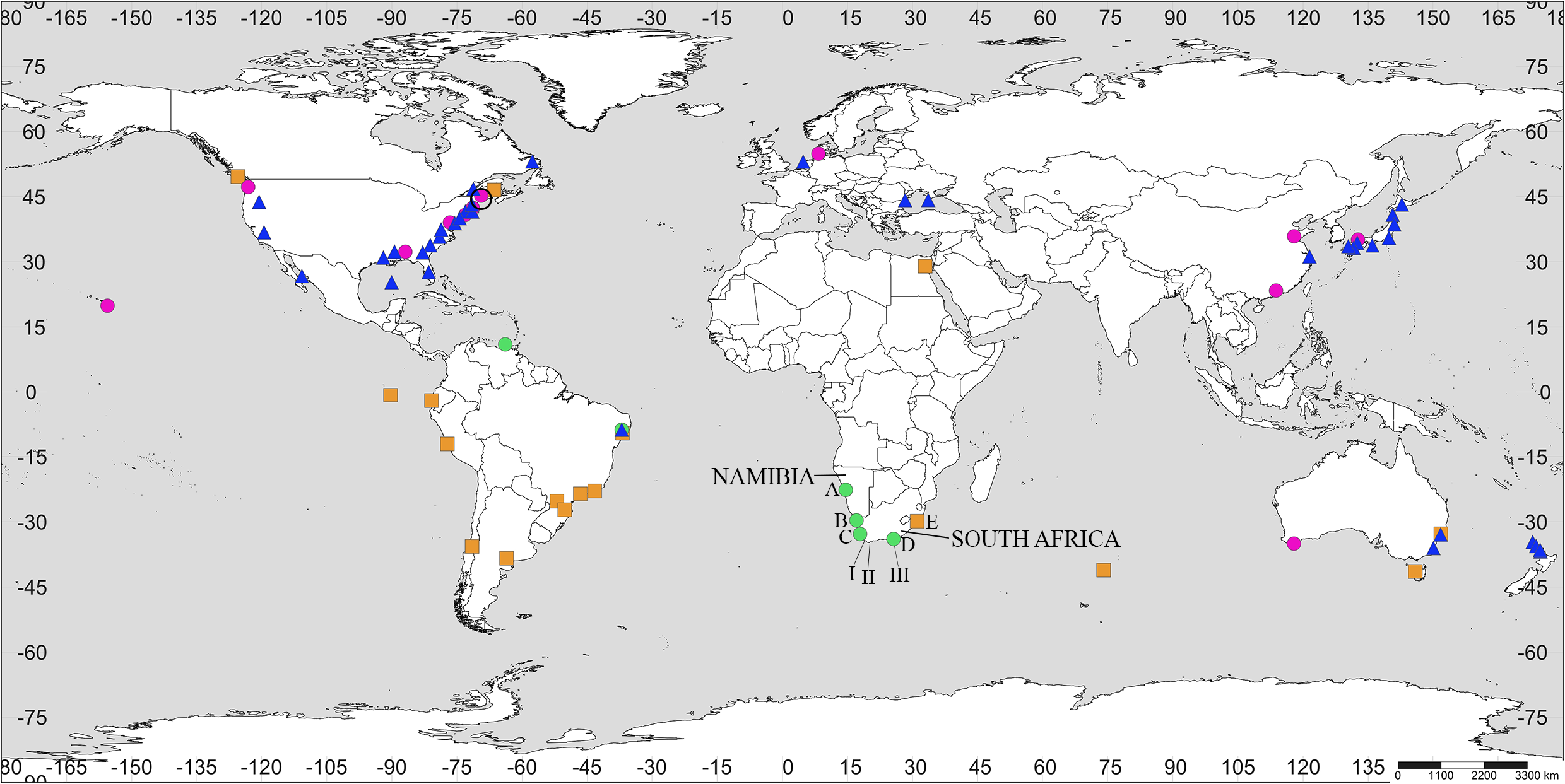
Distribution. Namibia: Swakopmund; South Africa: Paternoster and Kleinzee on the west coast (Simon 2015; Williams 2015); and Port Elizabeth, Nelson Mandela Bay on the east coast ( Simon 2011; this study).
Globally: USA: West coast: Washington State ( Martinelli et al. 2020) and Gulf of California in Mexico ( Blake 1969, 1971; Foster 1971); East coast: Maine, Massachusetts, Maryland ( Blake 1969, 1971; Foster 1971; Rice et al. 2018) and New York ( Martinelli et al. 2020); Gulf coast: Alabama ( Rice et al. 2018) and the Gulf of Mexico ( Blake 1969, 1971; Foster 1971); Australia ( Blake & Kudenov 1978; Sato-Okoshi & Abe 2013), Canada ( Blake 1969, 1971; Foster 1971), China (Sato-Okoshi et al. 2013; Ye et al. 2017), Japan ( Sato-Okoshi 1999; Sato-Okoshi & Abe 2012, 2013), Hawaii ( Rice et al. 2018), New Zealand ( Read 2010), Romania, Ukraine ( Surugiu 2005, 2012) and the Wadden Sea ( Waser et al. 2020).
Ecology. In South Africa, P. websteri is currently only found boring into shells of cultured oysters, C. gigas ( Simon & Sato-Okoshi 2015; Williams 2015; Williams et al. 2017). P. websteri is a pest of commercial molluscs in most locations where it has been reported ( Simon & Sato-Okoshi 2015), and also abundant in intertidal and shallow waters ( Blake & Evans 1973). Polydora websteri is not host specific and creates U-shaped burrows that induce the formation of mud-blisters by molluscs such as Argopecten irradians Lamarck, 1819 ( Lauckner 1983) , Crassostrea cf. brasiliana Lamarck, 1819 ( Barros et al. 2017; Bonifácio 2009), C. gigas ( Read 2010; Rice et al. 2018), Crassostrea hongkongensis Lam & Morton, 2003 ( Ye et al. 2017), Crassostrea rhizophorae Guilding, 1828 ( Barros et al. 2017; Bonifácio 2009), Crassostrea virginica Gmelin, 1791 ( Loosanoff & Engle 1943; Rice et al. 2018; Martinelli et al. 2020), Crepidula fornicata Linnaeus, 1758 ( Blake 1971) , Euspira heros (Say, 1822) ( Blake 1971) , Littorina littorea Linnaeus, 1758 ( Blake 1971) , Mytilus edulis Linnaeus, 1758 ( Blake & Evans 1973), Mytilus galloprovincialis Lamarck, 1819 ( Surugiu 2005, 2012), Nucella lapillus (Linnaeus, 1758) ( Blake 1971) , Ostrea angasi Sowerby, 1871 ( Nell 2001) , Pinctada fucata Gould, 1850 ( Simon & Sato-Okoshi 2015), Pinctada imbricata Röding, 1798 ( Díaz-Díaz & Liñero-Arana 2003), Placopecten magellanicus Gmelin, 1791 ( Blake 1969, 1971), Patinopecten yessoensis Jay, 1857 ( Bower et al. 1992), Saccostrea cucullata Born, 1778 ( Skeel 1979) and Saccostrea glomerata Gould, 1850 (as Saccostrea commercialis Sato-Okoshi et al. 2008 ), and also been found in limestone ( Surugiu 2005, 2012).

No known copyright restrictions apply. See Agosti, D., Egloff, W., 2009. Taxonomic information exchange and copyright: the Plazi approach. BMC Research Notes 2009, 2:53 for further explanation.
|
Kingdom |
|
|
Phylum |
|
|
Class |
|
|
Order |
|
|
Family |
|
|
Genus |
Polydora websteri Hartman
| Rodewald, Nicola, Snyman, Reinette & Simon, Carol A. 2021 |
Polydora haswelli:
| Sato-Okoshi, W. & Okoshi, K. & Shaw, J. 2008: 495 |
Polydora websteri:
| Sato-Okoshi, W. & Abe, H. 2013: 1280 |
| Surugiu, V. 2012: 50 |
| Read, G. B. 2010: 9 |
| Surugiu, V. 2005: 67 |
| Sato-Okoshi, W. 1999: 832 |
| Radashevsky, V. I. & Williams, J. D. 1998: 212 |
| Handley, S. J. & Bergquist, P. R. 1997: 191 |
| Blake, J. A. & Kudenov, J. D. 1978: 258 |
| Blake, J. A. 1971: 6 |
| Foster, N. M. 1971: 26 |
| Blake, J. A. 1969: 815 |
Polydora websteri:
| Loosanoff, V. L. & Engle, J. B. 1943: 70 |