
Mesenchytraeus nivalis, Torii, Takaaki, 2015
|
publication ID |
https://doi.org/10.11646/zootaxa.4000.4.6 |
|
publication LSID |
lsid:zoobank.org:pub:DEB97452-A6E8-4AA9-A248-A65FD50D203F |
|
DOI |
https://doi.org/10.5281/zenodo.5202350 |
|
persistent identifier |
https://treatment.plazi.org/id/03DC87B4-D12F-FFF9-069E-9472ED2BF87B |
|
treatment provided by |
Plazi |
|
scientific name |
Mesenchytraeus nivalis |
| status |
sp. nov. |
Mesenchytraeus nivalis View in CoL sp. nov.
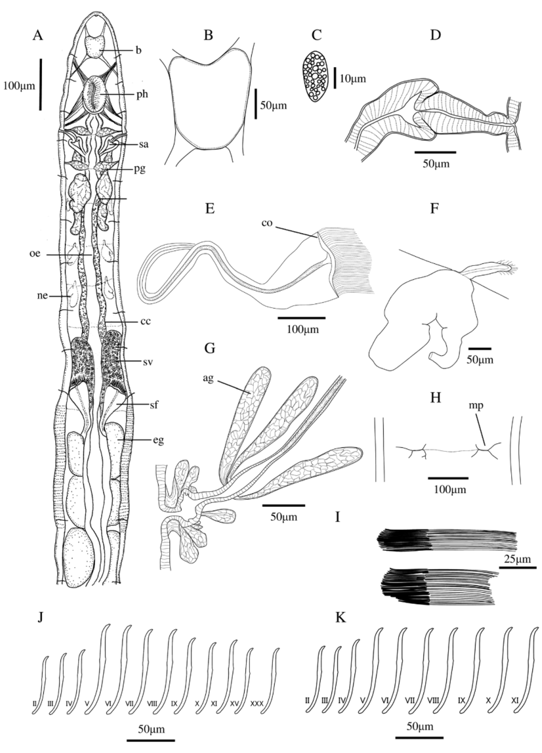
(Japanese name: Yuki-himemimizu, new) ( Figure 1 View FIGURE 1 )
Type material. Holotype: One whole-mounted mature specimen, Mihon-en, Ozegahara mire, Katashina-mura, Tone-gun, Gunma Prefecture, Japan, N36°54’54.8’’, E139°11’53.7’’, 4 June 2006, T. Torii (NSMT-An 463). Paratypes: Five whole-mounted mature specimens, same data as holotype (NSMT-An 464–467).
Further material. Twenty one whole-mounted mature specimens, same data as holotype, in the author’s collection.
Description. Color whitish or pare yellow. Live length 9–18 mm, fixed length 7–15 mm. Width at clitellum 300–450 µm. Segments 47–54. Chaetae sigmoid and simple-pointed, with nodulus at about 2/5 from the distal tip ( Fig. 1 View FIGURE 1 J,K), distal end thinner than proximal. Chaetal formula 2–4 – 2,3: 3,4 – 2,3. Ventral chaetae of II shortest, largest in V or VI, from VII on chaetal length gradually reduced. Lateral chaetae of II and III shortest, other precliteller chaetae all of same length, even in immature specimens. Both ventral and dorsal postcliteller chaetae slightly thinner and shorter than in preclitellar region. Chaetae of XII lacking in mature specimens. Clitellum elevated conspicuously, from mid XI to XIII, girdle-shaped, absent between male pores. Hyalocytes and granulocytes irregularly arranged, hyalocytes in contact with each other.
Prostomium rounded. Head pore present as a longitudinal slit, located at tip of prostomium. Epidermal gland cells invisible. Brain ( Fig. 1 View FIGURE 1 A,B) in I, II, dorsoventrally compressed and narrow, trapezoidal, concave anteriorly in living and fixed specimens, straight posteriorly, approximately 1.5 times as long as wide, length 165–180 µm, posterior width 88–94 µm. Pharyngeal pad thickened. No oesophageal appendages. Two pairs of primary pharyngeal glands in IV and V, not connected dorsally, attached to anterior face of respective posterior septa. Three pairs of secondary pharyngeal glands in V–VII, largest in VI, secondary glands not attached to septa. Transition of oesophagus to intestine gradual, no oesophageal or intestinal diverticula. Chloragogen cells light-brown granulated, beginning from V backwards, slightly smaller than coelomocytes. Blood pale-reddish. Dorsal blood vessel originating from intestinal sinus in XVI or XVII; anteriorly bifurcating beneath the front of the brain, circum-oesophageal connectives merging ventrally in IV. Coelomocytes ( Fig. 1 View FIGURE 1 G) densely distributed, very abundant in preclitellar region mainly of V–IX, flattened oval or slightly spindle-shaped, nucleate, their granular cytoplasm appears whitish under the stereomicroscope with top light, ca. 21–25 µm long, ca. 1.2–2.0 times as long as wide, filled with distinct, globular vesicles, conspicuous in living specimens, not always preserved in fixed specimens. Preclitellar nephridia four or five pairs from 6/7 to 9/10 or 10/11, anteseptal part of nephridium funnel only, about 55–75 µm long. Postseptale about 185–220 µm long, bilobed. Shape of postclitellar nephridia more compact than preclitellar.
Testes and ovaries not observed. Sperm sack in segments X–XV. Naked sperm bundles freely distributed in the sperm sack, the spermatozoal heads attached to each other in parallel, and the spermatozoa heads clumped at one end and ordered tails extending to the other end. Each bundle ca. 88–140 µm long, and 17–25 µm wide at the head and 12–19 µm at the tail ( Fig. 1 View FIGURE 1 I). Sperm funnel ( Fig. 1 View FIGURE 1 E) 140–200 µm long and 80–140 µm wide when extended. Collar either slightly wider than or equal funnel diameter. Vas deferens stout, thick-walled, moderately long, in loose or tight irregular coils in XII to XIV; atrium fusiform, encircled by a set of 3 or 4 club-shaped large atrial glands ( Fig. 1 View FIGURE 1 G). Penial bulb with some accessory glands. Bursal slit transversal with bifurcations at the two ends, transverse component long ( Fig. 1 View FIGURE 1 H). No subneural glands. Egg sac unpaired in XIII–XV, well-developed, sometimes extending back to XVIII–XXI. Paired spermathecal pores midlateral at 4/5, attached to oesophagus in posterior of V. Spermatheca thick walled, with onion-shaped ampulla ( Fig. 1 View FIGURE 1 D), ectal duct ca 100–115 µm long and 27–45µm wide, with no ectal glands. Ectal duct length less than ampulla plus ental duct. Ental duct ca 40–70 µm long and 35–45µm wide. Sperm in the ampullar lumen. Ampulla only oval or marginally lobed in immature specimens.
Habitat. The specimens were present in streams, in wet soil, sphagnum mires and in the snow.
Etymology. Named “ nivalis ” (Latin, = snowy) after the snowy habitat where they were first found.
Remarks. The following combination of characters is useful to identify the new species: 1) no enlarged chaetae; 2) no spermathecal diverticula; 3) vas deferens with 3 or 4 club-shaped atrial glands; 4) sperm bundles in sperm sack; 5) dorsal blood vessel from XVI or XVII.
The new species resembles the Korean Mesenchytraeus longiductus Christensen & Dózsa-Farkas, 2012 and the Chinese Mesenchytraeus gigachaetus Shen, Chen & Xie, 2011 ( Shen et al. 2011, Xie 2012), Mesenchytraeus anisodiverticulus Shen, Chen & Xie, 2012 and Mesenchytraeus monodiverticulus Shen, Chen & Xie, 2012 ( Shen et al. 2012a, b). M. longiductus is similar regarding spermatheca, male glands and sperm funnels. M. gigachaetus , M. anisodiverticulus and M. monodiverticulus also have sperm bundles in the sperm sack and spermathecae with onion-shaped ampulla. Differences of these species to M. nivalis sp. nov. are as follows:
M. longiductus : Vas deferens with 2 atrial glands (3 or 4 atrial glands in M. nivalis ). Spermathecal ental ducts 150–280 µm long (40–70 µm long in M. nivalis ). No sperm bundles in the sperm sack (with sperm bundles in the sperm sack in M. nivalis ).
M. gigachaetus : Vas deferens without atrial glands (3 or 4 atrial glands in M. nivalis ). Spermatozoa ca. 165– 190 µm long (ca. 88–140 µm long in M. nivalis ). Collar of sperm funnel narrower than funnel body (collar either slightly wider than funnel body or as wide as funnel body in M. nivalis ).
M. anisodiverticulus: Body length 21.0– 24.5 mm (9–18 mm in M. nivalis ). Dorsal blood vessel origin in XXI– XXII (XVI or XVII in M. nivalis ). Vas deferens without atrial glands (3 or 4 atrial glands in M. nivalis ). Spermathecal ampulla possessing two asymmetrical diverticula (without ampulla in M. nivalis ).
M. monodiverticulus: Preclitellar nephridia six pairs (four or five pairs in M. nivalis ). Vas deferens without atrial glands (3 or 4 atrial glands in M. nivalis ). Spermathecal ental ducts 185–204 µm long (40–70 µm long in M. nivalis ). Spermathecal ampulla possessing one ear-shaped diverticula (without ampulla in M. nivalis ).
No known copyright restrictions apply. See Agosti, D., Egloff, W., 2009. Taxonomic information exchange and copyright: the Plazi approach. BMC Research Notes 2009, 2:53 for further explanation.
|
Kingdom |
|
|
Phylum |
|
|
Class |
|
|
Order |
|
|
Family |
|
|
Genus |