
Fauveliopsis confusa, Thiel & Purschke & Böggemann, 2011
|
publication ID |
https://doi.org/ 10.1080/00222933.2010.540046 |
|
persistent identifier |
https://treatment.plazi.org/id/03E787D5-FFEE-FFA8-807F-FC7C36F7FAD3 |
|
treatment provided by |
Carolina |
|
scientific name |
Fauveliopsis confusa |
| status |
sp. nov. |
Fauveliopsis confusa View in CoL sp. nov.
( Figures 1 View Figure 1 , 2 View Figure 2 C–F, 3, 4A–C)
Holotype
R / V “ METEOR ” 63 / 2 St. 89 ES-S, 0 ◦ 43 ′ N, 5 ◦ 31.3 ′ W to 0 ◦ 43 ′ N, 5 ◦ 31.2 ′ W, 20 March 2005, 5137– 5141 m; cs / 7 / 28 / 0.55 / 0.2 ( ZMH P-24873).
Paratypes
R / V “ METEOR ” 63 / 2 St. 89 ES-S, 0 ◦ 43 ′ N, 5 ◦ 31.3 ′ W to 0 ◦ 43 ′ N, 5 ◦ 31.2 ′ W, 20 March 2005, 5137– 5141 m; cs / 7.75 / c.28 / 0.5 / 0.1, cs / c.3.5 / c.22 / 0.5 / 0.15 (ZMH P-24874) – St. 89 ES-E, 0 ◦ 43 ′ N, 5 ◦ 31.3 ′ W to 0 ◦ 43 ′ N, 5 ◦ 31.2 ′ W, 20 March 2005, 5137– 5141 m; cs / 3 / 28 / 0.5 / 0.2 [regenerated] (ZMH P-24876).
Additional material
Twenty-nine complete specimens, 47 anterior fragments, 16 middle fragments, 26 posterior fragments (listed in Appendix 1).
Etymology
The species name was chosen because nearly all the observed specimens apparently have a retracted prostomium so there were no clear morphological signs from which to determine the anterior and posterior ends.
Diagnosis
Adult specimens with up to 31 chaetigers; one acicular + one capillary chaeta per ramus in all chaetigers; three to four short chaetigers in anterior region, telescope-like
middle region, expanded posterior region with short chaetigers and slightly smaller chaetae in the last few chaetigers.
Distribution
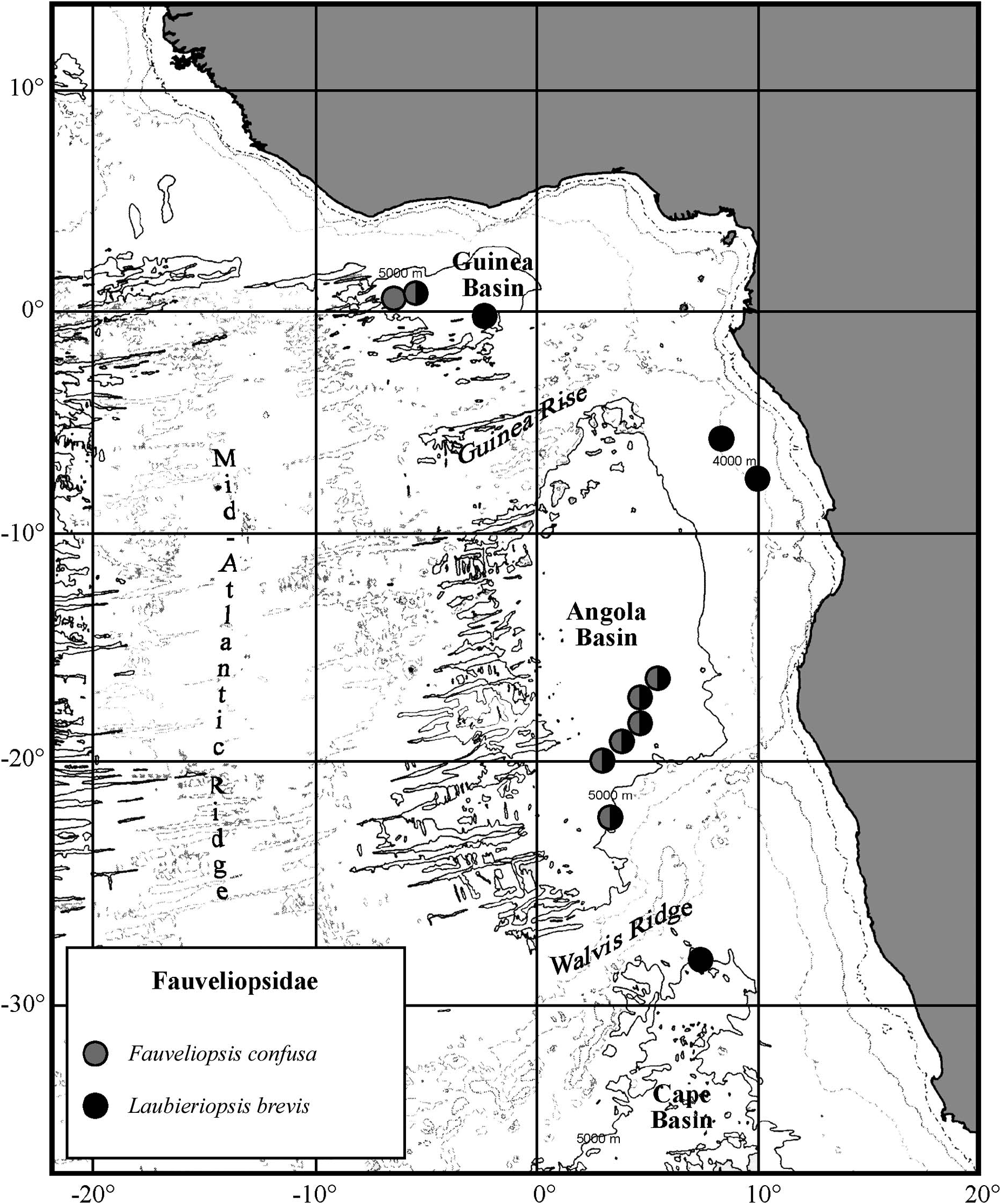
Angola and Guinea Basin; 5137–5496 m ( Figure 1 View Figure 1 ).
Description
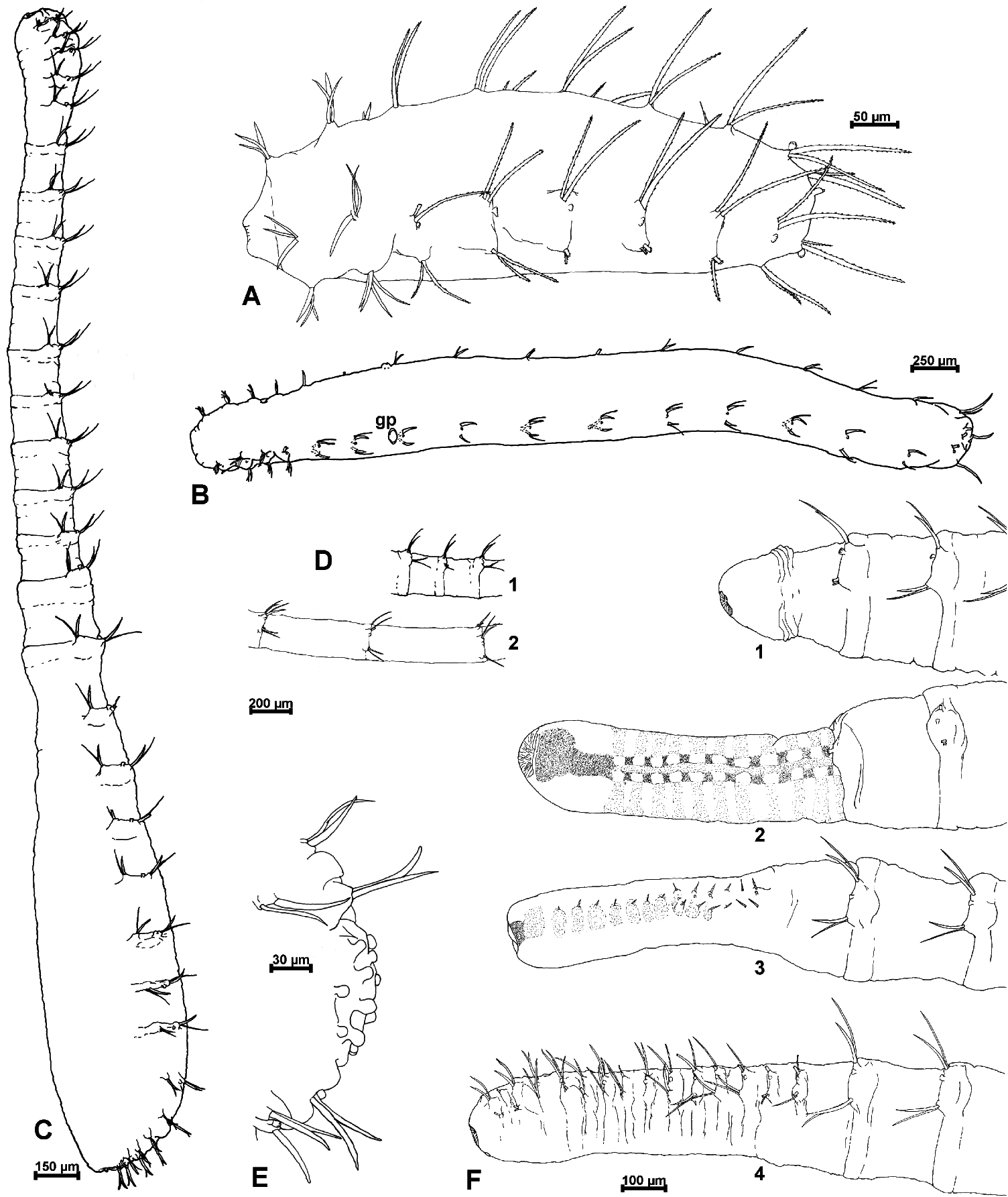
Complete specimens with 15–31 chaetigers; length of specimens highly variable as they are very expandable (shortest complete specimens 3 mm in length and with 15–29 chaetigers); body club-shaped, white, divided into three regions ( Figure 2C View Figure 2 ).
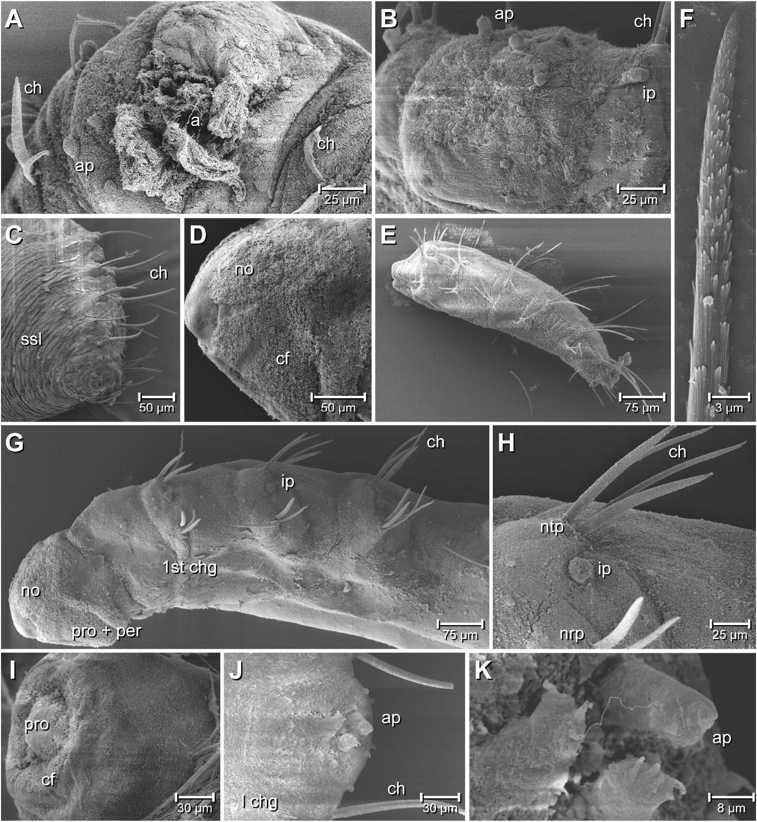
Prostomium and peristomium retracted, probably extendable; with prostomial ciliary field and nuchal organs ( Figure 3 View Figure 3 D–F).
Anterior region smooth, with three to four short chaetigers on a common elevation ( Figure 2C View Figure 2 ).
Middle region telescope-like; chaetigers highly contractible, varying in number, length and width; elongated segments at least two and a half times longer than contracted ones ( Figure 2D View Figure 2 ); segmental lines clearly apparent, broadest part of chaetigers on same level as parapodia on the anterior end of the segments; whole middle region very fragile with predefined breaking points between parapodia. ( Figure 2C, D View Figure 2 ).
Posterior region swollen and broadest part of the body, consisting of 7–13 chaetigers without segmental lines dividing the different segments, but with a varying number of secondary segmental lines within each segment; posterior four to five chaetigers very short, becoming abruptly smaller in diameter ( Figure 2C View Figure 2 , 4C View Figure 4 ). Pygidium with terminal anus, surrounded by many papillae ( Figures 2E View Figure 2 , 4 View Figure 4 A–C).
Parapodia biramous, in anterior region nearly totally reduced to chaetae, in middle region with weakly developed lobes, in last region chaetigers totally reduced to chaetae; each ramus with one slim capillary on outer side and one broader and shorter acicular spine on inner side of each ramus ( Figures 2 View Figure 2 C–F, 4C). Notopodia of anterior and middle regions located dorsolaterally, neuropodia nearly all located laterally; parapodia of posterior end located nearly all on dorsum ( Figures 2C View Figure 2 , 4C View Figure 4 ). Chaetae of last few chaetigers smaller than others. Small interramal papilla between noto- and neuropodia, located near notopodia ( Figure 2C, D, F View Figure 2 ).
No genital papilla observed and no ventral shield observed.
To determine the anterior end with certainty, semi-thin sections were cut to examine the central nervous system. The brain was found to be situated in the thinner end, which was therefore identified as the anterior region ( Figure 3 View Figure 3 ). The brain consists of an anterior part giving rise to two pairs of lobes extending posteriorly: the median lobes on the dorsal side ( Figure 3A,B View Figure 3 ) and the lateral lobes, located more laterally and slightly more ventrally ( Figure 3 View Figure 3 B–D). The neuropil is located anteriorly whereas the lobes are exclusively made up of somata of neurons. The ventral nerve cord ( Figure 3G, H View Figure 3 ) is well developed. It comprises fused cords as well as closely allied and indistinctly separated ganglia ( Figure 3G,H View Figure 3 ). Although bulging deeply into the body cavity on longitudinal sections ( Figure 3G,H View Figure 3 ) it actually has a basiepithelial position because the surrounding extracellular matrix is continuous with that of the epidermis. Septa between adjacent segments are well developed and clearly visible ( Figure 3G View Figure 3 ). The retracted prostomium bears a ventral ciliary field and the prominent nuchal organs ( Figure 3 View Figure 3 C–F). Oesophagus ( Figure 3D, E View Figure 3 ) and hind gut are clearly distinguishable ( Figure 3H View Figure 3 ). A large pair of retractor fibres attaches to the anterior end between the lateral and median lobes of the brain, runs posteriorly and ends at the first septum between chaetigers 2 and 3 ( Figure 3A View Figure 3 ). Additional, less prominent, retractors are present more ventrally ( Figure 3C View Figure 3 ).
Remarks
A few specimens were found with a possibly extended prostomium; however, this feature could not be recognized under a compound microscope with certainty. The only morphological indications allowing determination of the anterior and posterior ends without dissection are the papillae surrounding the pygidium ( Figures 2E View Figure 2 , 4 View Figure 4 A–C). Semi-thin sections clearly allowed the identification of the thinner end as the anterior end with the brain ( Figure 3 View Figure 3 A–F).
Some specimens were found that seemed to regenerate their anterior end, caught in different regenerating stages. Apparently they first grow out an unsegmented tube-like end and after this the chaetae are formed, proceeding from the regenerating area to the terminal end, so the most terminal chaetae appear to be the youngest ( Figure 2F1–4 View Figure 2 View Figure 1 View Figure 3 View Figure 4 ). A few specimens were found in which the posterior end was probably regenerated because they only have three or four chaetigers on both sides of the clearly segmented middle region.
Fauveliopsis confusa sp. nov. probably feeds on bacteria growing on sediment grains and detritus, in view of the fact that the gut contents comprise sediment in the posterior part of several specimens.
Discussion
The only species of Fauveliopsis McIntosh, 1922 described with two chaetae per ramus in all chaetigers are F. armata Fauchald and Hancock, 1981 , F. brevipodus ( Hartman, 1967) and F. fauchaldi Katzmann and Laubier, 1974 . In contrast to F. confusa , F. armata has clearly thicker and strongly curved acicular chaetae at both ends compared with those of the middle region, the anterior region has a rugose epithelium, the segmental lines of the middle region are indistinct and parapodial lobes are most fully developed in the posterior region, which has more chaetigers and is longer than the middle region ( Fauchald and Hancock 1981; Blake and Petersen 2000). In contrast to F. confusa , F. brevipodus has a laterally located pair of papillae at the posterior end, the first neuropodial acicular chaetae are clearly thicker and more curved than the acicular chaetae of the other chaetigers, all adult specimens apparently have 28 chaetigers and the whole body-shape differs greatly ( Hartman 1967). Fauveliopsis fauchaldi has a cylindrical body-form without very short chaetigers at the posterior end ( Katzmann and Laubier 1974). The drawings of F. scabra Hartman and Fauchald, 1971 look similar to some of the specimens of F. confusa but F. scabra has up to two acicular chaetae, combined with up to three capillary chaetae per ramus ( Hartman and Fauchald 1971).

| R |
Departamento de Geologia, Universidad de Chile |
| V |
Royal British Columbia Museum - Herbarium |
| ZMH |
Zoologisches Museum Hamburg |
No known copyright restrictions apply. See Agosti, D., Egloff, W., 2009. Taxonomic information exchange and copyright: the Plazi approach. BMC Research Notes 2009, 2:53 for further explanation.
|
Kingdom |
|
|
Phylum |
|
|
Class |
|
|
Order |
|
|
Family |
|
|
Genus |