
Megacormus xichu, González-Santillán & González-Ruíz & Escobedo-Morales, 2017
|
publication ID |
https://doi.org/10.11646/zootaxa.4299.2.3 |
|
publication LSID |
lsid:zoobank.org:pub:CA08CC9A-58D6-454D-A150-22CE260F3DFC |
|
DOI |
https://doi.org/10.5281/zenodo.6021218 |
|
persistent identifier |
https://treatment.plazi.org/id/03FE2938-0104-8708-A0ED-4D9F065BFAFC |
|
treatment provided by |
Plazi |
|
scientific name |
Megacormus xichu |
| status |
sp. nov. |
Megacormus xichu View in CoL sp. nov.
( FigureS 1–13 View FIGURE 1 View FIGURE 2 View FIGURE 3 View FIGURE 4 View FIGURE 5 View FIGURE 6 View FIGURE 7 View FIGURE 8 View FIGURE 9 View FIGURE 10 View FIGURE 11 View FIGURE 12 View FIGURE 13 ; Table 1)
Zoobank: urn:lSid:zoobank.org:act:37F20C1D-CCB5-444B-A24C-3183CB377ED3
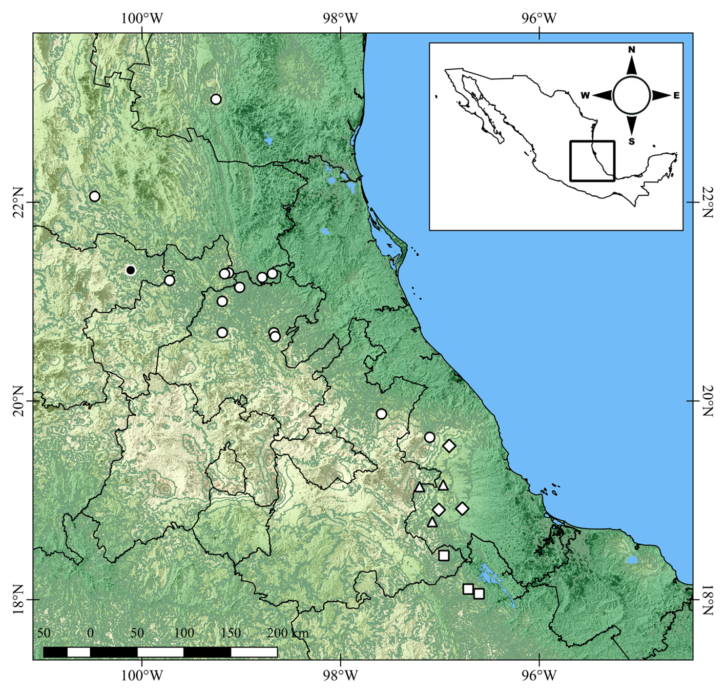
Type material. MEXICO: Guanajuato: Municipio de Xichú : Holotype: male ( CNAN-T 1169 ), 4 maleS and 16 femaleS paratypeS ( CNAN-T 1170 ), Charco Azul, El Ocotero, ca. 4 km NE of Xichú , 21°18'54'' -100°06'38'', 2283 m, 29.viii.2015, J.M. González-Ruíz and G.D. García-AriaS, under rockS, along a trail margin and SlopeS with dominant oakS and few Scattered pine treeS; 5 male and 5 female paratypeS ( CNAN-T 1171 ), 2 maleS and 2 femaleS paratypeS ( AMNH), 2 maleS and 2 femaleS paratypeS ( CAS), 21°19'01'' -100°06'49'', 14-16.iii.2016, Arthropod claSS ITESI.
Etymology. The SpecieS name iS a noun in appoSition derived from the type locality. In the original Chichimeca language the name of the SpecieS meanS “blinded man”.
Diagnosis. Megacormus xichu sp. nov. iS cloSely related to M. granosus and M. segmentatus by the preSence of Straight cutting margin of the pedipalp chela fingerS and the abSence of proximal notch, median lobe and gap when fingerS cloSe. Megacormus xichu sp. nov. may be diStinguiShed from M. granosus by the preSence of a deep SulcuS, completely Separating diStal and median lamella of the pectineS in both SexeS, inStead of having marginal and median lamella fuSed without SulcuS (female) or veStigially developed (male), and by the preSence of four trichobothria in each of the SerieS et and em on the pedipalp patella, inStead of three in each SerieS. Megacormus xichu sp. nov. iS diStinguiShed from M. segmentatus by the denSely granular carapace, pedipalp dorSal femoral and patellar, and prolateral leg SurfaceS, inStead of Scattered fine granulation. The maleS of Megacormus xichu sp. nov. are readily diStinguiShed from the maleS of M. gertschi and M. grubbsi becauSe theSe SpecieS have the cutting margin of pedipalp chela fingerS Scalloped, bearing prominent proximal notch and median lobe, creating a gap when fingerS cloSe.
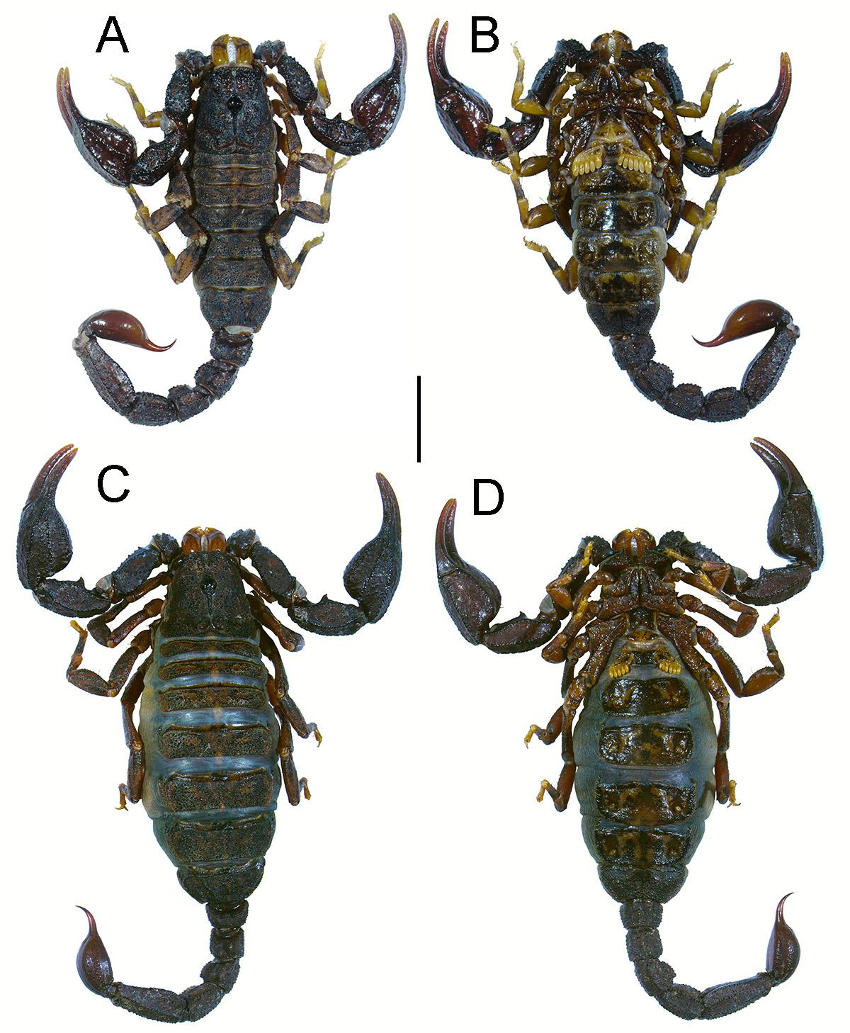
Description. The following deScription iS baSed on the type SerieS and includeS both SexeS, variation due to Sexual dimorphiSm iS indicated when neceSSary.
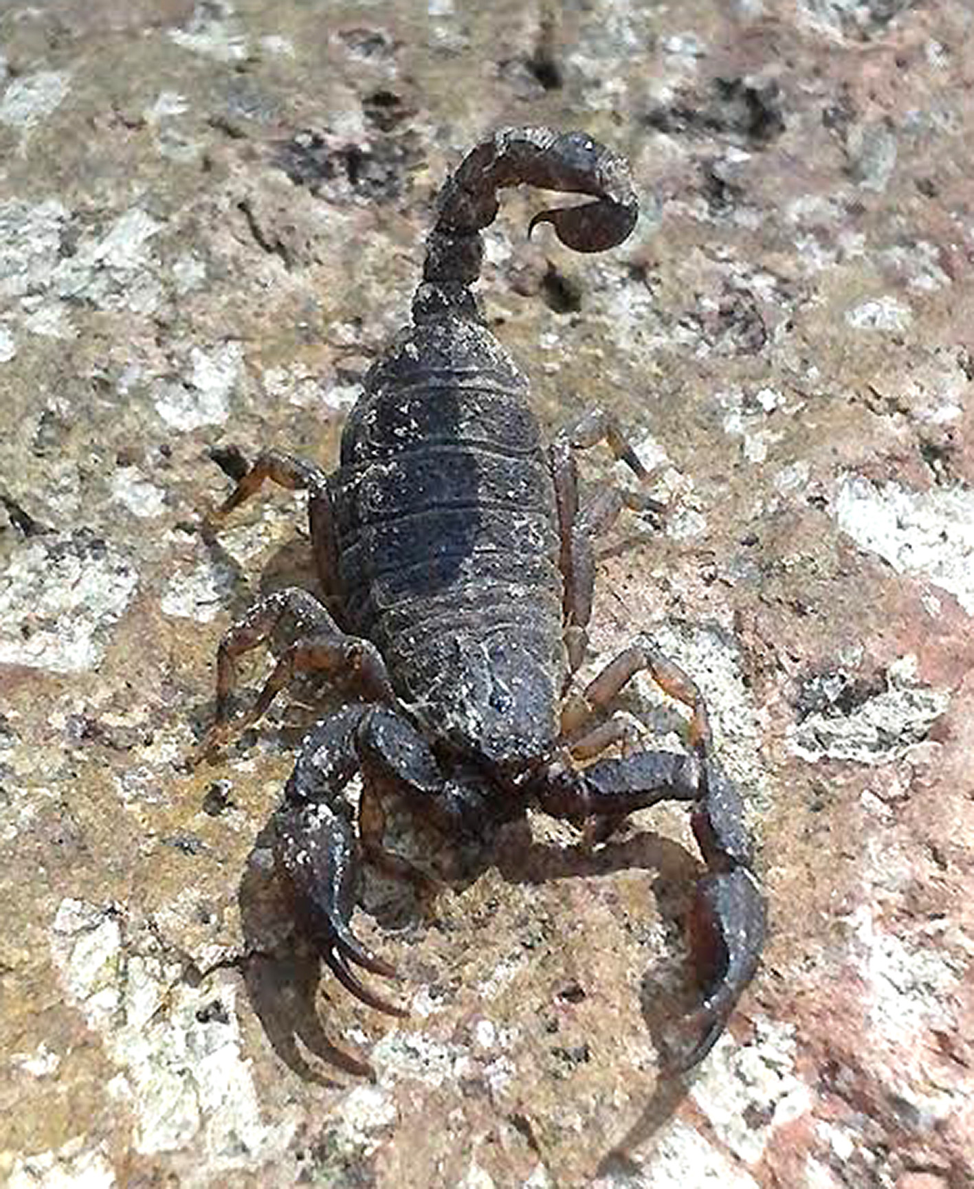
Color and infuscation: BaSe color yellowiSh to orange. Carapace, tergiteS, prolateral Surface of legS, coxoSternal region, SterniteS, genital operculum, marginal and median lamellae of the pectineS, metaSomal SegmentS and telSon , with denSe, marbled infuScation ( FigureS 2 View FIGURE 2 , 3 View FIGURE 3 ); Sternum and baSal piece of pectineS denSely infuScate. Chelicerae manuS baSe color yellowiSh with reticulated longitudinal infuScation; fingerS moderately infuScate proximally. Pedipalp baSe color orange with fuScouS markingS, all carinae denSely infuScate. All trichobothrial baSeS with a bright yellowiSh areola. Retrolateral Surface of all legS yellowiSh. SpiracleS light beige. Pectinal teeth whitiSh to light beige. TelSon veSicle, ventral Surface with three broad bandS of infuScation flanking two Submedian bandS of orange yellowiSh baSe color, dorSal Surface entirely (male) or predominantly (female) infuScate. AculeuS baSe faintly infuScate (female) or not infuScate (male), reddiSh brown diStally.
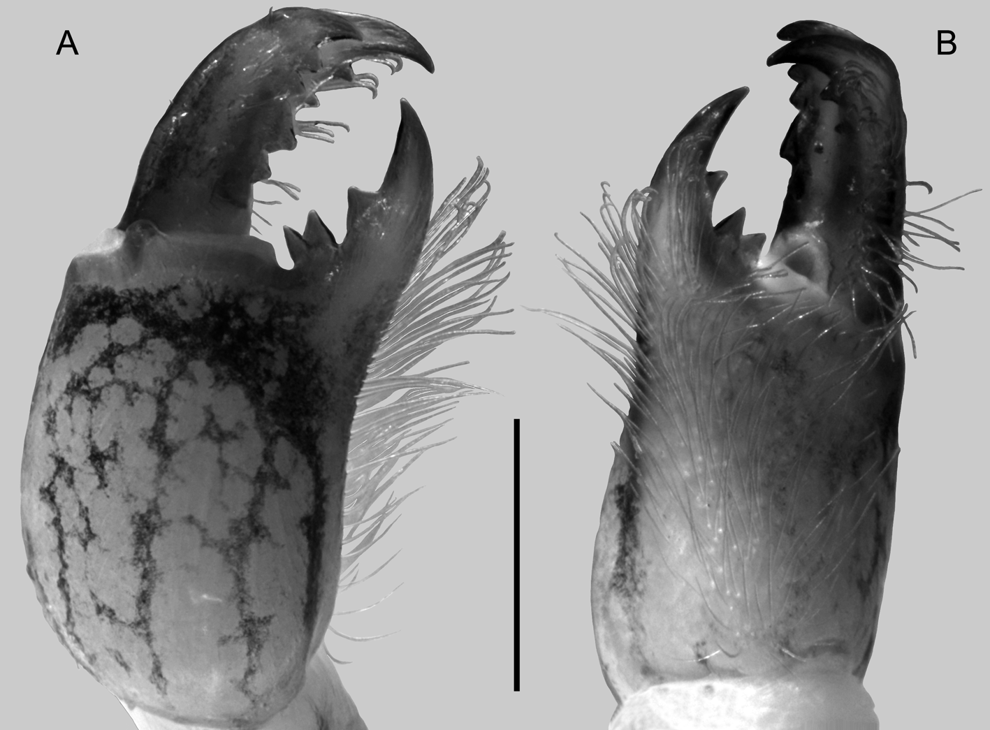
Chelicerae: ManuS dorSal Surface Smooth, luStrouS, with three macroSetae diStally, decreaSing in Size from median to lateral Surface. Movable finger, prolateral margin with baSal, two SubdiStalS, and diStal teeth Similar in Size, median tooth larger than the otherS; retrolateral margin diStal half with two to four Smaller, conical, Spaced, teeth. Fixed finger margin with baSal and median teeth joined in a bicuSpid tooth, SubdiStal tooth poSitioned midpoint of finger; diStal tooth enlarged and Sharp ( Figure 4 View FIGURE 4 ); ventral Surface of manuS, fixed and movable finger with a denSe tuft of Setae with curved tipS. Serrula abSent.
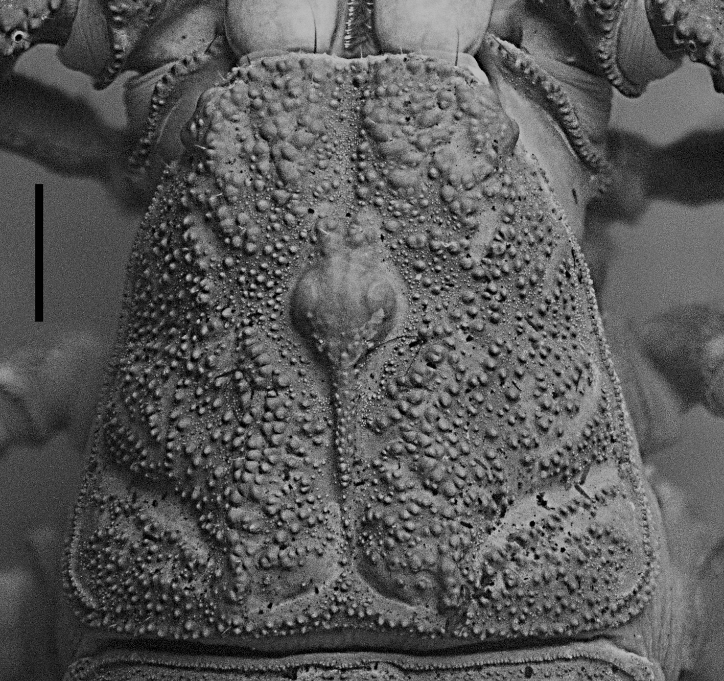
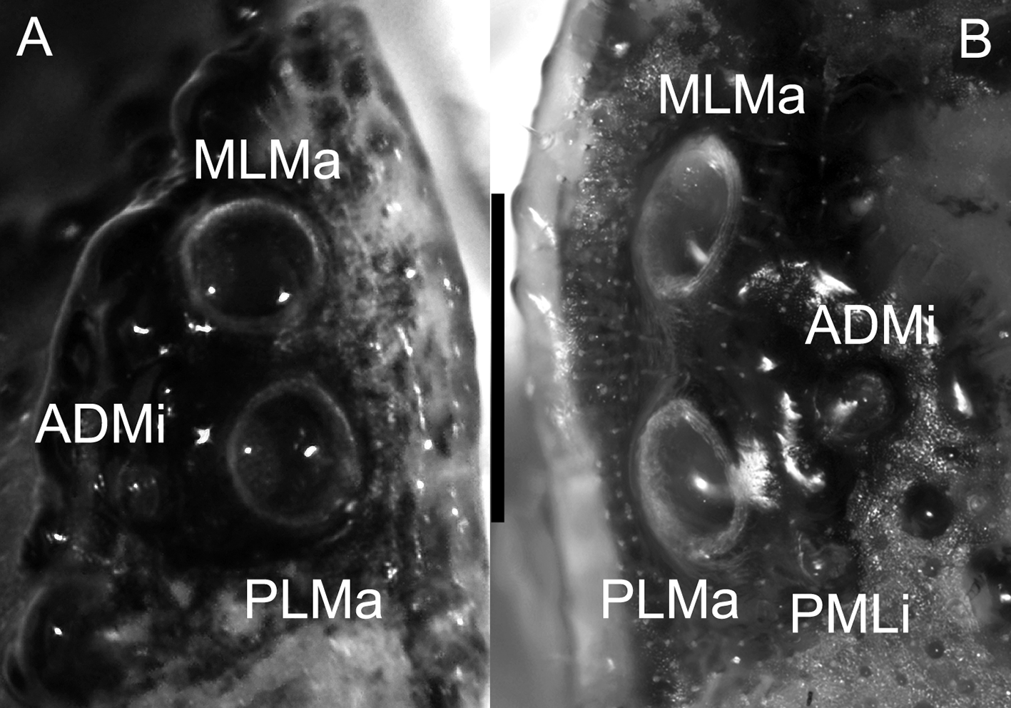
Carapace: Length (1.1/1.0) timeS greater than poSterior width. Surface Shagreened, with enlarged, Scattered granuleS covering entire SurfaceS ( Figure 5 View FIGURE 5 ). Anterior margin with one or two pairS of macroSetae; emarginated, bilobed, median notch veStigial, lateral marginS curved, Such that lateral ocelli are placed in anterolateral poSition. Three pairS of lateral ocelli conforming to Type 3B ( Loria & Prendini, 2014), PLMa, MLMa of equal Size, ADMI half the Size of prior ocelli (male, 14 and 15 timeS obServed in dextral and SiniStral SideS, reSpectively; female, 18 and 17 timeS obServed in dextral and SiniStral SideS, reSpectively, Figure 6 View FIGURE 6 ). Few SpecimenS with Type 4C, beSideS aforementioned ocelli, there iS a reduced to veStigial PDMi ocelluS (male, three timeS obServed Symmetrically in both dextral and SiniStral SideS; female, one and two timeS obServed in dextral and SiniStral SideS, reSpectively). Median ocular tubercle raiSed, Situated in anterior half of carapace. Superciliary carinae Strongly granular (male) or Smooth (female), lower than median ocelli. Anteromedian SulcuS deep and broad, with Scattered granuleS; poSteromedian with a granular carina and a deep, broad depreSSion poSteriorly; anterolateral, Shallow and narrow; poSterior tranSverSe deep.
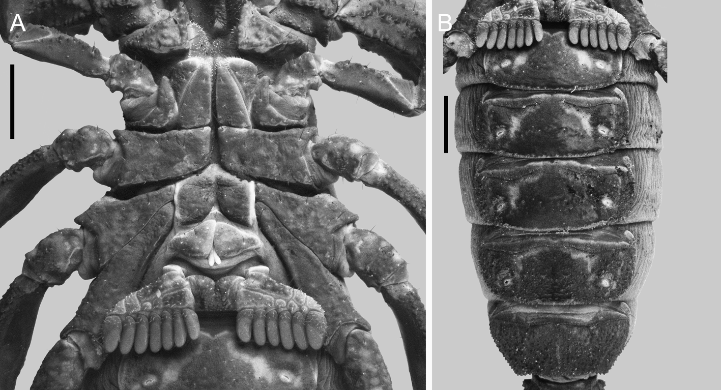
Coxosternal region: Sternum pentagonal, Subequilateral, length equal to the anterior width, with four microSetae. Median SulcuS of Sternum deep, with anterior and poSterior (female) or only poSterior margin broadened (male). Coxae I–IV SurfaceS with Scattered granuleS and marginS weakly granular, moStly Smooth; coxa II, prolateral Subproximal margin with one (female) or two (male) oblique Slit-like StructureS, adjacent to a moderate (female) or low (male) granular protuberance; coxae II–IV, prolateral carinae weakly granular ( Figure 7 View FIGURE 7 A); coxa IV twice the length of coxa II.
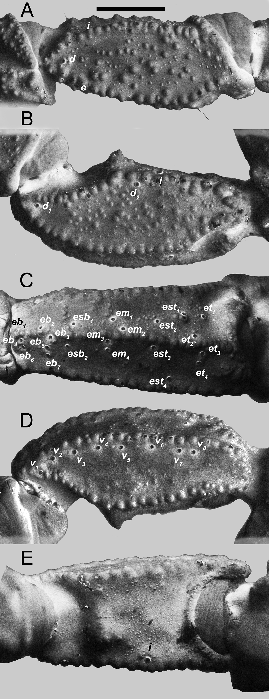
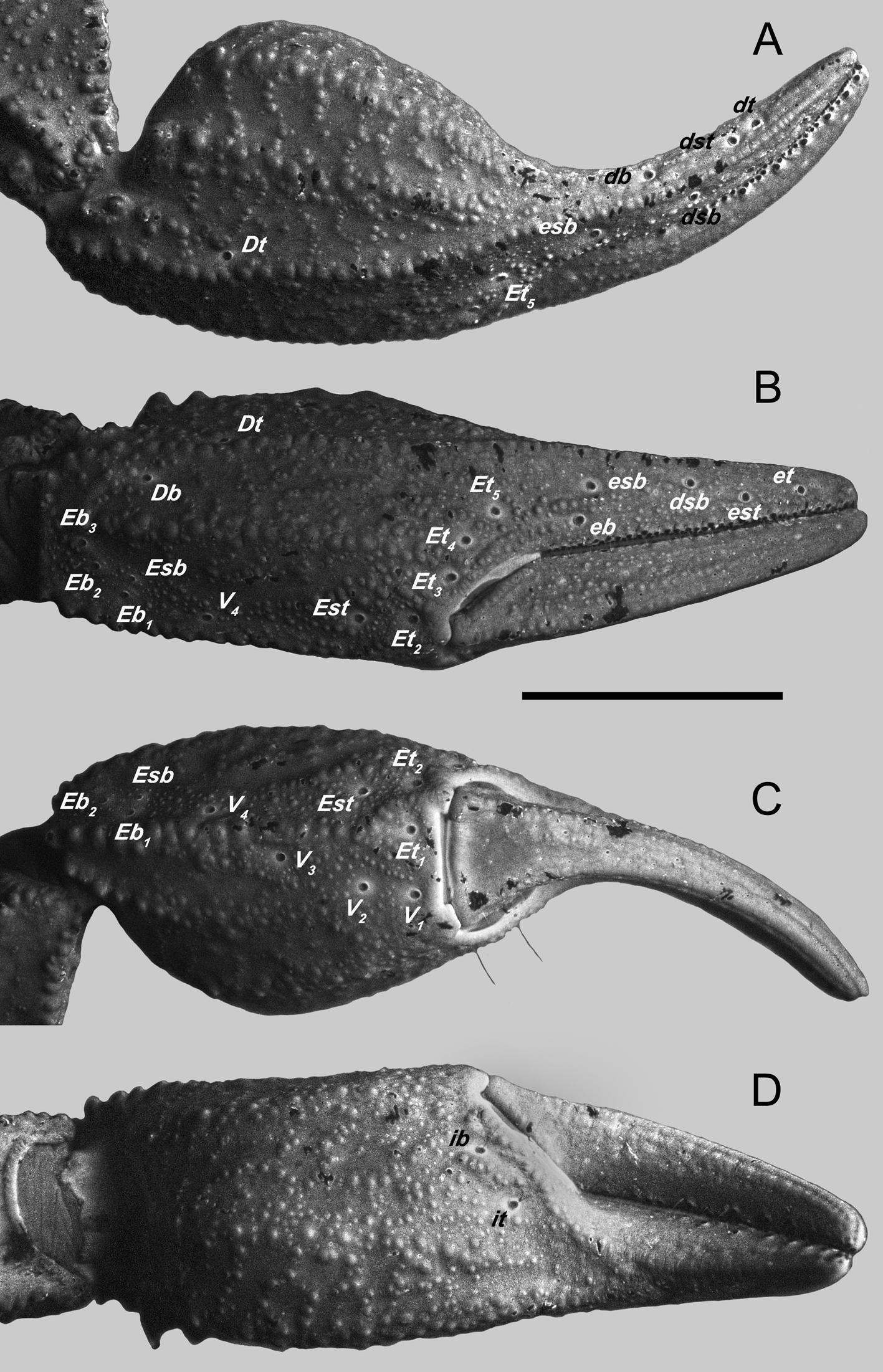
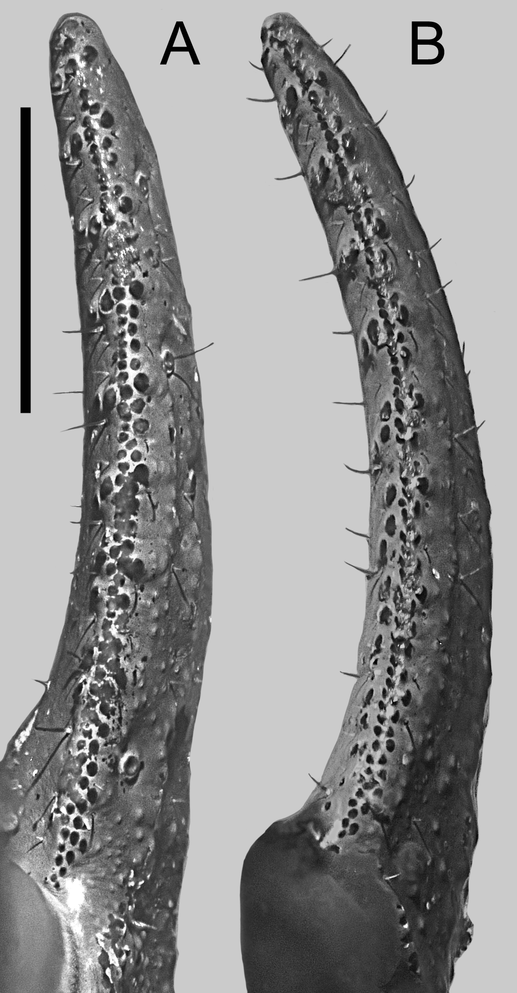
Pedipalps: Femur, length (2.1/1.8) timeS greater than width; prolateral, dorSal ( Figure 8 View FIGURE 8 A) and retrolateral intercarinal SurfaceS Shagreened, ventral Surface with a cluSter of fine granuleS medially; dorSal prolateral, dorSal retrolateral, ventral prolateral and ventral retrolateral carinae complete, irregularly granular; retrolateral dorSoSubmedian carina complete, weak proximally, becoming Strongly granular diStally; retrolateral ventral and ventral median carinae veStigial, conSiSting of few granuleS proximally; ventral retroSubmedian carina incomplete, with few Scattered enlarged granuleS on proximal half; prolateral ventral carina veStigial, with three median granuleS; prolateral ventroSubmedian carina incomplete, with Small granuleS on proximal third. Patella, width (2.1/ 1.8) timeS greater than femur width; dorSal intercarinal SurfaceS Shagreened; prolateral, retrolateral, and ventral SurfaceS SparSely coarSely granular; dorSal prolateral, dorSal retrolateral, ventral prolateral, ventral retroSubmedian and retrolateral median carinae complete, granular ( Figure 8 View FIGURE 8 B–E); retrolateral dorSoSubmedian carina veStigial; prolateral proceSS compriSing a large Spiniform tubercle, prolateral median carina abSent ( Figure 8 View FIGURE 8 B, D, E). Chela, length (2.4/2.5) timeS greater than femur length and (1.9) timeS greater than patella length, width (2.0/1.6) timeS greater than femur width and (1.6/1.3) greater than patella width; dorSal intercarinal SurfaceS Shagreened, with denSe field of minute and coarSe granuleS SubdiStally, other SurfaceS with Scattered minute and coarSe granuleS ( Figure 9 View FIGURE 9 ); dorSal retrolateral carina complete, granular, extending to proximal four-fifthS of the fixed finger, becoming weaker and Smooth diStally; dorSal retroSubmedian acceSSory carina veStigial, irregularly granular, reStricted to the level of trichobothrium Dt; dorSal median carina complete, Strongly coState-granular, with an enlarged proximal tubercle; dorSal retroSubmedian carina finely granular, with an enlarged proximal tubercle, running at the Side of the dorSal median carina; dorSal proSubmedian and dorSal prolateral carinae, fuSed on poSterior third of the manuS, granular, the latter extending to proximal Second third of fixed finger; retrolateral dorSal carina veStigial; retrolateral dorSoSubmedian veStigial, reStricted to Short diStal row of coarSe granuleS between trichobothria Et4 and Et5; retrolateral median carina complete, granular, ending at the level of trichobothria Et3 and Et4; retrolateral Subventral acceSSory and retrolateral Subventral carinae veStigial, reStricted to a Short diStal row of coarSe granuleS converging towardS trichobothrium Et2, commonly merging to ventral retrolateral carinae; retrolateral ventral carina veStigial; ventral retrolateral carina complete, granular; ventral median carina incomplete, SparSely granular, becoming weak medially and merging with a field of granuleS diStally; ventral retrolateral and ventral median carinae forming an acute angle proximally, becoming parallel medially to diStally; ventral prolateral and prolateral ventral carinae complete, fuSed moniliform; prolateral dorSal carina, veStigial; prolateral ventral acceSSory incomplete, reStricted to midpoint of the manuS aS a Short granular row; prolateral median incomplete, irregular row of coarSe granuleS reStricted to proximal half ( Figure 9 View FIGURE 9 D). Pedipalp fixed and movable fingerS cutting edge without notcheS, lobeS, and gap when fingerS cloSe ( FigureS 9 View FIGURE 9 B, 10A, B); dentate margin compriSing multiple rowS of prolateral, median and retrolateral denticleS; prolateral, prolateral acceSSory, retrolateral and median denticleS aligned in an oblique row angling retrolaterally in poSition III–VI. Fixed finger ( Figure 10 View FIGURE 10 A) median row compriSing Seven denticle SubrowS with one denticle in poSition I, three to Seven in poSition II–VII; flanked by a two- or three-denticle retrolateral acceSSory median Subrow, abSent in poSition I; median SubrowS divided by Six or Seven retrolateral denticleS, denticleS in poSition VII/VIII–X indiStinguiShable in Size from denticleS of median SubrowS; median acceSSory SubrowS divided by five or Six Subpaired retrolateral acceSSory denticleS, abSent in poSition I and undiStinguiShable in Size from denticle of median acceSSory denticleS SubrowS in poSitionS VII/VIII–X; flanked by Six or Seven prolateral denticleS and a Subpaired prolateral acceSSory denticle in poSition III–VI, abSent in poSition I and II, and veStigial or abSent in poSition VII–X ( Figure 10 View FIGURE 10 A). Movable finger ( Figure 10 View FIGURE 10 B) median denticle row compriSing Seven median denticle SubrowS, one in poSition I, three to Seven in poSitionS II–IX, abSent on X; flanked by a two- or three-denticle median acceSSory Subrow, abSent in poSition I; median SubrowS divided by Seven or eight retrolateral denticleS, indiStinguiShable in Size from denticleS of median SubrowS in poSition IX and X, and median acceSSory SubrowS divided by Seven Subpaired retrolateral acceSSory denticleS, abSent in poSition I, undiStinguiShable in Size from denticleS of median acceSSory SubrowS in poSition VII–X; flanked by eight prolateral denticleS, poSition III–X Subpaired by prolateral acceSSory denticleS, lower and leSS defined in poSitionS VIII–X ( Figure 10 View FIGURE 10 B). Trichobothrial pattern Type C, neobothriotaxic. Femur, trichobothria d, e, and i poSitioned proximally, equidiStant; d on dorSal intercarinal Surface, e on dorSal prolateral carina, i ventral to the dorSal prolateral carina ( Figure 8 View FIGURE 8 A). Patella, trichobothria d1 and d2 on dorSal intercarinal Surface, proximal and medial reSpectively; i on prolateral diStal half, ventral to the dorSal prolateral carina; eb1, eb2, esb1, em1, em2, est1, est2, et1, and et2 on intercarinal Surface between dorSal retrolateral and retrolateral median carinae, eb3–7, esb2, em3, em4, est3, est4, et3, et4 on intercarinal Surface between retrolateral median and ventral retroSubmedian carinae; esb2 petite; v 1– v 8 proximal to ventral retroSubmedian carina ( Figure 8 View FIGURE 8 C); variation of ventral trichobothrial count per patella aS followS: one with five, eight with Six, 15 with Seven and one with eight trichobothria (male) or one with five, Seven with Six, 44 with Seven and one with eight trichobothria (female); variation of retrolateral trichobothrial count per patella aS followS: em SerieS: one with three, 27 with four trichobothria (male) or 50 with four and one with five trichobothria; est SerieS: one with three and 27 with four (male) or 52 with four trichobothria (female); et SerieS: one with two, 27 with four (male) or Six with three and 48 with four trichobothria (female). Chela, trichobothrium Db on retrolateral Surface between the dorSal retrolateral and retrolateral dorSal carinae; Dt on dorSal Surface at diStal end of dorSal retroSubmedian acceSSory carina ( FigureS 9 View FIGURE 9 A, B); db, dst, and dt on dorSal Surface, dsb on retrolateral Surface at denticle poSition VII; SerieS eb–et on retrolateral Surface, eb between poSition VIII and IX, esb between poSitionS VII and VIII, est between poSitionS IV and V, et at poSition III ( Figure 9 View FIGURE 9 A, B); Eb1–Eb3 on retrolateral Surface, between retrolateral median and ventral retrolateral carinae; Esb petite, proximal to Eb1; Et1 on ventral Surface cloSe to retrolateral condyle, Et2–Et5 on retrolateral Surface, Et2–Et4 on diStal margin of manuS, Et4 not petite (See remarkS for a diScuSSion), Et5 on baSe of fixed finger; V1–V3 on ventral Surface, equidiStant, V4 on ventral retrolateral carina; ib and it on diStal margin of manuS ( Figure 9 View FIGURE 9 B, C).
Legs: BaSitarSi, retrolateral ventral Spinule row incomplete in legS I and II, prolateral ventral row abSent in legS I and II; retrolateral ventral and prolateral ventral Spinule rowS abSent in legS III and IV; retrolateral dorSal row compriSing enlarged Scattered SpinuleS on legS I and II, abSent in III and IV; macroSetal countS on legS I–IV, reSpectively: dorSal 3:3:3:3, retrolateral dorSal 2:2:2:2, retrolateral ventral 4:4:4:4, prolateral ventral 4:4:4:4, all macroSetae pigmented; dorSal and retrolateral dorSal macroSetae arranged in two Separate parallel rowS in each leg. TelotarSi I–IV, each with Single irregular ventromedian row of Scattered SpinuleS and one ventrodiStal Spinule, flanked by prolateral and retrolateral rowS of five macroSetae each. UngueS Short and curved.
Genital operculum: Wider than long, with four (male) or Six–eight (female) pairS of macroSetae; ScleriteS divided longitudinally except for the anterior two thirdS (male) or fuSed longitudinally by a looSe pleura folding into a valve covering the genital opening (female). Genital papillae preSent, protruding poSteriorly (male) or abSent (female).
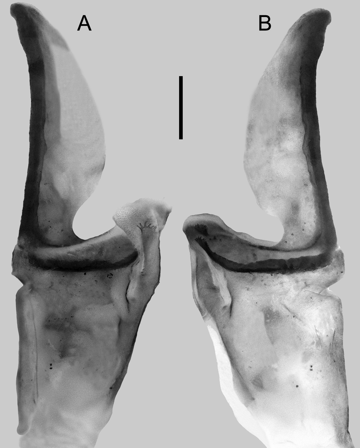
Hemispermatophore: DiStal lamina 1.1 timeS the length of trunk; tapering diStally, baSal conStriction welldeveloped ( Figure 11 View FIGURE 11 ). DorSal and ventral troughS Strongly Sclerotized, merging into a complete, thick, tranSverSe plate, Separating lamina and trunk. Marginal terminuS of dorSal and ventral troughS with Spiculated proceSSeS of irregular SpineS. Hemi-mating plug gelatinouS. Sperm duct formed by a Spicule-coated membrane connected to the Spiculated proceSSeS of the dorSal and ventral troughS and to the crown-like proceSS. Trunk broad proximally, tapering diStally; crown-like proceSS relatively long, with row of Six to eight irregular SpinuleS on diStal margin; truncal flexure and dorSal axial carinae well-developed ( Figure 11 View FIGURE 11 ).
Pectines: BaSal piece with two (male) or three pairS (female) of macroSetae, proximal Surface Smooth, iSoSceleS trapezoidal (male) or broader (female). Marginal and median lamellae with three pieceS each. Fulcra abSent. Pectinal teeth countS variation per pectine (dextral/SiniStral): one with five, five with Six, Seven with Seven and one with eight pectineS/three with Six, eight with Seven teeth (male); one with five, four with Six, 11 with Seven and one with eight pectineS/three with Six, 13 with Seven teeth (female). PectineS relatively Short, marginal lamella Shorter than coxa IV margin ( Figure 7 View FIGURE 7 ).
Tergites: I–VI, intercarinal SurfaceS Shagreened, denSely covered with minute and coarSe granuleS, ( Figure 3 View FIGURE 3 A, C); acarinated expect for dorSal median carina veStigial on I–III and moderate on IV–VI; VII, dorSal lateral carina veStigial; dorSal median carina veStigial, reStricted to anterior margin; dorSal Submedian carina complete, oblique, Strongly coState-granular; dorSal Sublateral carinae complete, coarSely granular.
Sternites: III–VI, SurfaceS Smooth medially, Shagreened laterally ( Figure 7 View FIGURE 7 B); SpiracleS minute, Slightly longer than wide in III and IV but rounded in V and VI; Sternite V, with diStinct hyaline glandular area poSteromedially ( Figure 7 View FIGURE 7 B), (male) or weakly developed (female). Sternite VII, intercarinal SurfaceS Shagreened; ventral median carina coState; ventrolateral carina veStigial with three or four granuleS.
Metasoma: Length (1.3/1.0) timeS greater than meSoSomal length ( Table 1); SegmentS I–V length (0.6/0.5), (0.8), (0.9), (1.0), (2.1) timeS greater than width; Segment V width (1.1/1.3) timeS greater than telSon width; dorSal intercarinal SurfaceS of SegmentS I–III with Scattered enlarged granuleS, III and IV weakly granular ( Figure 12 View FIGURE 12 A), V Shagreened (male), or I–IV weakly granular, V mate (female); lateral and ventral SurfaceS Shagreened ( Figure 12 View FIGURE 12 B, C); dorSal lateral and ventral lateral carinae complete, Strongly Serrate on Segment I–V; lateral median carina complete, Serrate on I–IV, granular, reStricted to proximal half on V; lateral inframedian carina complete, with Scattered, coarSe granuleS on I, incomplete, granular, reStricted to poSterior half on II and III, veStigial and reStricted to a poSterior marginal tubercle on IV, abSent on V; ventral Submedian carina veStigial, reStricted to a poSterior, marginal, paired tubercle on I–IV, abSent on V; ventral median carina complete, Strongly Serrate on I–V, bifurcated diStally on V ( Figure 12 View FIGURE 12 C); other carinae abSent. MacroSetal countS on carinae of SegmentS I–V, reSpectively: dorSal lateral, 0:0:0:0:0; lateral median, 0:1:1:1:1; lateral inframedian, 0:0:0:0:0; ventral lateral, 1:2:2:2:5; ventral Sublateral, 0:0:0:0:0; ventral Submedian, 2:2:2:2:3.
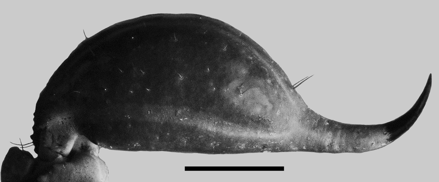
Telson: VeSicle globoSe, Slightly elongated, length 1.7/2.0 timeS greater than width ( Table 1); dorSal Surface Smooth, ventral Surface moStly Smooth with Scattered coarSe granuleS (male), or veSicle elongated with Scattered coarSe granuleS on entire Surface (female); carinae obSolete, without macroSetae except a pair at the baSe of the poSition of the Subaculear tubercle, which iS obSolete, annular ring moderately developed ( Figure 13 View FIGURE 13 ). AculeuS, elongated, laterobaSal microSerration abSent, venom delivery openingS Slit-like, paired.
REMARKS: Loria and Prendini (2014) determined that M. gertschi exhibitS a pattern for the lateral ocelli Type 4B, baSed on a juvenile Specimen. Their figure 3J ShowS the following ocelli: MLMa, PLMa, and ADMi each conSpicuouSly viSible under the microScope, but a draStically reduced PDMi ocelluS. We obServed in M. xichu sp. nov. the Same poSition and number of ocelli in few SpecimenS: per Side of the carapace (dextral/SiniStral) three in both SideS (maleS) or one/two in each Side (female). In contraSt, moSt SpecimenS Studied herein exhibited Type 3B with 14/15 (maleS) or 18/17 (female) in each Side of the carapace ( Figure 6 View FIGURE 6 ). Additionally, there were one male and two femaleS with only the MLMa and PLMa ocelli preSent, which correSpond to Type 2a. Finally, we obServed minor variation in Symmetry of lateral ocelli, only one male and two femaleS were aSymmetric. Until more data iS available for the other SpecieS, the lateral ocelli TypeS propoSed by Loria and Prendini (2014) Shall be evaluated to adapt thiS character SyStem to the variation obServed in Megacormus and other taxa.
MoSt ScorpionS with Type C trichobothrial pattern ( Vachon 1974) exhibit trichobothrium Et4 petit, a landmark that haS Served to homologize contiguouS trichobothria ( Vachon 1974, SiSSom, 1990, Soleglad & Fet 2001, 2003, Vignoli & Prendini 2009, Prendini et al, 2010). Unlike moSt SpecieS with Type C trichobothrial pattern, the two New World euScorpiid genera Plesiochactas Pocock, 1900 and Megacormus preSent, in moSt SpecieS, a trichobothrium Et4 normally developed, i.e. not petite. ThiS iS preSent in all SpecieS of Megacormus including M. xichu sp. nov. ( Figure 9 View FIGURE 9 B; Soleglad 1976 pp. 271, 275, 283, FigureS 34, 47, 72; SiSSom 1994 p. 267 FigureS 4 View FIGURE 4 , 7 View FIGURE 7 ; González-Santillán & Alvarez-Padilla 2015 p. 81 Figure 15) aS well aS in Plesiochactas dilutus ( Soleglad 1976 p. 291 Figure 91), but Et4 iS petit in P. mitchelli ( Soleglad 1976 p. 296 Figure. 123, Zarate-Galvez & Francke 2009 p. 343 Figure 12 View FIGURE 12 ). Unfortunately, in the third SpecieS, Plesiochactas vazquesi Trujillo & ArmaS 2012 , the trichobothrial pattern waS not completely illuStrated and thuS the condition of that trichobothrium iS unknow for that SpecieS. Future StudieS would have to eStabliSh whether thiS character State iS taxonomically informative and to what extent.

No known copyright restrictions apply. See Agosti, D., Egloff, W., 2009. Taxonomic information exchange and copyright: the Plazi approach. BMC Research Notes 2009, 2:53 for further explanation.
|
Kingdom |
|
|
Phylum |
|
|
Class |
|
|
Order |
|
|
Family |
|
|
Genus |