
Protoperidinium euxinum Krachmalny, 2023
|
publication ID |
https://doi.org/ 10.11646/phytotaxa.598.3.4 |
|
DOI |
https://doi.org/10.5281/zenodo.7975689 |
|
persistent identifier |
https://treatment.plazi.org/id/09666B09-135F-FFEF-F8AF-FE6D2394FBCC |
|
treatment provided by |
Plazi |
|
scientific name |
Protoperidinium euxinum Krachmalny |
| status |
sp. nov. |
Protoperidinium euxinum Krachmalny sp. nov.
( Fig. 2 View FIGURE 2 , A–E)
Division Dinoflagellata (Butschli 1885) Fensome et al. 1993.
Class Dinophyceae Pascher 1914.
Order Peridiniales Haeckel 1894 .
Family Protoperidiniaceae Balech 1988 nom. cons.
Genus Protoperidinium Bergh 1881 .
Diagnosis:— Cells medium-sized (length 88.6±7.2 µm, min. 73.3 µm, max. 99.0 µm; width 45.4±4.0 µm, min. 38.8 µm, max. 51,6 µm; n=20), pentagonal, elongated, slightly compressed dorsoventrally. Epitheca conical, with straight or concave lateral sides, tapers into a narrow and relatively long apical horn. Epitheca larger than the hypotheca, “meta–penta” type. The plate 2a asymmetrical, laterally compressed. Cingulum ascending by about one-half of its height, shallow and bears lists. Hypotheca inverse trapeziform, with large slightly divergent antapical horns bearing wide membranes. Sulcus straight, broad, shallow, slightly invades the epitheca, reaches the antapex. The sulcal area bordered. Thecal surface with fine reticulations and numerous pores. Cells colorless or light brown. Nucleus spherical, located in the center of the epitheca.
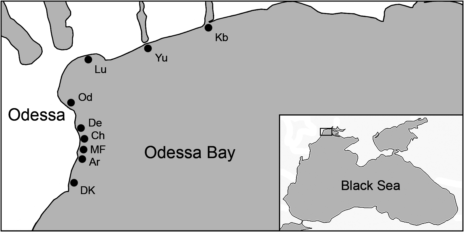
Holotype:— Fig. 2 View FIGURE 2 , A–Е. The specimen on which the illustration is based was collected in the northwestern part of the Black Sea (Odessa Bay ) in August 2020, at 46°26′28.25′′N, 30°46′25.52′′E ( Fig. 1 View FIGURE 1 : MF, Maliy Fontan). GoogleMaps
Etymology:— The specific epithet was chosen in order to emphasize the locality of this species—the Black Sea, which was called in ancient times “Pontus Euxinus”.
Paratype:— The phytoplankton sample was preserved in 4% formaldehyde solution and is stored at the Institute for Evolutionary Ecology of the National Academy of Sciences of Ukraine (Kiev, Ukraine).
Ecology and distribution:— Protoperidinium euxinum reaches large numbers in Odessa Bay (northwestern part of the Black Sea, sampling sites are shown in Fig. 1 View FIGURE 1 ) during the warm period of the year. Water temperature and salinity were 20° C and 18 psu, respectively. Occurs together with Protoperidinium steinii (Jørgensen) Balech and P. mediterraneum (Kofoid) Balech.
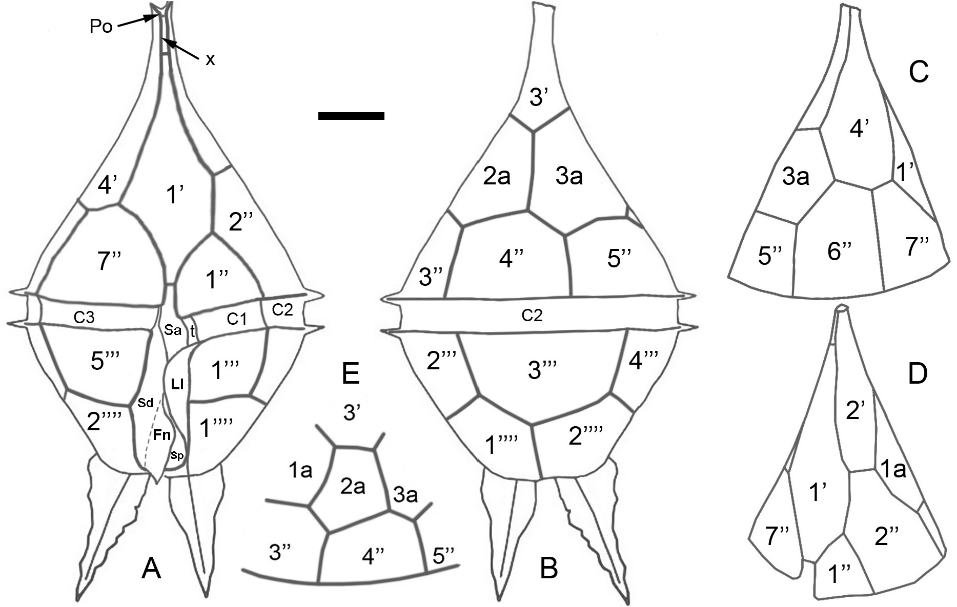
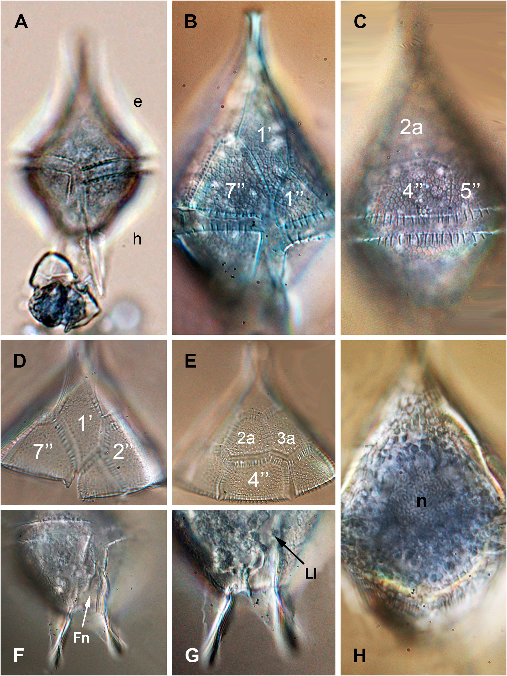
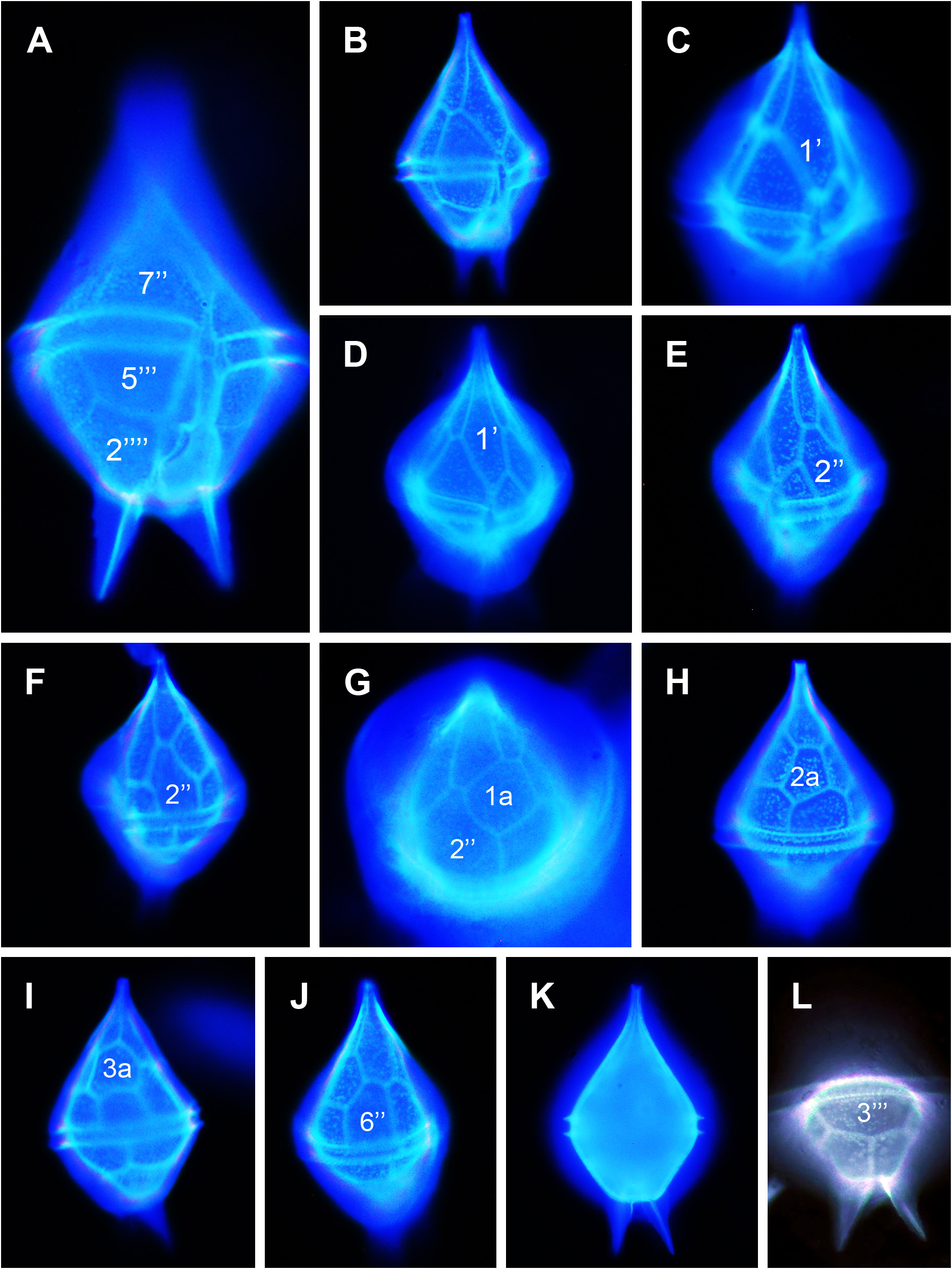
Description:— Cells of Protoperidinium euxinum are medium-sized (73.3–99.0 µm in length and 38.8–51.6 µm in width), shaped like elongated pentagons ( Fig. 2 View FIGURE 2 , A–B; Fig. 3 View FIGURE 3 , A; Fig. 4 View FIGURE 4 , A, B, K), slightly compressed dorsoventrally ( Fig. 4 View FIGURE 4 , B, E, F, J). The epitheca is conical, with straight or slightly concave lateral sides ( Fig. 2 View FIGURE 2 , C, D; Fig. 3 View FIGURE 3 , B, C; Fig. 4 View FIGURE 4 , A–K), tapers into a narrow and relatively long apical horn ( Fig. 2 View FIGURE 2 , A, B). The apical pore plate (Po) connects with the plate 1’ through a small and narrow plate “x” ( Fig. 2 View FIGURE 2 , A). The epitheca is larger than the hypotheca, the plate arrangement is “meta–penta”; plates 1’ and 2a are pentagonal ( Fig. 2 View FIGURE 2 , A, E; Fig. 3 View FIGURE 3 , B, D, E; Fig. 4 View FIGURE 4 , D, H). The plate 1a is also pentagonal ( Fig. 4 View FIGURE 4 , G). The plate 2a is asymmetrical, laterally compressed, the length of the suture between plates 1a and 2a is greater than between 3’ and 2a ( Fig. 2 View FIGURE 2 , E; Fig. 3 View FIGURE 3 , E; Fig. 4 View FIGURE 4 , H). The plate 3a is hexagonal ( Fig. 4 View FIGURE 4 , I). The cingulum is ascending by about one-half of its height, shallow, composed of plates C1–C3 and transitional plate “t”, and bears lists with pronounced ribs ( Fig. 2 View FIGURE 2 , A, B; Fig. 3 View FIGURE 3 , B, C; Fig. 4 View FIGURE 4 , A). The sulcal area is bordered by lists ( Fig. 3 View FIGURE 3 , F, G). The sulcus is straight, broad, shallow, slightly invades the epitheca (plate Sa), reaches the antapex, composed of the plates Sa, Sd, Ss, Sm and Sp. The right sulcal plate (Sd), in its apical part, tapers into a very narrow protrusion that overlaps the cingulum and reaches 7’’. The left side of Sd has a well-developed fin (Fn). The plate Ss is covered by the left sulcal list (Ll), which is attached to the plates 1’’’ and 1’’’’ and merges with the left antapical horn. The small median sulcal plate (Sm) and partially the posterior sulcal plate (Sp) are positioned under the fin of the plate Sd ( Fig. 2 View FIGURE 2 , A; Fig. 3 View FIGURE 3 , F; Fig. 4 View FIGURE 4 , A). The right sulcal list is underdeveloped. The hypotheca is inverse trapeziform, with large slightly divergent antapical horns bearing membranes; the right horn is more tilted to the side ( Fig. 2 View FIGURE 2 , A, B; Fig. 3 View FIGURE 3 , A, G; Fig. 4 View FIGURE 4 , A, K, L). Thecal surface with fine reticulations and numerous pores ( Fig. 3 View FIGURE 3 , B–E). Cells are colorless or light brown. The nucleus is spherical, located in the center of the epitheca ( Fig. 3 View FIGURE 3 , H).
No known copyright restrictions apply. See Agosti, D., Egloff, W., 2009. Taxonomic information exchange and copyright: the Plazi approach. BMC Research Notes 2009, 2:53 for further explanation.
|
Kingdom |
|
|
Phylum |
|
|
Class |
|
|
Order |
|
|
Family |
|
|
Genus |