
Paraprionospio, CAULLERY, 1914
|
publication ID |
https://doi.org/ 10.1111/j.1096-3642.2007.00323.x |
|
publication LSID |
lsid:zoobank.org:pub:824A2B6D-5F84-4310-9524-76B0843871EB |
|
persistent identifier |
https://treatment.plazi.org/id/0A0A87A5-FFEB-E006-0524-4713FAA5FC74 |
|
treatment provided by |
Felipe |
|
scientific name |
Paraprionospio |
| status |
|
GENUS PARAPRIONOSPIO CAULLERY, 1914 View in CoL
Type species: Prionospio pinnata Ehlers, 1901 , designated by Caullery, 1914: 358.
Diagnosis: Prostomium fusiform with rounded, truncated or bluntly pointed anterior end, extending posteriorly as a faintly raised ridge to setiger 1. Peristomium fused with achaetous segment, forming conspicuous lateral wings enfolding prostomium. A pair of palpi grooved and with membranous basal Character numbers (1–28) correspond to Table 1. A ‘?’ denotes missing data, and ‘–’ a non-applicable character.
sheath. Three pairs of branchiae on setigers 1–3, all with densely packed lamellar plates attached serially on inner to posterior face of shaft except in basal region and distal tip. Transverse ridge between branchial bases on setiger 1. Notopodial and neuropodial capillaries limbate and granulated in anterior setigers replaced by non-limbate, slender capillaries without granulation in middle and posterior setigers. Neuropodial hooks geniculate appearing from setiger 9, accompanied by alternating capillaries and one or two sabre setae. Notopodial hooded hooks not as geniculate as neuropodial hooks, appearing from middle body region. Notopodial and neuropodial hooks with primary and secondary hoods. Shallow ventral furrow running longitudinally from middle to end of body. Membranous dorsal crests appearing from setiger 21 present or absent. Eversible proboscis bilobate. Muscular gizzard present.
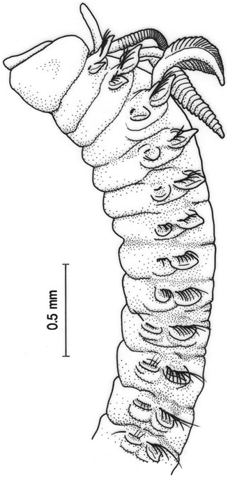
PARAPRIONOSPIO AFRICANA AUGENER, 1918
( FIG. 1 View Figure 1 )
Prionospio africana Augener, 1918 View in CoL
Material examined: Seven paratypes of Prionospio africana collected from the following localities in West Africa: HZM v.1559, Drewin, 33 m deep; HZM v.1561, Cape Lopez, 3 m ; HZM v.1563, Nyanga-Fluss, 11 m ; HZM v.1565, Landana; HZM v.1566, Lagos, 15 m ; HZM v.1714, Whydah, 13 m.
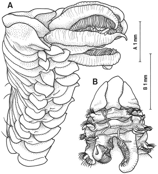
Description of paratypes: Paratypes, all incomplete with up to 47 setigers measuring 32 mm long and 2.5 mm wide (setiger 5, excluding parapodia). Prostomium fusiform with bluntly pointed anterior end ( Fig. 1A, B View Figure 1 ). Eyes not visible. Palpi missing. Peristomium with no marginal papilla on lateral sides. No pigmented patch on peristomium. Three pairs of branchiae on setigers 1–3. Each pair of branchiae approximately equal length (v.1559; Fig. 1A View Figure 1 ) or first branchiae slightly larger and third branchiae slightly smaller (v.1561). Branchiae bearing lamellae on branchial shaft except on its base and distal tip. Each branchial lamella consisting of two plates on entire shaft. First pair of branchiae without protuberances on anterior face in basal region of branchial shaft. Branchial shafts stout and two branchiae uniting at bases forming a transverse ridge on setigers 1–3 ( Fig. 1B View Figure 1 ). A filament present at base of third branchia ( Fig. 1B View Figure 1 ). Notopodial postsetal lamellae elongate subtriangular on setiger 1, foliaceous and distally pointed on setigers 2–4, becoming low, rounded back to setiger 13 ( Fig. 1A View Figure 1 ); thereafter, lamellae elevated, becoming lanceolate posteriorly. Neuropodial postsetal lamellae on setigers 1– 3 lanceolate, distally pointed; lamella on setiger 4 cordate, distally pointed; thereafter, lamellae becoming low rounded ( Fig. 1A View Figure 1 ), and reduced to a low ridge from about setiger 13. Notopodial setae anteriorly bilimbate capillaries only, replaced gradually by long slender non-limbate capillaries posterior to about setiger 20. Neuropodial setae in anterior setigers bilimbate capillaries only, replaced by 11–17 hooded hooks bearing 3–5 pairs of apical teeth above main fang and secondary hood, alternating capillaries and 1–2 punctate sabre setae from setiger 9. Alternating capillaries on setiger 9 limbate and short, capillaries on setiger 10 and succeeding setigers non-limbate and slender. Notopodial hooded hooks appearing from setigers 35–41, numbering 2–4. Notopodial hooks with 3 pairs of apical teeth and secondary hood. Membranous dorsal crest connecting notopodial postsetal lamellae present on setigers 21–36, accompanied by semi-transparent, dorsal cuticle bearing circular concavities. Ventral bilobed flap absent. Interparapodial pouches absent. Muscular gizzard present between setigers 9–14.
Remarks: The description of Paraprionospio africana was originally given by Augener (1918) based on specimens collected from West Africa as having a pointed prostomium. The shape of the prostomium, however, is not reliable for distinguishing species, because it often varies widely within a species (e.g. rounded, truncated, bluntly pointed). Kirkegaard (1959) synonymized Paraprionospio africana with Paraprionospio pinnata . The former, however, differs from the latter in having bifoliate lamellae of branchiae, a ridge between branchial bases on setigers 2–3, a filament at the base of the 3rd branchia, limbate capillaries in the 9th neuropodium and dorsal crests on setigers 21–36.
Distribution: West Africa.
PARAPRIONOSPIO ALATA ( MOORE, 1923)
( FIGS 2–7 View Figure 2 View Figure 3 View Figure 4 View Figure 5 View Figure 6 View Figure 7 )
Prionospio alata Moore, 1923: 185–186 View in CoL
Prionospio treadwelli Hartman, 1951 View in CoL (new name for Prionospio plumosa Treadwell, 1931 View in CoL , preoccupied) Paraprionospio tamaii Delgado-Blas, 2004 View in CoL Paraprionospio yokoyamai Delgado-Blas, 2004 View in CoL
Material examined: Prionospio alata , holotype ( USNM 17369): off Point Pinos Lighthouse, Monterey Bay, California, 102 m deep. Prionospio treadwelli , holotype ( USNM 19598): Chesapeake Bay. Five specimens from 99 paratypes ( UMML 22.717) of Paraprionospio tamaii: Lake Worth , Florida (26°36′N, 80°03′W). Paraprionospio yokoyamai : holotype ( USNM 90709), between Dry Tortugan National Park and Naples, Florida (25°46′N, 82°24′W), 26 m; three paratypes ( USNM 1020531), in front of Cabo Catoche, Quintana Roo, Caribbean Sea (22°15′N, 86°36′W), 76 m. A specimen ( RBCM 974-00591-043) incomplete with 24 setigers measuring 12 mm long and 1.3 mm wide, Mouth of Barkley Sound, British Columbia (48°49.6′N, 125°24.6′W), 88 m; three specimens ( RBCM 981-00222-02) all incomplete with 41–96 setigers measuring 32–68 mm long and 1.8–2.0 mm wide, Kyuquot Sound, British Columbia (50°2′N, 127°19′W), 60–70 m; 72 specimens (OMNH-Iv 4860), all incomplete with up to 35 setigers, measuring 18 mm long and 1.1 mm wide, Chesapeake Bay, coll. D. M. Dauer.
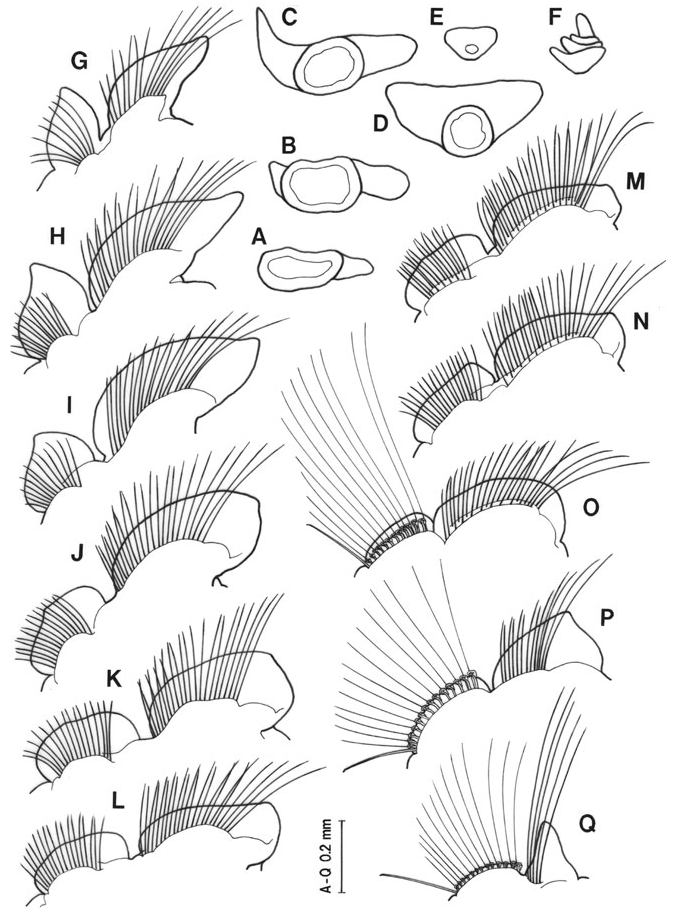
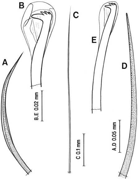
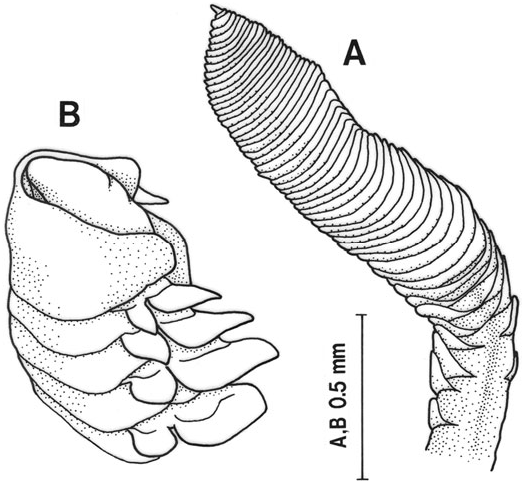
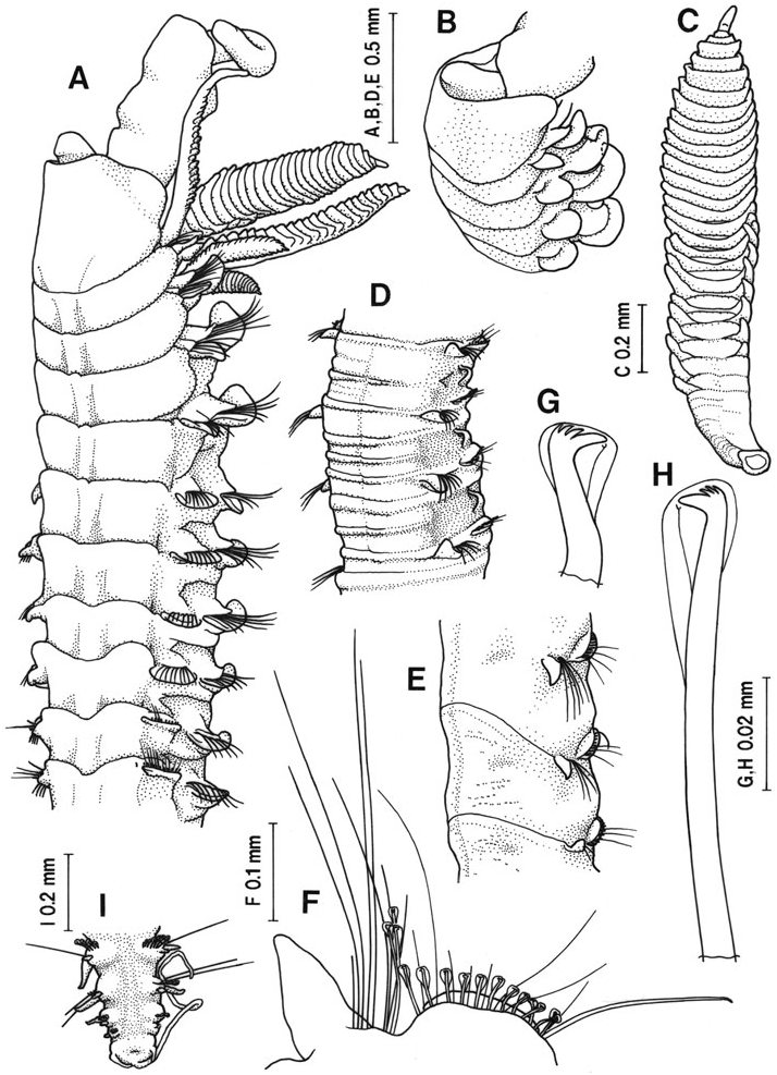
Description: Prostomium fusiform with bluntly pointed or round anterior end. Two pairs of brown or black eyes in trapezoidal arrangement usually visible through cuticle of prostomium. Palpi with basal sheath. Peristomium without pigment spots and marginal papilla. Three pairs of branchiae on setigers 1–3. First pair longest, third pair shortest. All branchiae bearing lamellae; one or two lamellae in proximal region of branchial shaft consisting of single plate ( Fig. 2A View Figure 2 ); succeeding lamellae consisting of two distinct plates, one round and the other triangular ( Fig. 2B, C View Figure 2 ); in middle and distal regions two plates united completely, showing flabellate ( Fig. 2D–F View Figure 2 ). No processes along anterior face of first branchiae. No slender filament at base of third branchia. Notopodial postsetal lamellae elongate subtriangular on setigers 1–3 ( Fig. 2G–I View Figure 2 ), becoming low, rounded back to about setiger 11 reducing in size ( Fig. 2J–O View Figure 2 ); thereafter, elevated posteriorly ( Fig. 2P View Figure 2 ), becoming triangular from about setigers 21 ( Fig. 2Q View Figure 2 ). Neuropodial postsetal lamella on setiger 1 lanceolate, distally pointed ( Fig. 2G View Figure 2 ); lamella on setiger 2 largest, subovoid, acuminate ( Fig. 2H View Figure 2 ); lamellae becoming low, rounded posterior to setiger 3 ( Fig. 2I–N View Figure 2 ), and reduced to low ridge posterior to setiger 9 ( Fig. 2O–Q View Figure 2 ). Notopodial setae anteriorly bilimbate capillaries only, replaced gradually by long slender non-limbate capillaries posterior to about setiger 17. Neuropodial setae in anterior setigers bilimbate capillaries only ( Fig. 3A View Figure 3 ), replaced by hooded hooks ( Fig. 3B View Figure 3 ), slender nonlimbate capillaries ( Fig. 3C View Figure 3 ), and one to two punctate sabre setae ( Fig. 3D View Figure 3 ) from setiger 9 ( Fig. 2O View Figure 2 ). Notopodial hooded hooks ( Fig. 3E View Figure 3 ) beginning between setigers 29–46. Neuropodial hooded hooks with 3–4 pairs of apical teeth above main fang and secondary hood; notopodial hooks with 2–3 pairs of apical teeth and secondary hood. Dorsum of setigers 12–17 transverse series of lighter coloured, slightly raised ridges, 2–3 ridges per setiger. Membranous dorsal crests connecting notopodial postsetal lamellae present on setigers 21–28, accompanied by semi-transparent, dorsal cuticle bearing circular concavities ( Fig. 4A View Figure 4 ). Ventral bilobed flap on setiger 8 absent. Interparapodial pouches absent. Pygidium with median long cirrus and two lateral short cirri. Muscular gizzard present between setigers 7–9.
Type specimens synonymized with Paraprionospio alata : Holotype of Paraprionospio alata incomplete with 50 setigers, measuring 29 mm long and 1.6 mm wide ( Fig. 4 View Figure 4 ). Eyes not visible. Palpi missing. First and third branchiae on the right side missing. Second branchia on the right side well developed, bearing 40 lamellae, although the tip missing; lamellae consist of two plates basally; thereafter all lamellae flabellate ( Fig. 4B View Figure 4 ). First and second branchiae on the left side appear in process of regeneration. Notopodial hooded hooks appear on setiger 46. Muscular gizzard present in setigers 8–9.
Holotype of Prionospio treadwelli incomplete with 46 setigers, measuring 15 mm long and 0.6 mm wide ( Fig. 5 View Figure 5 ). Eyes not visible. Palpi missing. Second branchia on the right side and third branchia on the left side well developed, bearing flabellate lamellae. Other branchiae appearing in process of regeneration. Notopodial hooded hooks from 45 setigers. Membranous dorsal crests on setigers 21–28, accompanied by semitransparent dorsal cuticle.
Two paratypes of Paraprionospio tamaii complete with 85–98 setigers, measuring 23–24 mm long and 0.7–0.8 mm wide, and three paratypes incomplete with 62–82 setigers, measuring 16–29 mm long and 0.7–0.9 mm wide. Eyes inconspicuous. A pair of yellowish-brown pigment patches present on widest part of prostomium. First pair of branchiae longest extending posteriorly to about setiger 5, third pair shortest extending to about setiger 7. All branchiae bearing lamellae; in proximal region of first branchia, two lamellae consisting of one triangular plate; succeeding several lamellae consisting of two plates; thereafter two plates connecting at their bases; in middle and distal regions, two plates united completely, showing flabellate-shape ( Fig. 6A View Figure 6 ). Notopodial postsetal lamellae elongate subtriangular on setigers 1–3, lamellae oval on setiger 4 ( Fig. 6B View Figure 6 ). Neuropodial postsetal lamellae lanceolate on setiger 1, subovoid, acuminate on setiger 2, round cordate on setiger 3 and low, rounded on setiger 4. Notopodial hooded hooks beginning between setigers 31 and 46. Well-developed membranous dorsal crests on setigers 21–28, accompanied by semi-transparent dorsal cuticle.
Holotype of Paraprionospio yokoyamai complete with 72 setigers, measuring 21 mm long and 0.6 mm wide (setiger 5, excluding parapodia); posterior three setigers regenerating. One paratype complete with 77 setigers, measuring 22 mm long and 0.7 mm wide and the other two paratypes incomplete, measuring 0.6– 0.7 mm wide. Prostomium with round anterior end. Two pairs of black eyes in trapezoidal arrangement present on prostomium. Two paratypes bearing yellowish-brown pigment patches on lateral sides of prostomium. First branchiae longest extending posteriorly to about setiger 5, third pair shortest extending to about setiger 7. All branchiae bearing lamellae; in proximal region of first branchia, two to three lamellae consisting of one triangular plate; succeeding two lamellae consisting of two plates; thereafter two plates connecting at their bases; in middle and distal regions, two plates united completely, showing crescent- to flabellate-shape ( Fig. 7A View Figure 7 ). Notopodial postsetal lamellae elongate subtriangular on setigers 1–3, subtriangular to elongate oval on setiger 4 ( Fig. 7B, C View Figure 7 ). Neuropodial postsetal lamellae lanceolate on setiger 1, subovoid, acuminate on setigers 2–3, and low, rounded on setiger 4 ( Fig. 7B, C View Figure 7 ). Notopodial hooded hooks beginning between setigers 29 and 37. Low and comparatively inconspicuous membranous dorsal crests on setigers 21–28, accompanied by semitransparent dorsal cuticle ( Fig. 7D View Figure 7 ).
Remarks: Prionospio alata was described by Moore (1923) as a new species based on specimens collected from southern California. The original description noted that neuropodial hooks are present from setiger 18 and the notopodial lamellae in the anterior setigers are broad-tipped. However, the observation on the type specimen revealed that neuropodial hooks appear from setiger 9 and the anterior notopodial lamellae are bluntly pointed. This species should be retained as a valid species belonging to the genus Paraprionospio in having peristomial wings enfolding the prostomium and three pairs of branchiae on setigers 1–3. Parapri- onospio alata can be differentiated from Paraprionospio pinnata by the possession of a membranous dorsal crest on setigers 21–28. Paraprionospio alata differed also from Paraprionospio inaequibranchia by the absence of a filament on setiger 3 and interparapodial pouches.
Treadwell (1931) described Prionospio plumosa as a new species based on specimens collected from Chesapeake Bay. Hartman (1951) gave a new name for this species as Prionospio treadwelli , because the specific name is preoccupied by Prionospio plumosa Sars, 1872 . She described Prionospio treadwelli as possessing four pairs of branchiae. Observations on the holotype of Prionospio treadwelli , however, showed that the type specimen is similar to Paraprionospio alata in having three pairs of branchiae and a membranous dorsal crest on setigers 21–28. Observations on other specimens collected from Chesapeake Bay showed also that the specimens agree with Paraprionospio alata except in having four eyes. Clear eyes were not visible from the type specimens of Paraprionospio alata and Prionospio treadwelli , although Treadwell (1931) reported two pairs of reddish eyes on the prostomium of Prionospio treadwelli . Such observations suggest that the pigments of eyes had faded during preservation. The similarity of the characters between Para- prionospio alata and Prionospio treadwelli suggests that the two species are conspecific.
Delgado-Blas (2004) reported two new species, Paraprionospio tamaii and Paraprionospio yokoyamai , from the Grand Caribbean region. Paraprionospio tamaii was described as having dorsal crests on setigers 20–29, notopodial lamellae oval on setigers 1–2, reniform on setiger 4, neuropodial lamellae obovate on setiger 3, and branchiae with two kinds of lamellae, whereas Paraprionospio yokoyamai was described originally as having no dorsal crests, notopodial lamellae triangular and elongate on setigers 1–2, subtriangular on setiger 4, neuropodial lamellae cordate on setiger 3, and branchiae with three kinds of lamellae. The present examination, however, revealed that the two species have dorsal crests on setigers 21–28. Taking the variability in the shape of parapodial lamellae within the species ( Fig. 7B, C View Figure 7 ) into consideration, the differences in shape of notopodial and neuropodial lamellae on setigers 1–4 between the two species seem to be insignificant. Branchiae of Paraprionospio yokoyamai are slender, whereas those of Paraprionospio tamaii are thick. However, the arrangement of lamellar plates along the branchial shaft in the two species is similar; i.e. in the proximal region, lamellae consist of a single plate, succeeding lamellae consist of two plates, and thereafter the two plates connect with each other at their bases and are crescent- to flabellate-shaped from middle to distal regions ( Figs 6A View Figure 6 , 7A View Figure 7 ). Such morphological features and other characters (e.g. no papilla on the peristomium, no filament on setiger 3, no ventral flap on setiger 8, no interparapodial pouches, non-limbate slender capillaries on 9th neuropodium) suggest that both Paraprionospio tamaii and Paraprionospio yokoyamai are conspecific with Paraprionospio alata .
Distribution: British Columbia; California; Chesapeake Bay; Gulf of Mexico.
( FIG. 8 View Figure 8 )
Paraprionospio sp. form CI, Yokoyama & Tamai, 1981: 311–313, fig. 5.
Paraprionospio coora Wilson, 1990 View in CoL
Material examined: Thirty-five specimens (OMNH-Iv 4861–4872) collected from the following localities in Japan: off Tokai (36°28′N, 140°37–38′E), Ibaraki Pref., 15–18 m, coll. Ibaraki Fish. Exp. Stn; Tsuruga Bay (35°41–45′N, 135°4–5′E), 27–43 m, coll. I. Hayashi ; Obama Bay (35°31′N, 135°43′E), 14 m, coll. I. Hayashi GoogleMaps ; off Takahama (35°35′N, 135°30′E), Wakasa Bay , 60 m, coll. H. Yokoyama GoogleMaps ; Maizuru Bay (35°29– 30′N, 135°20–22′E), 13 m, coll. I. Hayashi ; off the mouth of Yura River (35°32–37′N, 135°17–19′E), 20– 60 m, iv.1973 – iii.1975, coll. H. Yokoyama ; Gokasho Bay (34°17′N, 136°40′E), 12 m, coll. H. Yokoyama GoogleMaps ; Owase Bay (34°4′N, 136°14′E), 23 m, coll. H. Yokoyama GoogleMaps ; Kata Bay (33°58′N, 136°13′E), 29 m, coll. H. Yokoyama GoogleMaps ; Tanabe Bay (33°42–43′N, 135°21–22′E), 18–34 m, coll. H. Yokoyama ; Usuki Bay (33°9′N, 131°49′E), 18 m, coll. H. Yokoyama GoogleMaps ; off the mouth of Sendai River (31°51′N, 130°10′E), Kagoshima Pref., 25 m, coll. H. Yokoyama. GoogleMaps
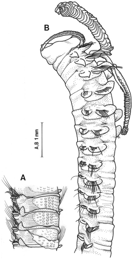
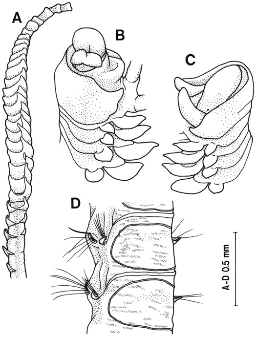
Description: Body up to 70 mm long, 2.1 mm wide with 120 setigers. Prostomium fusiform with round or bluntly pointed anterior end, extending posteriorly as a faintly raised ridge to the first setiger ( Fig. 8A View Figure 8 ). Two pairs of black, small eyes in trapezoidal arrangement. Brown pigment patch occasionally present on lateral side of peristomium. Faint groove just posterior to pigmented area on peristomium. Small papilla on posterior margin of peristomium. A pair of grooved palpi with membranous basal sheath ( Fig. 8A View Figure 8 ). Three pairs of branchiae on setigers 1–3. First branchiae usually longest extending posteriorly to about setiger 6, or first and second branchiae approximately equal length, third pair shortest extending to about setiger 6. In basal region of first branchial shaft, 2–9 tubercular or conic processes placed along anterior face ( Fig. 8B View Figure 8 ). Processes occasionally found on well-developed second branchial shaft. All branchiae bearing lamellar plates; in proximal region of branchial shaft, two lamellae consisting of single triangular plate; succeeding lamellae consisting of two plates; in middle and distal regions, two plates united completely, showing flabellate shape ( Fig. 8C View Figure 8 ). No slender filament at base of third branchia. Notopodial postsetal lamellae on setigers 1–3 long, foliaceous and distally pointed; posterior to setiger 4, becoming rounded and reducing in size; posterior to setiger 10, elevated increasingly, showing triangular to lanceolate ( Fig. 8A, D, E View Figure 8 ). Neuropodial postsetal lamellae of setiger 1–3 lanceolate; posterior to setiger 4, lamellae becoming rounded and reducing to low postsetal ridges by about setiger 9. Anterior setae all limbate capillaries bearing granules. Posterior to about setiger 16, notopodial limbate capillaries replaced by slender, non-limbate capillaries. Neuropodial hooded hooks with 2–4 pairs of apical teeth above main fang and striate secondary internal hood ( Fig. 8F View Figure 8 ) from setiger 9, accompanied by alternating non-limbate slender capillaries and one to two granulated sabre setae ( Fig. 8E View Figure 8 ). Notopodial hooded hooks with 2–3 pairs of apical teeth above main fang and striate secondary internal hood ( Fig. 8G View Figure 8 ) appearing between setigers 24 and 54. No ventral bilobed flap on setiger 8. Interparapodial pouches present in some specimens, appearing between setigers 7 and 9 and disappearing between setigers 14 and 39 ( Fig. 8D View Figure 8 ). Dorsum of setigers 4–11 faintly biannulated. Dorsum of setigers 12–17 transverse series of lighter coloured slightly raised ridges, three ridges per setiger. Membranous dorsal crests and semi-transparent dorsal cuticle absent. Pygidium with a long median anal cirrus and two short, lateral cirri ( Fig. 8H View Figure 8 ). Muscular gizzard present in setigers 7–8.
Remarks: Japanese specimens closely agree with Paraprionospio coora , which was described for specimens from Australia by Wilson (1990), but have 2–4 pairs of apical teeth in hooded hooks rather than only two pairs of apical teeth. A variable number of apical teeth were found within individual specimens from Japan, suggesting that this is not a stable character to be used in distinguishing species. Japanese specimens therefore are referred to Paraprionospio coora . This species is recorded from Japan for the first time.
Distribution: Western Japan, more southern than 37°N; East China Sea ( Tamai, 1981), New South Wales to Tasmania in Australia.
PARAPRIONOSPIO CORDIFOLIA SP. NOV.
( FIG. 9 View Figure 9 )
Paraprionospio sp. form B, Yokoyama & Tamai, 1981: 309–311, fig. 4.
Paraprionospio sp. form B, Yokoyama, 1996.
Material examined: Holotype (OMNH-Iv 4873): entire specimen, female, 93 setigers, 29 mm long, 0.7 mm wide at setiger 5, off the mouth of Yura River (35°32′N, 135°18′E), Wakasa Bay , mud, 20 m deep, 20.viii.1973, collected by H. Yokoyama. Six paratypes (OMNH-Iv 4874–4879): off the mouth of Yura River (35°32–33′N, 135°17–19′E), 20–30 m, iv.1973 – iii.1975, coll. H. Yokoyama [see Yokoyama & Hayashi (1980) for the sampling stations of the type specimens and environmental factors]. Eighty-three specimens (OMNH-Iv 4880–4887) collected from the following localities along the Japanese coast: Tsuruga Bay (35°40′N, 136°1′E), 17–31 m, coll. I. Hayashi; Obama Bay (35°40–42′N, 136°3–4′E), 17–31 m, coll. I. Hayashi; Maizuru Bay (35°28–29′N, 135°19–24′E), 6–17 m, coll. I. Hayashi; Gokasho Bay (34°20′N, 136°43′E), 12 m, coll. Yokoyama; off Tanagawa (34°21′N, 135°11′E), Osaka Bay , 8 m, coll. H. Yokoyama; off Kobe (34°40′N, 135°17′E), Osaka Bay, 17 m, coll. R. Yamanishi; Ohmura Bay (32°59′N, 129°53′E), 19 m, coll. H. Yokoyama; off Ohmuta Ariake Sound (33°2′N, 130°21′E), 5 m, coll. H. Yokoyama. Seven specimens collected from Hong Kong by P. Shin. GoogleMaps
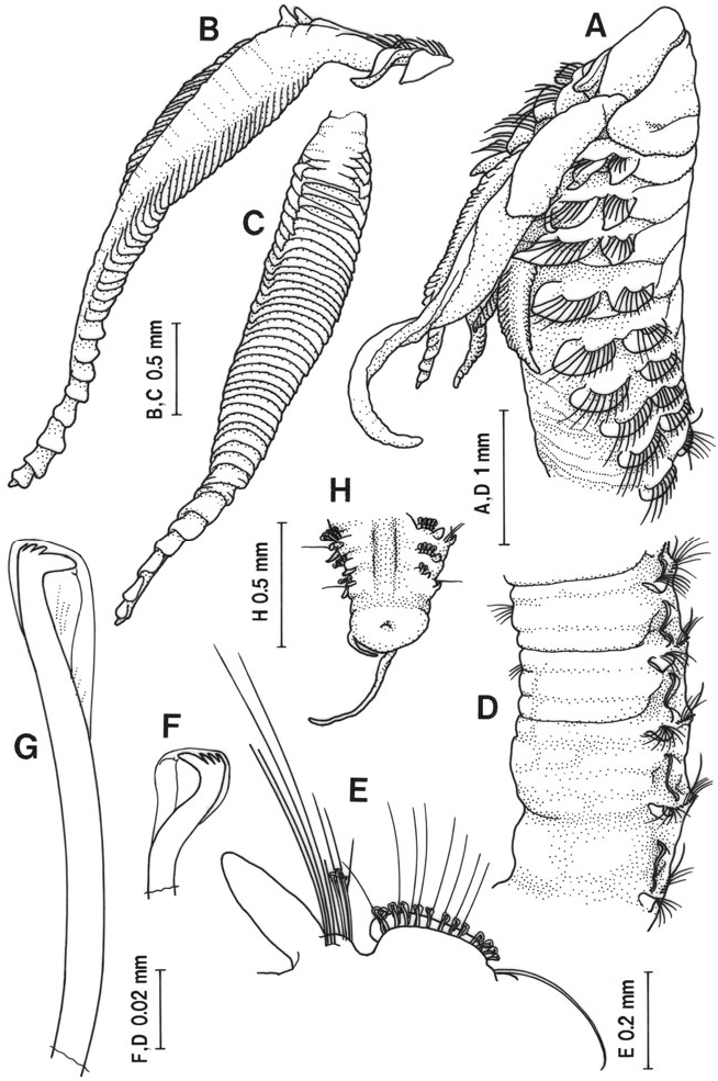
Description: Body up to 37 mm long, 1.0 mm wide (setiger 5, excluding parapodia) with 94 setigers. Prostomium fusiform with round or bluntly pointed anterior end ( Fig. 9A, B View Figure 9 ). Two pairs of black, small eyes in trapezoidal arrangement. A pair of yellowishbrown pigment patches occasionally visible between the two pairs of eyes. No papilla on posterior margin of peristomium. No pigment patch on peristomium. A pair of grooved palpi with membranous basal sheath ( Fig. 9A View Figure 9 ). Three pairs of branchiae on setigers 1–3. First branchiae longest extending posteriorly to about setiger 5, third pair shortest extending to about setiger 7. All branchiae bearing lamellae; in proximal region of branchial shaft, two lamellae consisting of one plate; succeeding lamellae consisting of two triangular plates; in middle and distal regions, two plates united completely, showing flabellate-shape ( Fig. 9C View Figure 9 ). A slender filament at base of third pair of branchiae. Notopodial postsetal lamellae lanceolate on setigers 1–2; posterior to setiger 3, lamellae becoming rounded and reducing in size to about setiger 15; thereafter elevated posteriorly, becoming triangular to lanceolate ( Fig. 9A, B, D–F View Figure 9 ). Neuropodial postsetal lamellae of setigers 1–3 lanceolate, becoming rounded and reducing to low postsetal ridges posteriorly. Anterior ventral margin in setiger 8 protruding to form a bilobed membranous flap; on 2–3 succeeding setigers anterior ventral margin projecting slightly, but not forming such conspicuous structure ( Fig. 9A View Figure 9 ). Interparapodial pouches ( Fig. 9D View Figure 9 ) occurring usually from setiger 9, occasionally from setiger 8 or 10, through variable number of setigers (up to setiger 42). Dorsum of seti- gers 8–17 with transverse series of lighter coloured slightly raised ridges, 2–3 ridges per setiger; colouration distinct on setigers 13–16 ( Fig. 9D View Figure 9 ). Membranous dorsal crests absent, instead, a faint ridge between both notopodial postsetal lamellae on setigers 21–36 present, accompanied by semi-transparent dorsal cuticle bearing circular convexities ( Fig. 9E View Figure 9 ). Anterior setae all limbate capillaries bearing granules. Notopodial limbate capillaries replaced by slender, nonlimbate capillaries posterior to middle body region. Neuropodial hooded hooks with 3–4 pairs of apical teeth above main fang ( Fig. 9G View Figure 9 ) from setiger 9, accompanied by alternating non-limbate slender capillaries and one to two granulated sabre setae ( Fig. 9F View Figure 9 ). Notopodial hooded hooks with 2–3 pairs of apical teeth above main fang ( Fig. 9H View Figure 9 ) appearing between setigers 36 and 42. Pygidium with a long median anal cirrus and two short, lateral cirri ( Fig. 9I View Figure 9 ). Muscular gizzard present in setigers 7–9.
Remarks and etymology: Paraprionospio cordifolia is characterized by having a ventral bilobed flap on setiger 8, which is not found in any other Paraprionospio species. The species name (= heart-shaped leaf) is derived from this structure.
Distribution: Western Japan, more southern than 36°N; East China Sea ( Tamai, 1981); Hong Kong.
No known copyright restrictions apply. See Agosti, D., Egloff, W., 2009. Taxonomic information exchange and copyright: the Plazi approach. BMC Research Notes 2009, 2:53 for further explanation.
|
Kingdom |
|
|
Phylum |
|
|
Class |
|
|
Order |
|
|
Family |
Paraprionospio
| Yokoyama, Hisashi 2007 |
Paraprionospio sp.
| Yokoyama H & Tamai K 1981: 311 |
Paraprionospio sp.
| Yokoyama H & Tamai K 1981: 309 |
Prionospio alata
| Moore JP 1923: 186 |