
Mesomyzostoma okadai
|
publication ID |
https://doi.org/ 10.1080/00222933.2015.1056266 |
|
DOI |
https://doi.org/10.5281/zenodo.5672670 |
|
persistent identifier |
https://treatment.plazi.org/id/2F2FE66B-9404-FFD8-55F7-B97718A5FB3C |
|
treatment provided by |
Plazi |
|
scientific name |
Mesomyzostoma okadai |
| status |
sp. nov. |
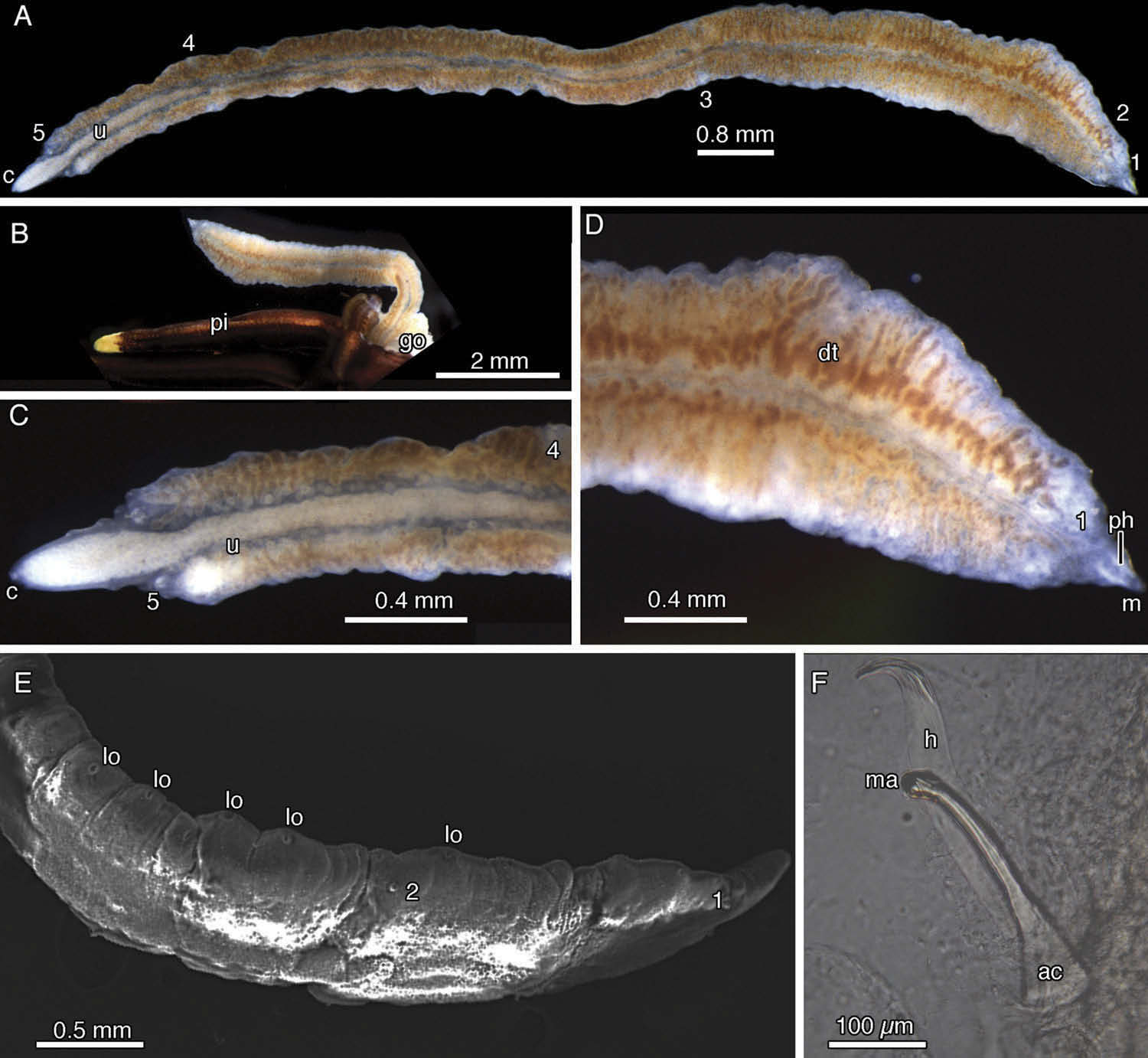
Mesomyzostoma okadai Rouse, Lanterbecq, Summers and Eeckhaut sp. nov.
( Figure 5 View Figure 5 )
Mesomyzostoma n. sp. 2 in Lanterbecq et al. (2006, 2009) Mesomyzostoma n sp. 2 in Summers and Rouse (2014)
Material examined
Off Misaki Marine Biological Station, Sagami Bay (Japan), 35°9.788ʹ N, 139°36.732ʹ E, 10 – 15 m depth. Specimens from seven A. japonica individuals. Collector: Greg Rouse, 13 – 28 May 1998. One to five per host. Holotype (SIO-BIC A4079) in 70% ethanol following glutaraldehyde fixation. Paratypes: SIO-BIC A4080, A4081, A4082, seven intact or incomplete paratypes in 70% ethanol following formalin fixation; SIO-BIC A4083, A4084, two specimens in 70% ethanol following glutaraldehyde fixation; SIO-BIC A4085, one specimen fixed and preserved in 95% ethanol; SIO-BIC A4086, two specimens used for SEM observations; SAM E3406, one complete in 70% ethanol following formalin fixation. One incomplete specimen, from paratype lot SIO-BIC A4082, dissolved in bleach for observation of parapodial hook apparatus and another, originally from paratype lot SIO-BIC A4085, digested for DNA extraction and molecular phylogenetic analyses.
Etymology
Named for Dr Yaichiro Okada (deceased) in recognition of his important early work on Japanese myzostomes.
Diagnosis
Mesomyzostoma with elongated, flat, ribbon-like, crenulate body. Muscular pharynx present; eversibility unknown. No marginal cirri. Five chaetigers along body, chaetae close to trunk margin but parapodia absent or very low and then only anteriorly. Emergent hooks small with thin shaft, tip curving to 90° with respect to shaft. No replacement hooks. Aciculae as long as emergent hooks, but twice as thick at the base. Manubrium flag-like, developed on one side. Numerous ovoid lateral organs located on humps all along body margin. Mouth at extreme anterior end, cloaca posteriorly. Penes absent, but seminal vesicles at third chaetiger. Hermaphrodite. Parasite of crinoid coelom.
Description
Holotype 12 mm long, 0.8 mm wide and about 0.2 mm thick. Body flat, curled dorsally with anterior end sharper than posterior end, latter slightly broken. Body margin crenulate with about 37 lateral organs visible on left and 33 on right side. Openings of lateral organs circular, c.40 µm in diameter, often located on small humps 80 µm high and spaced regularly at 200 – 400 µm intervals. Pharynx in pointed, anterior tip of body, 300 µm long ( Figure 5 View Figure 5 D). Mouth oval, 50 µm in diameter. First two chaetigers with low parapodia, others, if present, not observed. First chaetiger located 0.2 mm from anterior end, second 1 mm behind first. Remaining chaetigers distributed along body.
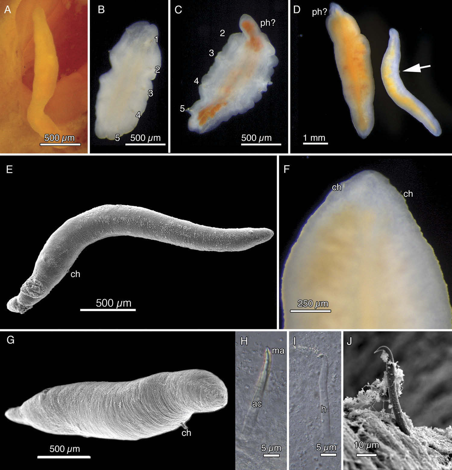
Entire paratypes ranging from 2 to 14 mm long and 0.8 to 1.5 mm wide ( Figure 5 View Figure 5 A). Most paratypes with anterior and posterior ends very pointed, with mouth and cloaca respectively ( Figure 5 View Figure 5 A, C, D). All paratypes with crenulate trunk margin due to presence of hump-shaped lateral organs ( Figure 6 View Figure 6 E). Lateral organs present all along trunk margin, spaced 100 μm (in contracted portion of body) to 400 μm apart, specimens having 20 – 70 pairs ( Figure 5 View Figure 5 A, D, E). Five chaetigers but only up to two pairs of low parapodia present, both of latter located anteriorly. Parapodia as small humps 0.1 mm high ( Figure 5 View Figure 5 E). Each emergent chaeta with an acicula, but no replacement hook ( Figure 5 View Figure 5 F). Emergent hook up to 0.4 mm long, shaft moderately thick, with distal third bowed slightly outward, tip curving 90° ( Figure 5 View Figure 5 F). Aciculae of similar size to emergent hooks but, being c.80 µm thick at base, twice as thick as hooks. Manubrium visible as small projection on one side ( Figure 5 View Figure 5 E).
Individuals found in coelom of pinnules close to gonads and in coelom of arms ( Figure 5 View Figure 5 B). Live specimens with translucent margin, yellowish ovaries, orange digestive diverticula. Individuals in life less translucent than M. katoi , but still allowing observation of some internal organs ( Figure 5 View Figure 5 A – D). Some specimens with pair of white seminal vesicles at level of third chaetiger, full of sperm/spermatophores.
Remarks
Mesomyzostoma okadai sp. nov. is found in the same host ( A. japonica ) and same geographic location as M. katoi . It also occurs in the same body region of the host and both species could be found in the same individual host. Mesomyzostoma okadai sp. nov. is morphologically distinct from M. katoi in its: (1) crenulate body margin from multiple lateral organs located on small humps; (2) its greater body size as adults and relative opacity. The two species are also very distinct in terms of DNA data, with M. okadai sp. nov. being the sister taxon to the other sequenced Mesomyzostoma species (see below, Figure 8 View Figure 8 ).

No known copyright restrictions apply. See Agosti, D., Egloff, W., 2009. Taxonomic information exchange and copyright: the Plazi approach. BMC Research Notes 2009, 2:53 for further explanation.