
Mesomyzostoma cf. reichenspergeri Remscheid, 1918
|
publication ID |
https://doi.org/ 10.1080/00222933.2015.1056266 |
|
DOI |
https://doi.org/10.5281/zenodo.5672666 |
|
persistent identifier |
https://treatment.plazi.org/id/2F2FE66B-940F-FFDC-5478-BC2D1B07FDA9 |
|
treatment provided by |
Plazi |
|
scientific name |
Mesomyzostoma cf. reichenspergeri Remscheid, 1918 |
| status |
|
Mesomyzostoma cf. reichenspergeri Remscheid, 1918
( Figure 3 View Figure 3 )
Mesomyzostoma reichenspergi (sic) in Lanterbecq et al. (2006, 2009, 2010) Mesomyzostoma cf. reichenspergeri in Summers and Rouse (2014)
Material examined
North Point, Lizard Island, Great Barrier Reef (Australia), 14°38.655' S, 145°27.267ʹ E; 10 m depth. Eight specimens in Himerometra robustipinna ( Carpenter 1881) (Himerometridae) . Collector: Greg Rouse, 18 March 2000. One fixed in 3% glutaraldehyde and post-fixed in 1% osmium tetroxide and prepared for SEM observations (SIO-BIC A4073); one fixed in formalin and preserved in 70% ethanol and described here below (SAM E3410); three (one fixed in formalin and two in ethanol) (SIO-BIC A4074, A4075); two specimens dissolved in bleach for observations of parapodial hook apparatus; and one specimen digested for DNA extraction and molecular phylogenetic analyses. Madang Lagoon, Papua New Guinea 5.136° S, 145.81° E; 3 m depth. Three specimens in H. robustipinna . Collector: Greg Rouse and Mindi Summers, 30 November 2012. One fixed in formalin and two in ethanol (SIO-BIC A3680).
Diagnosis
Mesomyzostoma with elongated, somewhat flattened body with no introvert and no cirri. Five pairs of very small parapodia with emergent hooks located ventrally, close to trunk margin. Emergent hooks small with thin shaft, tip curving to 90° with respect to shaft. Replacement hooks present in some parapodia. Aciculae as long and wide as emergent hooks. Manubria small, developed on one side. No lateral organs or penes. Eggs filling dorsal side, spermatocysts found ventrally. Simultaneous hermaphrodites. Parasites of crinoid coelom.
Description
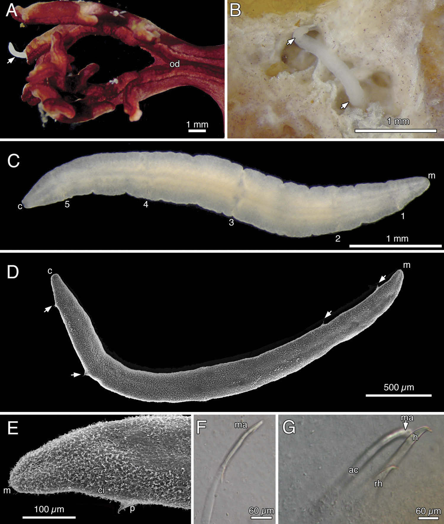
Ethanol-fixed specimen white, individuals in life opaque ( Figure 3 View Figure 3 A – C), observed in coelom of oral disc leading into gonads and coelom of arms ( Figure 3 View Figure 3 A). Specimen SAM E3410 with thin trunk 1.6 mm long and 0.4 mm wide. Body curled dorsally with mouth opening anterodorsally. No lateral organs visible. Other specimens 1.6 – 4.0 mm long ( Figure 3 View Figure 3 C). Parapodia visible on live specimens, difficult to observe on fixed specimens except with SEM ( Figure 3 View Figure 3 C, D). Parapodia low, 0.1 mm high, with one emergent hook ( Figure 3 View Figure 3 D, E). One acicula per parapodium, some with a replacement hook ( Figure 3 View Figure 3 F, G). Emergent hook up to 0.25 mm long, shaft moderately thick, with distal fifth bowed slightly outward, tip short and sharp, curving at 90° ( Figure 3 View Figure 3 E, G). Aciculae of same length and width as emergent hooks, c.30 µm wide at base. Manubrium developed on one side, small and rapidly digested by bleach ( Figure 3 View Figure 3 F). Replacement hook one-fifth as long as emergent hook ( Figure 3 View Figure 3 G). First to fifth pairs of parapodia located 0.3, 0.9, 1.7, 2.7 and 3 mm from mouth in a 3.5-mm-long specimen ( Figure 3 View Figure 3 C). Body margin without cirri ( Figure 3 View Figure 3 B – D), but body apparently covered with cilia ( Figure 3 View Figure 3 E). Lateral organs and penes absent. Cloaca at rear extremity.
Remarks
Remscheid (1918) obtained five specimens of M. reichenspergeri from Amphimetra discoidea A.H. Clark 1911 , [now recognized as Amphimetra tesselata ( Müller 1841) ] from off the Aru Islands (Indonesia). The specimens were from 0.26 to 1.91 mm long. A search was made by Dr Dieter Fiege, curator of marine invertebrates at the Senckenberg Museum in Frankfurt, to trace the material collected during the Merton Expedition. The material is not in the Senckenberg collections, although the Museum has material from other groups from this expedition, e.g. other polychaetes and cestodes. It is therefore probable that either Remscheid did not return the Mesomyzostoma specimens or they were lost during return or perhaps during the Second World War. Based on information given in Remscheid ’ s acknowledgements, it is highly probable that he worked at the University of Bonn, where Prof. R. Hesse followed Prof. H.L. Ludwig as head of the Institute of Zoology. Prof. Reichensperger worked on echinoderms with Prof. Ludwig and became head of the institute himself in 1928. Professors Strubell and Schmidt, both mentioned in Remscheid ’ s acknowledgements, also worked in Bonn. Unfortunately, the collections of the Institute of Zoology in Bonn were destroyed during the Second World War. Consequently, if the material should by chance have been deposited there, it is probably lost. However, owing to (1) the distance from both Lizard Island and Madang to the type locality of the Aru Islands; (2) the hitherto unrecognized diversity of Mesomyzostoma ( Summers and Rouse 2014) ; and (3) the fact that the host species recorded here is different to that named by Remscheid (1918), we hesitate to identify our material as M. reichenspergeri . Our specimens were reported as being parasites of Himerometra magnipinna in Lanterbecq et al. (2006, 2009 , 2010), but subsequent work on the hosts has shown that H. magnipinna is a junior synonym of H. robustipinna (Taylor et al. in prep.).
No known copyright restrictions apply. See Agosti, D., Egloff, W., 2009. Taxonomic information exchange and copyright: the Plazi approach. BMC Research Notes 2009, 2:53 for further explanation.