
Enchylea heteroducta Nielsen & Christensen, 1963
|
publication ID |
https://doi.org/ 10.11646/zootaxa.3647.2.4 |
|
publication LSID |
lsid:zoobank.org:pub:33866E2B-6B0F-4124-A6A6-2B057E642149 |
|
DOI |
https://doi.org/10.5281/zenodo.5612048 |
|
persistent identifier |
https://treatment.plazi.org/id/301187BC-2C01-FFE5-88B0-FD5A481AFC01 |
|
treatment provided by |
Plazi |
|
scientific name |
Enchylea heteroducta Nielsen & Christensen, 1963 |
| status |
|
Enchylea heteroducta Nielsen & Christensen, 1963 View in CoL
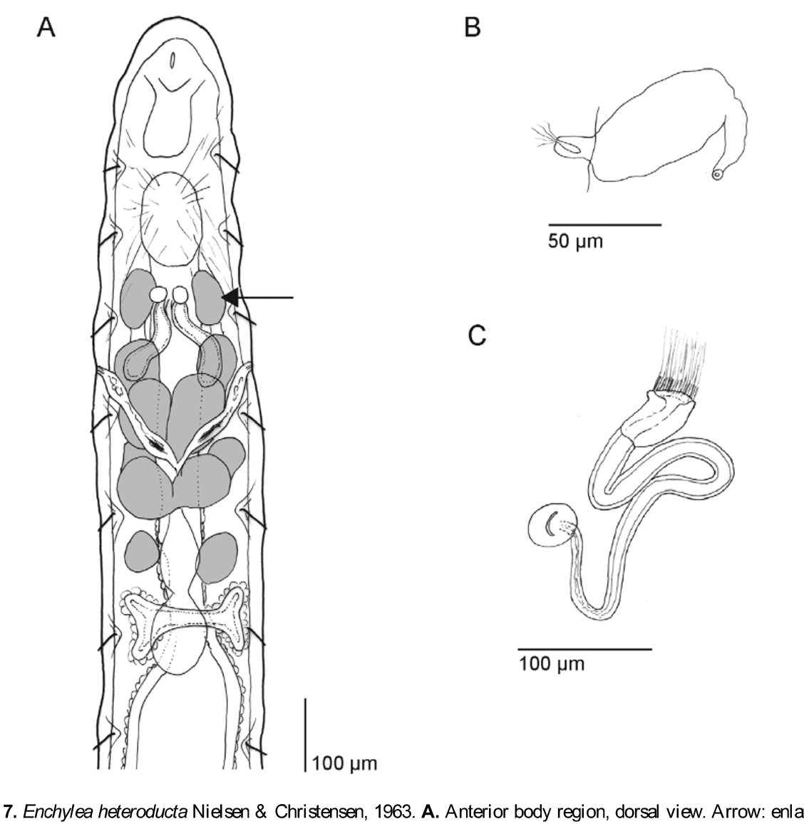
( Fig. 7 View FIGURE 7 A–C)
Enchylea heteroducta Nielsen & Christensen, 1963: 11 –12, Figs 14–17. Schmelz & Collado 2012: 54f.
Holotype and paratypes. Not designated in original description, not located, probably lost.
Investigated material. Three spms, 1 adult, 2 early-stage subadults. Portugal, Coimbra, in soil from the experimental field area of the Coimbra Agricultural School (ESAC), crop site ( Table 2 View TABLE 2 ); IV 2010. One of the subadults cut in halves: posterior half used for DNA extraction and sequence generation; anterior half as voucher in 100% ethanol. Rest fixed in Bouin's fluid, stained with paracarmine, whole-mounted in Canada balsam.
Description. Dimensions are from the single adult whole-mounted specimen, unless stated otherwise. Specific traits found only in one of the whole-mounted specimens are marked with "AD" (adult specimen) or "SA" (subadult specimen). Differences of the original description are inserted, marked by "NC".
Length c. 5 mm (viv), 2.5–4 mm (fix); diameter (fix) 0.22 mm at V, XX, 0.26 mm at XII. Segments 25 (N=2), NC: (27)–31–(35). Chaetae straight with weak ental hook, 2 and 3 per bundle, ventrally only 3. Formula 2,3 – 2,3: 3 – 3. In lateral bundles 2 chaetae mostly near clitellum. Chaetae absent at XII. Lateral chaetae in truly lateral position, not shifted dorsally. Chaetae mostly 40–50 µm long and 3–4 µm wide, largest in caudal segments (> 50 μm), smallest in II (c. 35: 3 µm); size differences between ventral and lateral chaetae not ascertained. Loose packages of detached chaetae present in the coelom in the adult specimen.
Head pore mid-dorsally at 0/I, a longitudinal slit, length c. 15 µm. Prostomium rounded, broadly conical, about as long as wide or high, shorter in fixed specimens. Epithelium without frontal recess, increasing in height towards the front, lined basally with musculature (not shown in Fig. 7 View FIGURE 7 A). No sensory papillae apparent (fix), no prostomial papillae seen. Fibers of subepithelial muscular lining extending across lumen of prostomium in various longitudinal, transverse and oblique directions (not shown in Fig. 7 View FIGURE 7 A). Peristomium c. 3/5 the length of following segments (II, III etc.). Epidermal gland cells not observed. No segmental dorsal pores. Body wall thin, c. 10 µm, up to 20 µm in anterior segments. Longitudinal muscle layer about as thick as layers of ring muscles, epidermis, and cuticle together. Cuticle very thin, ring muscles in irregular arrangement (optical section). Septa very thin, slightly thickened at 4/5–6/7.
Brain 85: 60 µm (fix), rounded or truncate posteriorly, sides converging anteriad, anteriorly concave; prostomial nerves with a pair of ganglia (SA), or ganglia not seen (AD). Ventral nerve cord medullar, slightly constricted and with fewer perikarya near the septa. Inner pair of postpharyngeal ganglia situated on top of oesophageal appendages; outer pair on afferent fascicles, enlarged and apparently with pharyngeal gland tissue in adult specimen ( Fig. 7 View FIGURE 7 A, arrow).
Pharyngeal pad comparatively large, 130 µm long and 90 µm wide in adult specimen (fix), heavily invested with musculature dorsally, fibers extend backwards into V. Oesophageal appendages in III–IV, a pair of short, unbranched, free-floating, sac-like tubes with wide lumen, attached separately and adjacently to oesophagus dorsally behind pharyngeal pad and below post-pharyngeal ganglia, extending obliquely backwards and downwards, blind-ending ventrally below pharyngeal gland lobes of IV (AD); maximum diameter 40 µm, estimated length c. 160 µm; epithelium 3–4 µm thick, not folded, smooth towards coelom, slightly rugose towards inner lumen. Pharyngeal glands in IV–VI, decreasing in size from IV to VI (AD) or of equal size in IV–VI (SA); dorsal lobes separate or with narrow dorsal connection, ventral lobes present from IV–VI, no secondary glands. Intestinal diverticula in VII, dorsally and laterally of oesophagus, with common unpaired opening into oesophagus dorsally, lateral wings widely connected dorsally of oesophagus, laterally flattened, extending forwards and backwards to the same degree; diverticular epithelium smooth, not folded, lined with blood sinus and chloragocytes, with histological properties different from adjacent oesophageal and intestinal epithelium: somewhat yellowish in whole mounts, not staining with paracarmine. Intestine widening strongly from c. 50 µm diameter at 6/7 to c. 150 µm in VIII; widening abrupt at 7/ 8 in contracted specimens. Pars tumida of midgut not distinguished. Intestine filled with amorphous humous substance mixed with sand grains up to 40 µm in diameter. Chloragocytes with dense layer from VI, covering also intestinal diverticula; cell diameter c. 10 µm (fix); in postclitellar segments cells often higher than wide (fix).
Blood colourless, dorsal blood vessel from VII, large and pulsating, bifurcating in I below brain into circumesophageal connectives, these uniting into ventral vessel in IV. Two pairs of commissural vessels seen in III posteriorly, branching off dorsal vessel close to each other. Preclitellar nephridia 6 pairs, from 4/5 to 9/10. Postclitellar nephridia from 13/14, absent at several positions around XVII–XX; length c. 80 µm; anteseptale with funnel only, postseptale laterally flattened, c. 1.5x as long as high, with well-developed interstitial tissue, efferent duct about half the length of postseptale, rising terminally, nephroporus with small terminal vesicle, conspicuous in vivo. Coelomocytes one type, mucocytes, flat, broadly oval or broadly pear-shaped, c. 20 µm long (viv), 16–22 µm long and 10–12 µm wide (fix), filled with small, slightly refractile vesicles (viv), nucleus and vesicles distinct (fix). Cells numerous, but not in dense aggregations.
Clitellum in XII and XIII (NC: XII–1 /2XIII), extending beyond chatae of XIII by 2–3 transverse cell rows; girdle-shaped, cells in c. 32 dense rows, hyalocytes evenly distributed on all sides, present also mid-ventrally; celldiameter c. 8–10 µm (fix), cells higher than wide laterally (c. 15 µm, fix). Testes and sperm funnels in XI, male pores and ovaries in XII. Testes and developing sperm enclosed by a common membrane, i.e. sperm developing in testis sacs and not freely in the coelom. Two testis sacs present, one on each side, not lobed, confined to lateral sides of XI, occupying up to one segment length. Spermatozoa c. 50 µm long, heads c. 12 µm (fix); lengths in vivo not measured. Sperm funnel small, 1.5– 2 x as long as wide (e.g. 60: 36 μm, fix), tapering distad, collar as wide as funnel body. Vas deferens short (estimated length c. 400 µm), not winded into coils, proximal region with wide ciliated lumen, tube diameter here c. 20 μm, tapering distad more or less continuously down to 7–8 µm near male pore. Tube walls c. 2 µm thick throughout, cilia in lumen conspicuous. Male copulatory organ with bursa, gland, and copulatory muscles. Gland rounded and compact, c. 50 μm long, 40 μm wide, 30 µm high (fix), bursal slit longitudinal, c. 25 µm long, transverse copulatory muscles only in immediate vicinity of male gland. Subneural glands absent.
Spermathecae as simple, mid-dorsally united tubes. Each tube with a double swelling, the ectal one possibly representing ectal duct, but cuticular lining not distinguished here and sperm present in both swellings. Ental swelling wider (c. 25 µm) than ectal swelling (c. 20 µm). No diverticula, no ectal glands. (NC: Ectal duct long and narrow, with distinct canal, sometimes with few separate gland cells near ectal pore). Ectal porus minute, at halflength between lateral chaetae of IV and V, slightly shifted ventrally. Two mature eggs in the adult specimen, each occupying about one segment length (NC: 1– 8 eggs at a time).
No intestinal or coelomic parasites observed.
Remarks. Enchylea heteroducta had been described from laboratory cultures of unknown origin (Nielsen & Christensen 1963). This is the first record of the species from a natural habitat; the note in Schmelz and Collado (2012) refers to the same record. Specimens underlying the original description had more segments (27–35), up to 8 mature oocytes at a time, and a slightly different spermatheca: There was an ectal duct at the same place where we found a sperm-containing swelling. For the time being we assume that these differences are intraspecifi c. Several structures differ in appearance between the adult and the subadult specimen; more material is necessary to decide which character state is the 'valid' one. The reduced size of the hindmost pair of pharyngeal glands in the adult specimen may be a case of degeneration, although it agrees with the original description.
Peculiar in this species are the combined presence, position and shape of oesophageal appendages and intestinal diverticula, and the short and stout vasa deferentia. Gut appendages are among the most important characters to distinguish genera among enchytraeids, therefore the new species was accomodated in a genus of its own. The oesophageal appendages are very similar to those in the Enchytraeus albidus group sensu Schmelz and Collado (2010), the only difference in the latter being their smaller size relative to the rest of the body and a thicker epithelium relative to the lumen diameter. Enchytraeus differs from Enchylea in the absence of intestinal diverticula, among other characters, and species of the E. albidus group are much larger than Enchylea , with body lengths around 3 cm. The intestinal diverticula in E. heteroducta are almost unique in the family: paired in VII laterally, broadly interconnected dorsally and with one common opening into the intestine. Perhaps they are better described in the singular form, as one dorsal diverticulum, bifurcating twice, first in lateral, then in antero-posterior direction. Such a structure is also described in Guaranidrilus sawayai Righi, 1973 , the only species in this predominantly neotropical genus where the diverticula are not separate. Guaranidrilus differs from Enchylea in the oesophageal appendages (rounded and compact bodies in VI, when present), in not more than 2 chaetae per bundle and in free spermathecae, among other characters.
Crop site Meadow site
* Enchytraeus bulbosus Nielsen & Christensen, 1963 x x * Enchytraeus dichaetus Schmelz & Collado, 2010 x x = Enchytraeus christenseni bisetosus Rota & Healy, 1994
1 Form without refractile granules in coelomocytes (Schmelz & Collado 2010), possibly comprising several species. 2 Form with straight canal of spermathecal ectal duct near ampulla (Schmelz 2003: 291f.).
3 Possibly new species but without reference material of sufficient quality.
4 Form with refractile granules in coelomocytes (Schmelz & Collado 2010), possibly comprising several species. 5 Form with spiral canal of spermathecal ectal duct near ampulla (Schmelz 2003: 291f.).
6 The recent split of M. argentea into four species (Rota 2012) could not be considered here.
Nielsen and Christensen (1963) suggested an affinity of Enchylea to Enchytraeus , because of the shape and position of the oesophageal appendages. The presence of testis sacs, one of the traits not mentioned originally but present in our material, supports their assumption. Testis sacs are known so far only in Enchytraeus and in Lumbricillus , the latter without oesophageal appendages. These two genera further agree with Enchlyea in the short nephridial anteseptale, which consists of not more than the funnel. However, Buchholzia is also a likely candidate. Species of this genus have oesophageal appendages and intestinal diverticula, and these are sufficiently similar to the respective structures in Enchylea to claim a homology. Noteworthy is the dorsal insertion of the intestinal diverticula, because lateral insertion is more common in enchytraeids (genera Henlea , Enchytronia , Guaranidrilus ). However, diverticula in Buchholzia are compact bodies in VIII without lateral wings and with narrow canals. Oesophageal appendages differ in details of shape (more sac-like) and insertion (laterally and more posteriorly in IV), but other details (simple tubes with wide lumen inserting in IV) are similar. Other structures differ from Enchylea: The nephridial anteseptale is with small parts of the nephridial body, testis sacs are absent, and there are two types of coelomocytes.
Further peculiarities in Enchylea , not shared by species of Enchytraeus or Buchholzia , are the short and stout vasa deferentia (present in Cernosvitoviella and some tropical Achaeta species), and the anterior location of the first nephridium (4/5, present also in some species of Henlea and Bryodrilus ). Enchytraeus and Buchholzia occupy different branches in the molecular phylogenetic trees generated by Erséus et al (2010). DNA has been extracted from one of the three specimens of Enchylea that we found, and future sequencing may help to clarify the phylogenetic relationships of Enchylea .
Enchylea heteroducta was found in low numbers (3 specimens among thousands of enchytraeids), which means that the preferred habitat type of the species may differ from the types represented by the sites sampled in this study. The high number of oocytes mentioned in the original description (up to 8 per individual) and its first appearance in laboratory cultures suggests a lifestyle similar to that of other easily culturable enchytraeid species, all of them belonging to Enchytraeus , r-strategists adapted to exploiting high amounts of all kinds of organic material. We therefore believe that higher densities of this interesting species will be found in mediterranean compost heaps.
No known copyright restrictions apply. See Agosti, D., Egloff, W., 2009. Taxonomic information exchange and copyright: the Plazi approach. BMC Research Notes 2009, 2:53 for further explanation.