
Fridericia sphaerica, Dózsa-Farkas, Klára, Felföldi, Tamás & Hong, Yong, 2015
|
publication ID |
https://doi.org/ 10.11646/zootaxa.4006.1.9 |
|
publication LSID |
lsid:zoobank.org:pub:E987B43A-E54A-4F64-9829-0B8EBE457E03 |
|
DOI |
https://doi.org/10.5281/zenodo.6100532 |
|
persistent identifier |
https://treatment.plazi.org/id/304F8793-6813-FFD4-FF3B-FE83FB9FADEA |
|
treatment provided by |
Plazi |
|
scientific name |
Fridericia sphaerica |
| status |
sp. nov. |
Fridericia sphaerica View in CoL sp. n.
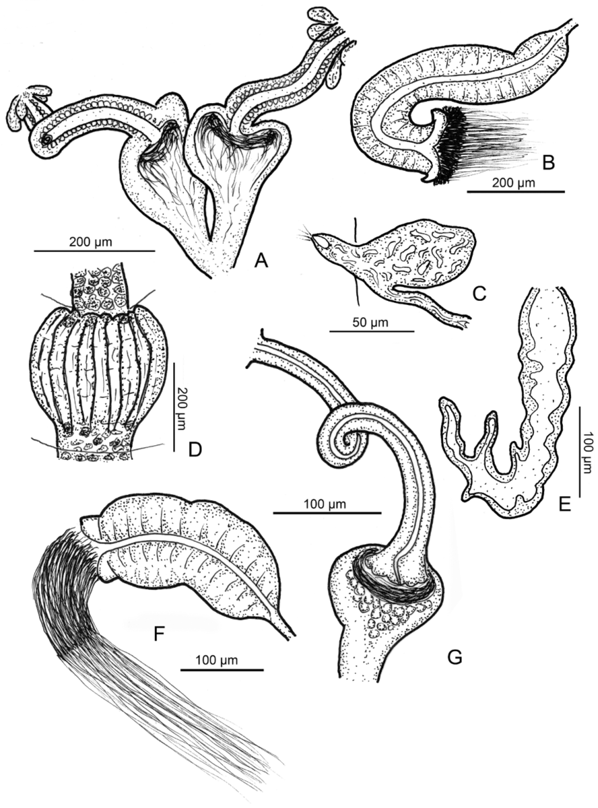
( Figures 1 View FIGURE 1 E–G, 2G–I, 4, 5)
Type material. Holotype. NIBRIV0000320518, slide No. 1114, adult specimen, stained whole mount. Type locality: site 1. College of Agriculture & Life Science, Chonbuk National University, Jeonju-si, Jeollabuk-do, Korea, 35º50'59.0"N 127º07'56.4"E, 55 m asl, soil and litter layers of woodland, leg.Y. Hong, 19.05.2014.
Paratypes. NIBRIV0000320519, NIBRIV0000320520, 2 adult specimens in 70 % ethanol from type locality, 19.05.2014. P. 105.1–105.11. 11 adult, fixed, stained whole mounts, slides No. 1003, 1109–1111, 1115–1117, 1123– 1125, 1174, from type locality. P.105.12. One adult, fixed, stained whole mount, slide No. 1101, site 7: Dongjinmyeon, Buan-gun, Jeollabuk-do, Korea, 35º46'04.6"N 126º43'14.8"E, 23 m asl, garden (planting garden tree), agronomical fields, leg. Y. Hong, 19.05.2014. P.105.13–105.14, slides No. 1167–1168, two fixed stained whole mounts from site 13: Gyeongju National Park Buddhist Center, Korea, 35º52'032"N 129º13'21"E, leg. Sándor Mahunka, 0 9.12.1990. P.105.15, six specimens in 70 % ethanol from site 1, P.105.16, six specimens in 70 % ethanol from site 7.
Further material examined. 8 specimens from sites 1, 6, and 7.
Etymology. Named after the shape of spermathecal ampulla ('sphaera' = sphere, Latin).
Diagnosis. The new species can be recognized by the following combination of characters: (1) large size (13– 21.5 mm long, 400–630 µm wide at clitellum in vivo, but the cuticle thin, <1 µm), segments 47–64; (2) maximum 4 (5) chaetae per bundle; (3) clitellum girdle-shaped, gland cells arranged in transverse rows, between the bursal (=penial) slits only granulocytes; (4) five preclitellar pairs of nephridia; (5) coelomo-mucocytes type c, lenticytes small and scarce; (6) chylus cells in XIII–XVI (2–3 segments); (7) seminal vesicle large; (8) subneural glands absent; (9) sperm funnel cylindrical, about 1/2 or 2/3 as long as body diameter, collar slightly narrower as funnel body; (10) spermathecae with very long ectal duct, without ectal glands, the spherical ampulla large (diameter 95– 150 µm, in vivo), separate openings into oesophagus.
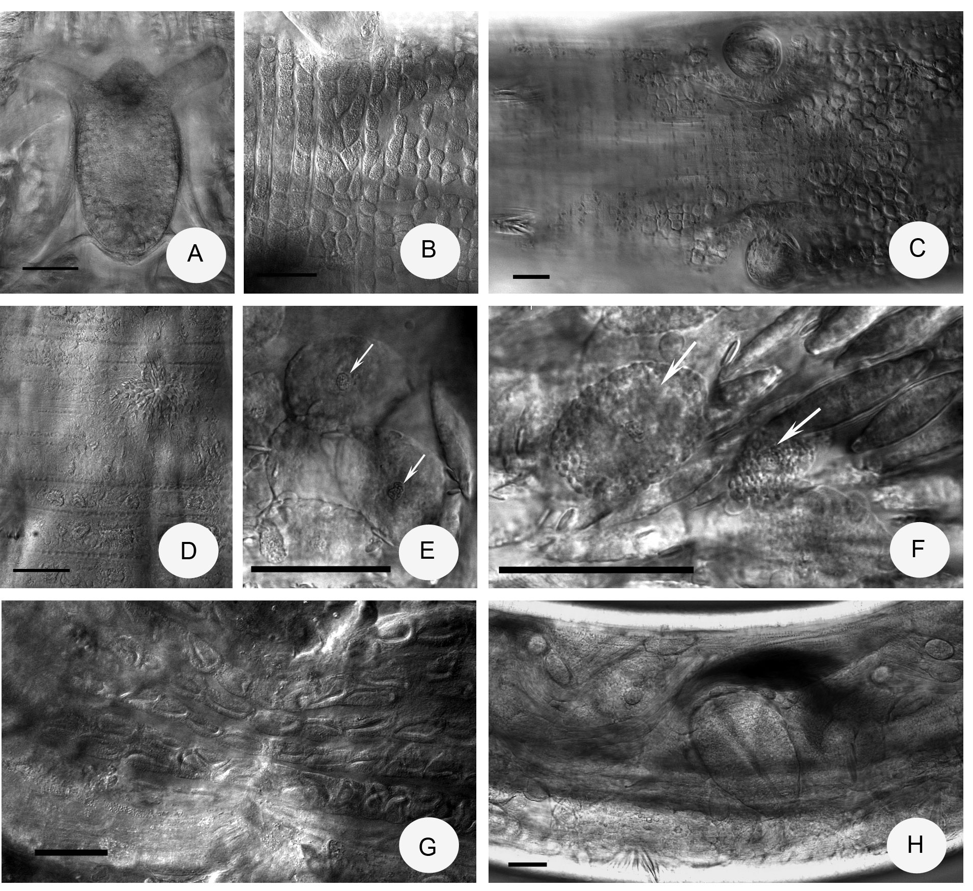
Description. Holotype 15.8 mm long, 410 µm wide at VIII and 420 µm at clitellum (fixed), segments 60. Body length of paratypes (11)– 13–21.5 mm, width 330–450 µm at VIII and 400–630 µm at clitellum, in vivo, length of fixed specimens 10–15.8 mm, width 420–580 µm at VIII and at clitellum, segments 47–64. Chaetal formula: (2)3,4 – 4,3,2: 3,4(5) – 4,3,(1)2. As in other Fridericia species, the chaetae within a bundle arranged in pairs with the outer pair and thicker than the inner pair (52– 63 x 5 Μm vs. 33– 35 x 3–4 Μm, preclitellar bundles). Lengths about the same in postclitellar segments but between XVIII–XX often only the two smaller chaetae present. Two chaetae per bundle from about XXI–XXVI; chaetae gradually increasing in size posteriad from 65 Μm to 90– 95 x 5 Μm in terminal segments. Detached chaetae often present in the coelom. Head pore at 0/I. Dorsal pores from VII. Epidermal gland cells arranged in 2–3 transverse rows per segments ( Fig. 4 View FIGURE 4 D). Clitellum in XII–1 /3XIII, girdle-shaped, hyalocytes and granulocytes arranged in dense rows dorsally ( Fig. 2 View FIGURE 2 G), between the bursal slits only granulocytes ( Fig. 4 View FIGURE 4 B,C). Body wall thick, 40–50 µm, cuticula thin (<1 µm).
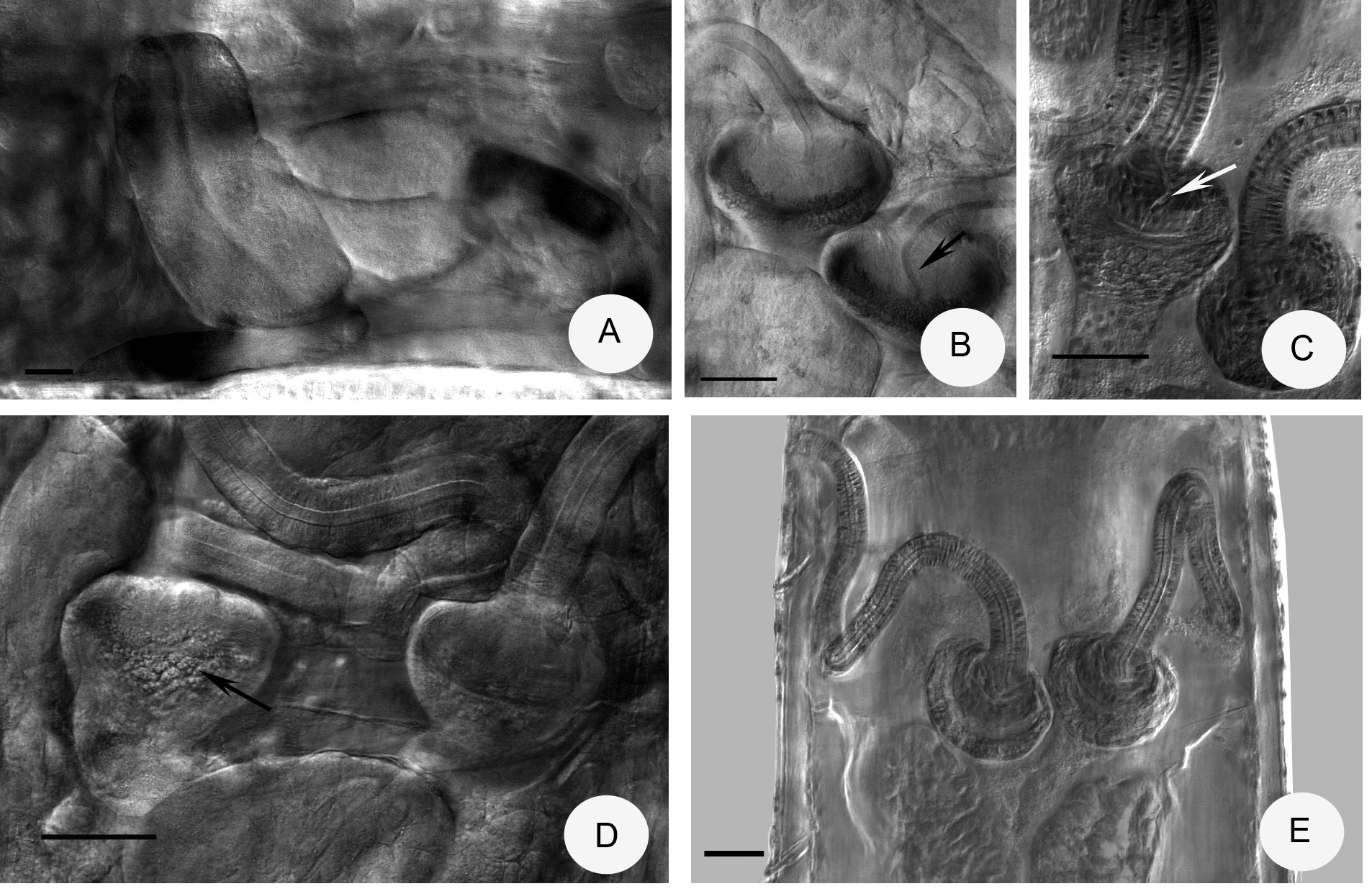
Brain egg-shaped ( Fig. 4 View FIGURE 4 A), 150–190 Μm long in vivo, about 2 times longer than wide. Oesophageal appendages ( Fig. 1 View FIGURE 1 E) with few short branches at the end. All pairs of pharyngeal glands united dorsally and with ventral lobes, lobes in VI longest. Chloragocytes from V. Dorsal vessel from XVII–XXIII (mostly XVIII–XX), blood colourless. Midgut pars tumida not seen. Five pairs of preclitellar nephridia from 6/7 to 10/11; length ratio anteseptale: postseptale 1: 1.2–1.3, midventral origin of efferent duct, anteseptale often brown in transmitted light. Coelomo-mucocytes with fine granular matrix and clearly visible nucleus, type c (sometimes a/c), length mostly 25–53 µm ( Fig. 4 View FIGURE 4 E), sometimes with coarser granulation ( Fig.4 View FIGURE 4 F), lenticytes small and scarce (4–8 Μm long, in vivo). Chylus cells ( Fig. 4 View FIGURE 4 G) between XIII–XVI, occupying 2–3 segments. Seminal vesicle large (X–XI or XI– XII). Sperm funnels cylindrical ( Figs. 1 View FIGURE 1 F, 2H, 4H, 5A), 200–370 µm long, in vivo and 1.5–2.7 times as long as wide. In fixed specimens the length of funnels 180–260 µm. Collar slightly narrower than funnel body. Spermatozoa long, length 400–600 µm, heads 150–170 µm, in vivo. Diameter of sperm ducts about 10 µm, in vivo. Male copulatory organs ( Fig. 4 View FIGURE 4 C) 100–220 µm long, 60–90 µm wide and 60–80 Μm high, in vivo (100–170, 50–90 and 40–90 µm, respectively fixed), the laterally bent bursal slits are longitudinal or H-shaped. No subneural glands. Ectal ducts of spermathecae very long, length 380–620 µm and width 24–35 µm, in vivo (300–500 µm long, and 20–25 µm wide, fixed), gland absent at the ectal opening ( Fig. 5 View FIGURE 5 E). Ampullae large, onion-shaped without diverticula, diameter 95–150 µm, in vivo (fixed 90–130 µm), ental bulbs (56–70 µm wide, in vivo), projecting into the lumina of ampullae, sperm in circles around the bulbs ( Figs. 1 View FIGURE 1 G, 2I, 5B). Ectal duct canal describing spiral loops inside the ental bulbs before entering the ampullae ( Figs. 1 View FIGURE 1 G, 2I, 5B,C), a characteristic trait similar to F. perrieri . Ampullae often with granular texture ( Fig. 5 View FIGURE 5 D). Distal and proximal parts of ampullae considerably set off by a constriction, the proximal part about 70–130 µm long, in vivo, separate openings into oesophagus. Spermathecae enlarged in one specimen: ectal ducts 621 µm long and 36 µm wide, diameter of ampulla 199 µm, ental bulb 90 µm wide, in vivo. One to three mature eggs at a time.
Distribution and habitat. In Korea, site 1, College of Agriculture & Life Science, Chonbuk National University, Jeonju-si, Jeollabuk-do, Korea, 35º50'59.0"N 127º07'56.4"E, 55 m asl, woodland. Site 7, Dongjinmyeon, Buan-gun, Jeollabuk-do, 35º46'04.6"N 126º43'14.8"E, 23 m asl, garden (planting garden tree), agronomical fields. Site 11, Sulchon-myeon, Muju-gun, Jeollabuk-do 35º59'28.7"N 127º50'46.8"E, 544 m asl, mixed forest. Site 1, collected in 2007, Soil and litter layers in College of Agriculture & Life Science, Chonbuk National University, Jeonju-si, Jeollabuk-do 35°50'51.1"N 127°08'0.3"E, maple trees cultivation of experimental farm (= Fridericia sp. 1 in Dózsa-Farkas & Hong 2010, Table 1). Site 13, Gyeongju National Park Buddhist Center 35º52'032"N 129º13'21"E.
Differential diagnosis. Among the previously described Fridericia species with more than 40 segments, onion-shaped spermathecal ampulla without diverticula and separate openings into oesophagus, five species ( F. paratalassia Schmelz, 2002 , F. peregrinabunda Michaelsen, 1913 , F. seoraksani Christensen & Dózsa-Farkas, 2012 , F. callosa ( Eisen, 1878) and F. tuberosa Rota, 1995 ) are similar to the new species ( Schmelz 2003; Christensen & Dózsa-Farkas 1999; Rota 1995; Christensen & Dózsa-Farkas 2012). The main differences are as follows: in F. parathalassia , the spermathecal ampullae are similarly large [70–100 Μm ( Schmelz 2003), 100–200 Μm ( Nielsen & Christensen 1959)], but there are only four pairs of preclitellar nephridia and subneural glands are present. The main differences of F. peregrinabunda to the new species are only two chaetae in the bundles and a smaller diameter of the spermathecal ampullae (60–70 Μm). These two species were found also together with F. seoraksani . F. seoraksani can be easily distinguished from F. sphaerica sp. n. by the following characters: by their smaller size (7.2–14.4 mm long, in vivo and 37–45 segments), by the smaller diameter of spermathecal ampulla (50–60 Μm) and by the smaller sperm funnel (160–190 Μm long, in vivo). F. callosa (as redescribed in Schmelz 2003) and the new species resemble each other in more traits (e.g. similar size, form of spermatheca, long spermatozoa, large seminal vesicle, the locality of chylus cells), but F. callosa has a thick cuticle (3–5 Μm, vs. less than 1 Μm in the new species) and never 4 chaetae in the dorso-lateral bundles. Moreover the chaetae are often absent in several bundles, the third pharyngeal glands are not united dorsally and the oesophageal appendages are much branched. In F. tuberosa diverticula-like protrusions may appear on the spermathecal ampullae, spermathecal ectal glands and subneural glands are present, and the coelomocytes are of type b.

No known copyright restrictions apply. See Agosti, D., Egloff, W., 2009. Taxonomic information exchange and copyright: the Plazi approach. BMC Research Notes 2009, 2:53 for further explanation.