
Henlea magnaampullacea, Dózsa-Farkas, Klára, Felföldi, Tamás & Hong, Yong, 2015
|
publication ID |
https://doi.org/10.11646/zootaxa.4006.1.9 |
|
publication LSID |
lsid:zoobank.org:pub:E987B43A-E54A-4F64-9829-0B8EBE457E03 |
|
DOI |
https://doi.org/10.5281/zenodo.6100530 |
|
persistent identifier |
https://treatment.plazi.org/id/304F8793-6817-FFDA-FF3B-FDF9FD81AB4E |
|
treatment provided by |
Plazi |
|
scientific name |
Henlea magnaampullacea |
| status |
sp. nov. |
Henlea magnaampullacea View in CoL sp. n.
( Figures 1 View FIGURE 1 A–D, 2A–F, 3)
Type material. Holotype. NIBRIV0000320515, slide No. 1126, adult whole mount stained with borax-carmine. Type locality: Site 1: College of Agriculture & Life Science, Chonbuk National University, Jeonju-si, Jeollabuk-do, Korea, 35º50'59.0"N 127º07'56.4"E, 55 m asl, soil and litter layers of woodland, leg.Y. Hong, 19.05.2014.
Paratypes. NIBRIV0000320516, NIBRIV0000320517, 2 adult specimens from type locality, in 70 % ethanol. P.103.1–4, slide No. 1005, 1121, 1122, 1127. Four adult whole mounts stained with borax-carmine from the type locality, 0 3.04.2014 and 19.05.2014. P.103.5–103.10. slide No. 1098, 1119, 1120, 1128–1130. Six specimens from site 7, Dongjin-myeon, Buan-gun, Jeollabuk-do, Korea, 35º46'04.6"N 126º43'14.8"E, 23 m asl, soil of agronomical fields, leg. Y. Hong, 19.05.2014. P.103.11. Six specimens in 70 % ethanol from type locality.
Further material examined. 12 and 10 specimens from sites 7 and 1, respectively.
Etymology. Named after the large spermathecal ampulla (magna = large, Latin).
Diagnosis. The new species can be recognized by the following combination of characters: (1) medium-sized stout worms ( 11–16 mm long and 600–800 Μm wide at clitellum in vivo, segments 47–58; (2) maximum 6 chaetae per bundle; (3) clitellum girdle-shaped: gland cells small in reticulate pattern dorsally and ventrally; (4) six preclitellar pairs of nephridia; (5) canals of the intestinal diverticula arranged longitudinally in VIII, dorsal vessel origin in IX; (6) coelomocytes large, rounded brownish, thickened at cell periphery; (7) seminal vesicle large; (8) sperm funnel large, cylindrical, collar wider as funnel body; (9) spermathecae with very large pear-shaped ampullae and without diverticula.
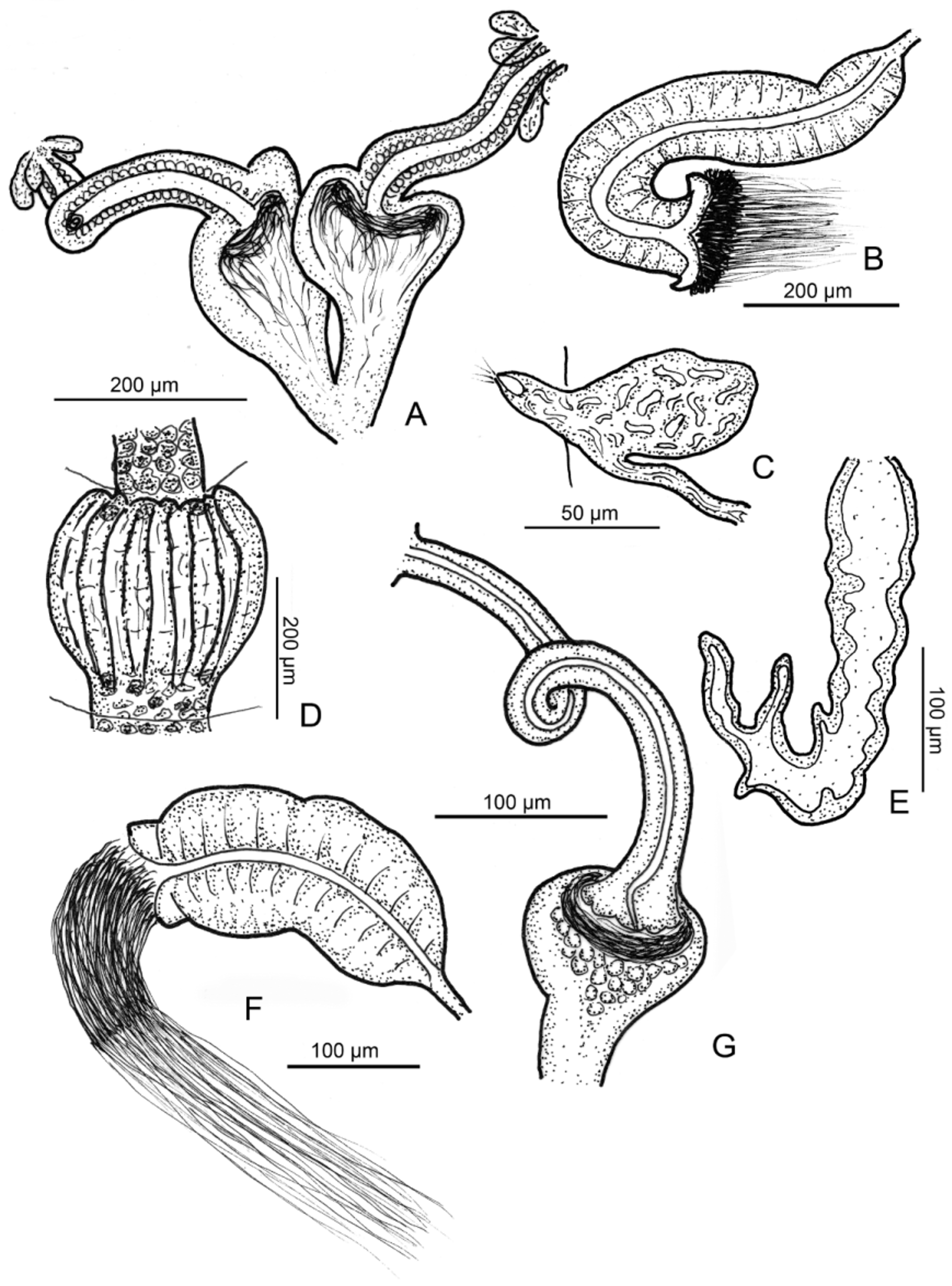
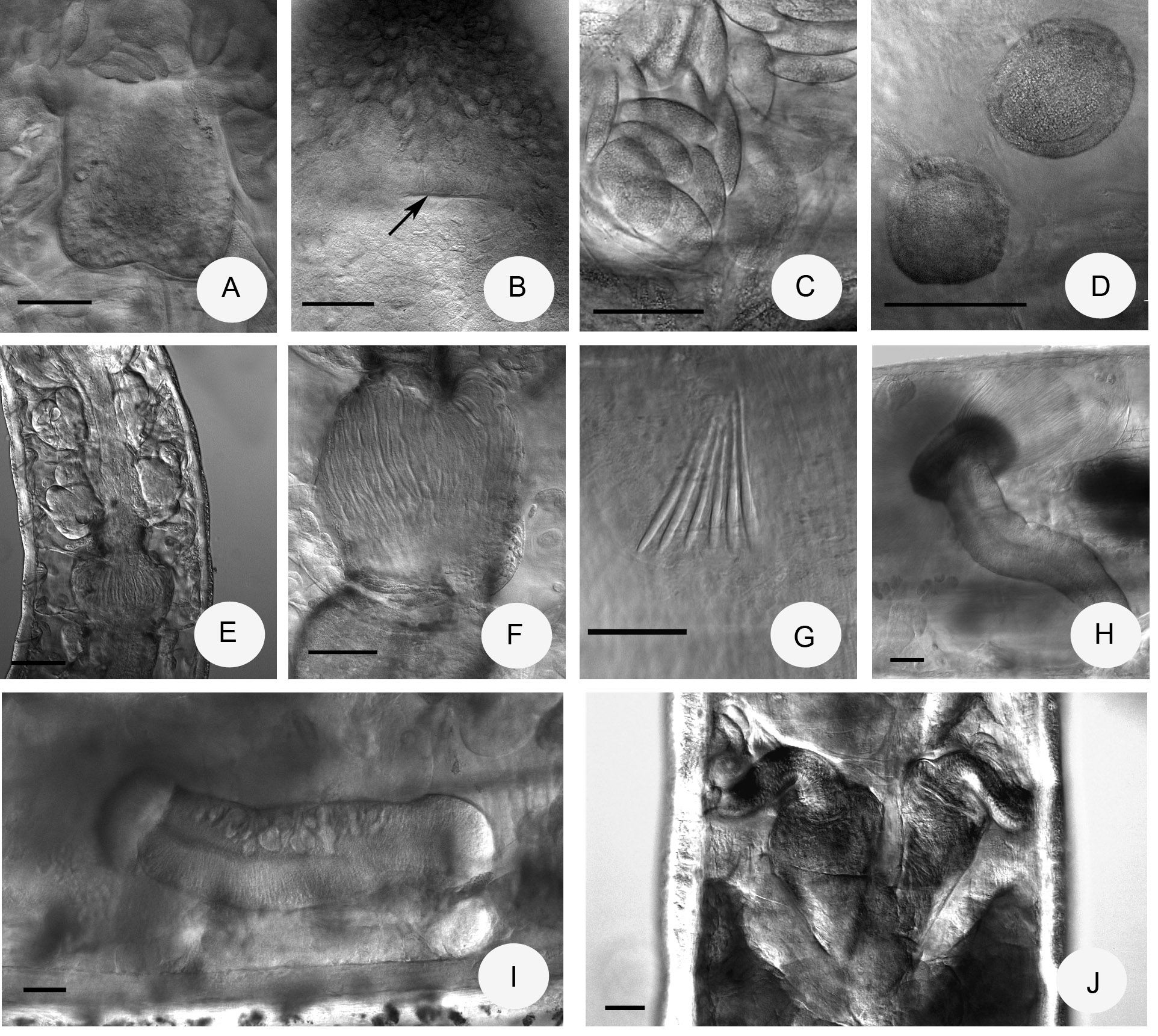
Description. Holotype 16.2 mm long, 590 µm wide at VIII and 620 µm at clitellum, in vivo ( 11.1 mm long, 490 µm wide at VIII and 660 µm at clitellum, fixed), 52 segments. Body length of paratypes 11–16 mm, width 520–700 µm at VIII and 530–800 µm at clitellum, in vivo. Length of fixed specimens 8.7–13.5 mm, width 400–670 µm at VIII and 400–730 µm at clitellum. Segments 47–58. Chaetal formula: 2,3,4,5 – 4,5,(3,2): 2,3,4,5,6 – 4,5,6,3. Chaetae straight, mostly unequal in size within a bundle; the largest 90–100 Μm long and 5 Μm wide, the smallest 63–70 Μm long and 4 Μm wide. Chaetae in XII absent. Head pore at 0/I, large transverse slit ( Fig. 3 View FIGURE 3 B). Epidermal gland cells arranged in 3–4 transverse rows per segment. Many glands also on the prostomium dorsally ( Fig. 3 View FIGURE 3 B). Clitellum girdle-shaped in XII–1 /2XIII, gland cells small in reticulate pattern ( Fig. 2 View FIGURE 2 C), also between the bursal slits ( Fig. 2 View FIGURE 2 D). Thickness of body wall about 40–60 µm, cuticle about 1 µm, in vivo. Brain concave posteriorly, about 150 Μm long, in vivo and slightly longer than wide ( Fig. 3 View FIGURE 3 A). Oesophagus in VI with a pattern of transverse streaks and one pair dorso-laterally and one pair ventro-laterally lobes of oesophageal appendages.
Pharyngeal glands all separate dorsally, the second and third pairs with ventral lobes ( Fig. 3 View FIGURE 3 E.). Chloragocytes from IV about 15–40 Μm long, in vivo (fixed 13–15 Μm). Dorsal vessel from IX, large heart-like pulsating expansion from IX to VII ( Fig. 2 View FIGURE 2 A), blood colourless. Intestinal diverticula forming a ring around intestine in VIII, consisting of 4 spherical diverticula, which unite proximally ( Figs. 2 View FIGURE 2 A,B, 3E). Canals of the diverticula arranged longitudinally and not including large hollows; especially well visible in subadult or juvenile specimens ( Fig. 3 View FIGURE 3 F). Intestinal epithelium behind clitellum with tall, hyaline type of cells ventrally and laterally from XXIV–XXV to XXXI–XXXII ( type 2 after Rota et al. 1998). Six pairs of preclitellar nephridia from 5/6 to 10/11 (in one case from 4/5–10/11, absent at 5/6), anteseptale small, efferent duct origin posteroventrally ( Fig. 1 View FIGURE 1 C). Coelomocytes large, rounded or ellipsoid ( Fig. 3 View FIGURE 3 C), with fine brown granula in the cytoplasma (coelomocyte aggregations dark brown in transmitted light), and a peripherally thickened cell border, distinct at high magnification ( Fig. 3 View FIGURE 3 D) (length 35– 60 Μm, in vivo, 20–30 µm, fixed).
Seminal vesicle large in XI or XI–XII. Sperm funnels cylindrical, large ( Figs. 1 View FIGURE 1 B, 3H–I), about 350–600 µm long and 4–6 times as long as wide, in vivo, about ¾ as long as body diameter. Funnel length in fixed specimens 230–550 µm, 2.5–4 times longer than wide. Collar wider as funnel body. Spermatozoa about 140–170 µm long, heads 40–70 µm, in vivo. Diameter of sperm ducts 12–13 µm, in vivo, 10–13 µm, fixed. Male copulatory organs ( Fig. 2 View FIGURE 2 D) 120–190 µm long, 100–145 µm wide and 80–110 Μm high, fixed, bursal slits H-shaped, modiolus well developed. Subneural glands absent. Spermathecae ( Figs. 1 View FIGURE 1 A, 2E–F, 3J) very prominent, large; pear-shaped ampullae without diverticula, 90–150 µm wide, up to 200 µm when full with sperm. Ectal duct about 160–300 µm long and 40–60 (80) µm wide, slightly tapering distally, canal wide (13–15 µm, in vivo), (170–260 µm long, 45–60 µm wide, diameter of canal 10–12 µm in fixed specimens). Ampullae about 250–280 µm long in vivo, merging entally and with joint opening into oesophagus in VI. At the orifice of spermathecal ducts 3–4 large glands (40–80 µm long, in vivo). 2–3 mature eggs at a time.
Distribution and habitat. In Korea site 1: College of Agriculture & Life Science, Chonbuk National University, Jeonju-si, Jeollabuk-do 35º50'59.0"N 127º07'56.4"E, 55 m asl), woodland, sites 6 and 7, Dongjinmyeon, Buan-gun, Jeollabuk-do 35º46'04.6"N 126º43'14.8"E, 23 m asl and 35º46'04.2" 126º43'15.6"E, 20 m a s l, agronomical fields, and planting garden tree.
Differential diagnosis. Henlea magnaampullacea sp. n. and 6 other Henlea species are characterized by intestinal diverticula with multitubular substructure in VIII, and the dorsal vessel origin in IX. Among these species, H. ventriculosa (d' Udekem, 1854), H. jutlandica Nielsen & Christensen, 1959 , H. andreae Rodriguez & Giani, 1986 , H. groenlandica Černosvitov, 1929 augm. Christensen & Dózsa-Farkas, 2006 and H. conchifera Christensen & Dózsa-Farkas, 1999 differ from the new species in the spermathecal ampullae which are only slightly wider than the ectal ducts ( Schmelz & Collado 2010; Christensen & Dózsa-Farkas 1999, 2006). H. irkutensis Burov, 1929 is much larger (60–90 segments, 23.6–55 mm) ( Burov 1929). H. magnaampullacea sp. n. is most similar to Henlea ochracea (Eisen, 1978) augm. Nurminen, 1973, because both are similarly large, have up to 6 chaetae per bundle, large sperm funnels and more robust spermathecae, but the collar of sperm funnels is not so high and bent backwards in the new species, and the spermathecal ampullae are abruptly widened, about 2–3 times wider than the diameter of ectal ducts, whereas in H ochracea the ampullae are about 1.5 times wider than the ectal ducts and the duct widens gradually into the ampullae (Dózsa-Farkas, personal observation, see Christensen & Dózsa-Farkas 1999, Fig 16). Based on the phylogenetic analysis ( Fig. 14 View FIGURE 14 ), individuals from the new species were separated from other Henlea species collected in Korea, and were also distantly related to H. ochracea individuals from Alaska (identified morphologically by Dózsa-Farkas). Therefore molecular taxonomical results supported the description of a new Henlea species.


No known copyright restrictions apply. See Agosti, D., Egloff, W., 2009. Taxonomic information exchange and copyright: the Plazi approach. BMC Research Notes 2009, 2:53 for further explanation.
|
Kingdom |
|
|
Phylum |
|
|
Class |
|
|
SubClass |
Oligochaeta |
|
Order |
|
|
Family |
|
|
Genus |