
Enchytraeus sp.
|
publication ID |
https://doi.org/ 10.1080/00222933.2022.2140085 |
|
DOI |
https://doi.org/10.5281/zenodo.7428372 |
|
persistent identifier |
https://treatment.plazi.org/id/3A40CF59-FF8C-FFC4-B3CF-43C1FE34DD70 |
|
treatment provided by |
Plazi |
|
scientific name |
Enchytraeus sp. |
| status |
|
Enchytraeus sp. , buchholzi group
( Enchytraeus buchholzi View in CoL ; Vejdovský, 1879)
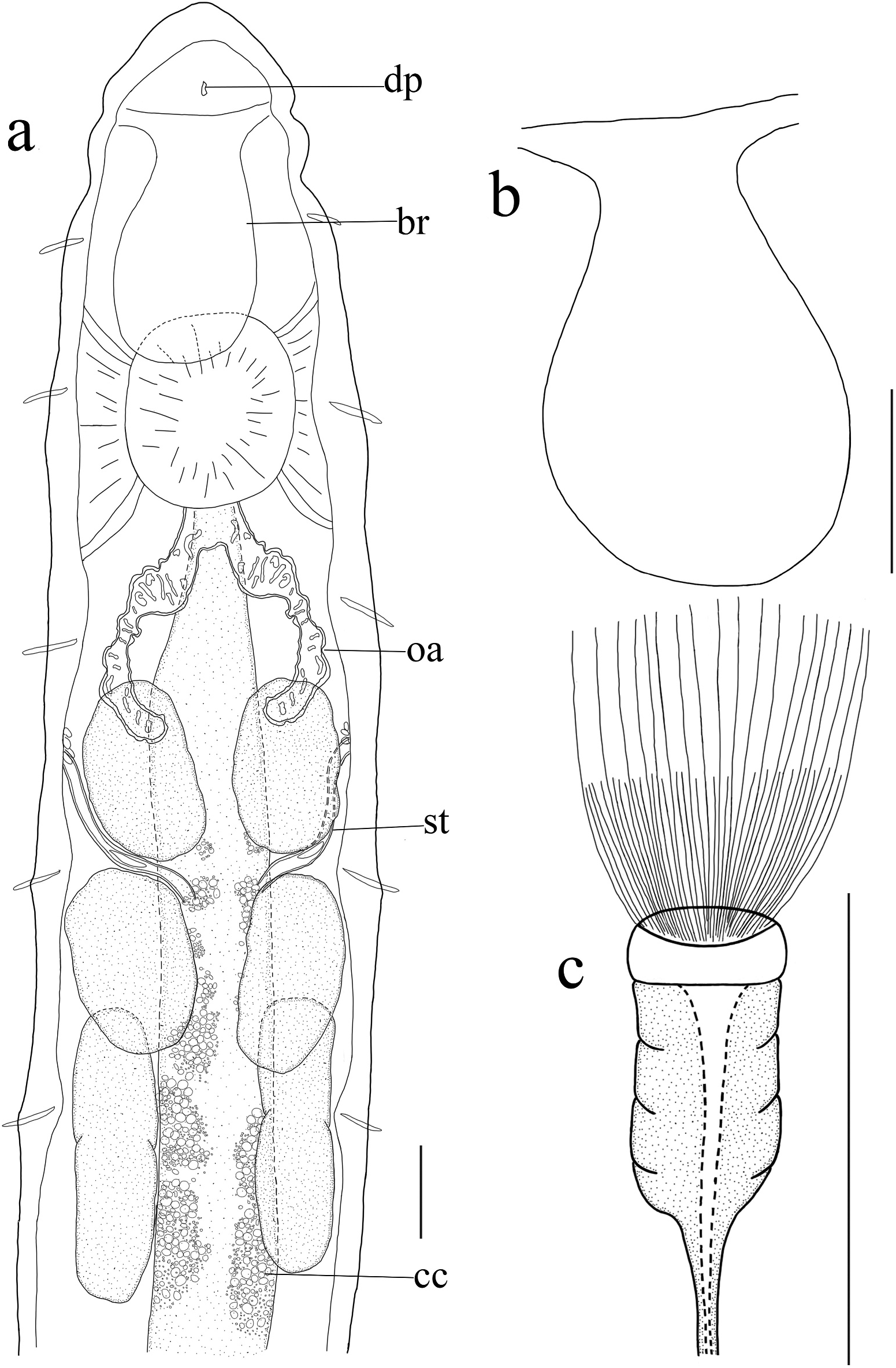
( Figures 11 View Figure 11 , 12 View Figure 12 )
= Enchytraeus florentinus Bell, 1947 View in CoL
= Enchytraeus polonicus Dumnicka, 1977 View in CoL
Enchytraeus buchholzi Vejdovsky, 1879 View in CoL ; Nielsen and Christensen, 1959; Dash 1970; Chalupský 1992; Rota and Healy 1994; Rota 1995; Schmelz et al. 2005; Schmelz and Collado 2010.
Material investigated
CJJ 95, one submature specimen from site 7, whole worm used for DNA extraction, preserved as total DNA. One submature specimen from site H and two submature specimens from site 7, preserved in 75% ethanol.
Description
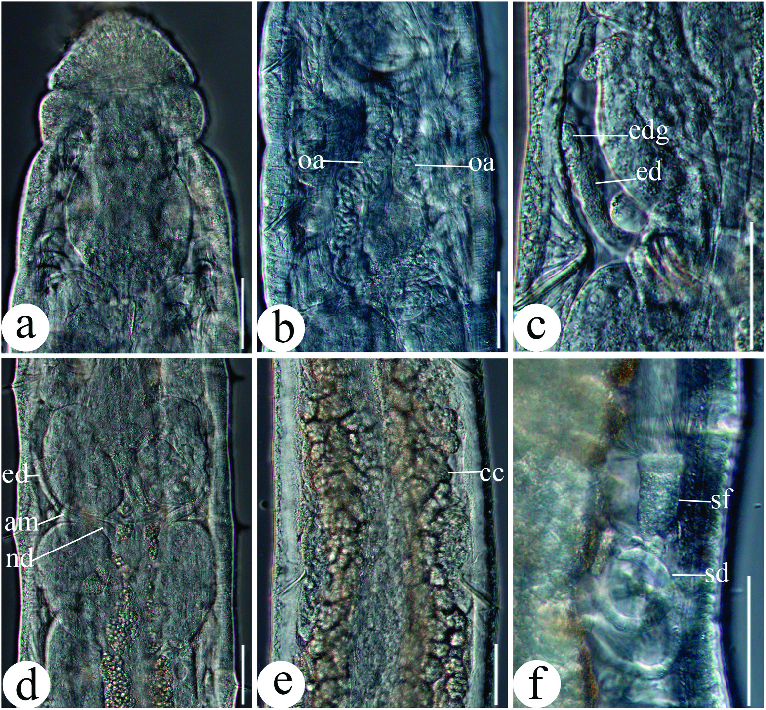
Small worms, yellowish white in stereomicroscope. Length ca. 3.3–5.0 mm (in vivo), 1.8– 2.8 mm, wide 122.5–180.0 μm in segment VII and 125–200 μm in clitellum. Number of segments 24–25. Chaetae straight, with ental hook, formula: 2–2, 3: (2) 3–3 (2). Head pore at 0/1, transverse slit ( Figure 11a View Figure 11 ). Epidermal gland cells grey, inconspicuous, 4–5 transverse rows per segment. Clitellum in XII–1/2XIII, saddle-shaped, hardly elevated, not fully developed (our specimens are all submature). Body wall ca. 15–26 μm thick (in vivo).
Brain trapezoidal, truncate anteriorly and round posteriorly, ca. 124 μm long, 46 μm wide anteriorly and 86 μm wide posteriorly (in vivo) ( Figures 11b View Figure 11 , 12a View Figure 12 ). Oesophageal appendages a pair of unbranched tubes with common opening leading dorsally into oesophagus in III, behind pharyngeal pad, tubes extending into IV with meandering cannel ( Figures 11a View Figure 11 , 12b View Figure 12 ). All three pairs of pharyngeal glands separated dorsally, pharyngeal gland in VI elongate, with a small constriction at septum ( Figures 11a View Figure 11 , 12d View Figure 12 ). Dorsal vessel from XII–XIII, blood colourless. Intestinal diverticula absent, gut widening gradually. Chloragocytes well developed, large and with retractile globules of differing size, present from V and forming a dense layer from segment VII onward, absent in XII ( Figures 11a View Figure 11 , 12e View Figure 12 ). Four pairs of preclitellar nephridia from 6/7–9/10, anteseptale with funnel only. Coelomocytes one type, mucocytes, with refractile granula.
Seminal vesicle absent. Spermatozoa sparse, ca. 37 μm long and head ca. 15 μm long (in vivo) ( Figures 11c View Figure 11 , 12f View Figure 12 ). Sperm funnel cylindrical, small, length about 1/4 body diameter (ca. 33 μm long and 17 μm wide, in vivo), with distinct midline, collar little wider than funnel body ( Figures 11c View Figure 11 , 12f View Figure 12 ). Vas deferens long, in tangled coils ventro-laterally, diameter ca. 6 µm (in vivo) ( Figure 12f View Figure 12 ). Male copulatory organ small, globular, not floppy. Spermathecae short, confined to V; ectal pores lateral at 4/5, surrounded by several glands at each pore, glands of varying size, sometimes stretching along ectal duct; ectal ducts ca. 52 μm long and 9 μm wide (in vivo); ampullae spindle-shaped, ca. 43 μm long (in vivo), almost with same diameter as ectal ducts; ental ducts short and narrow; spermathecae attached to oesophagus separately ( Figures 11a View Figure 11 , 12c, d View Figure 12 ). No mature eggs observed.
Remarks
Using the key for European species in Schmelz and Collado (2010), specimens key out as E. buchholzi Vejdovský, 1878 , based on the following characters: chaetal formula 2–2, 3: 3–3, oesophageal appendage with meandering canals, four pairs preclitellar nephridia, in 6/7 to 9/10, clitellum saddle-shaped, testis sac, sperm funnel and male glandular bulb small, spermathecal ectal glands covering only ectal part of ectal duct, ampulla small, ental duct present. Enchytraeus buchholzi is a species complex ( Rota and Healy 1994; Rota 1995; Schmelz et al. 2005). Descriptions of this species sensu lato usually distinguish two forms based on the texture of the coelomocytes: with or without refractile granula ( Schmelz et al. 2005). Specimens from Mount Fanjing have coelomocytes with refractile granules. All specimens are submature, i.e. without eggs or a fully developed clitellum. This record documents the presence of E. buchholzi sensu lato at Mt. Fanjing. It is unknown whether the specimens underlying this account belong to one or more species. In China, E. buchholzi has also been recorded fromMount Changbai, Jilin Province (J.J. Chen, unpublished data). The K2P distances based on COI sequences also confirm the complexity of E. buchholzi . The genetic distance between our specimens and E. buchholzi - Russia ( Erséus et al. 2010), E. buchholzi - Switzerland ( Vivien et al. 2015) and E. buchholzi - India (GenBank accession number: KX348269 View Materials ) are 18.7%, 20.7% and 19.9%, respectively. However, we failed to match molecular differences to morphological differences because of the lack of morphological descriptions.
No known copyright restrictions apply. See Agosti, D., Egloff, W., 2009. Taxonomic information exchange and copyright: the Plazi approach. BMC Research Notes 2009, 2:53 for further explanation.
|
Kingdom |
|
|
Phylum |
|
|
Class |
|
|
Order |
|
|
Family |
|
|
Genus |
Enchytraeus sp.
| Chen, Juanjuan, Schmelz, Rüdiger M., Zhang, Zuxu & Xie, Zhicai 2022 |
Enchytraeus polonicus
| Dumnicka 1977 |
Enchytraeus florentinus
| Bell 1947 |
Enchytraeus buchholzi
| Vejdovsky 1879 |
Enchytraeus buchholzi
| Vejdovsky 1879 |