
Guaranidrilus andreolii, Schmelz, Rüdiger M., Collado, Rut & Römbke, Jörg, 2011
|
publication ID |
https://doi.org/ 10.5281/zenodo.203260 |
|
DOI |
https://doi.org/10.5281/zenodo.5611821 |
|
persistent identifier |
https://treatment.plazi.org/id/520EAA7D-D64D-4503-FF40-8094FAAEFE76 |
|
treatment provided by |
Plazi |
|
scientific name |
Guaranidrilus andreolii |
| status |
|
Guaranidrilus View in CoL , descriptions of new species
Guaranidrilus andreolii sp. nov. ( Figs 1 View FIGURE 1 , 8 View FIGURE 8 A)
Holotype. MZUSP 1217, stained whole mount, Antonina, Cachoeira , 25°15'22.1''S, 48°40'15.8''W, young secondary forest on Cambisol [site 16], 30 m a.s.l., litter / top soil (0–5 cm), Mar 2004, leg. R.M. Schmelz.
Paratypes. MZUSP, 32 specimens, stained whole mounts:
MZUSP 1218: 7 specimens, Antonina, Cachoeira , 25°15’11“S, 48°40’22“W and 25°14’38“S, 8°40’10“W, medium secondary and old-growth forest on Cambisol [sites 10, 12, 13], 60 / 140 m a.s.l., litter / top soil (0–5 cm), May 2003, leg. R.M. Schmelz, J. Römbke.
MZUSP 1219, 21 specimens, same data as holotype.
MZUSP 1220, 4 specimens. Antonina, Cachoeira , young secondary forest on Gleysol [site 27], 25°19'32"S, 48°41'24"W, 20 m a.s.l. Litter / top soil (0–5 cm), Oct 2004, leg. B. Förster, R.M. Schmelz.
UFPR, 19 specimens, ethanol-preserved; Guarequeçaba, Itaqui, medium and old-growth forest:
UFPR OL-1, 3 specimens, 25°14'45.3''S, 48°30'19.4''W, and 25°14'51.1''S, 48°29'31,4''W, 8–27 m a.s.l., medium forest [sites 38, 39], Sep. 2007, leg. P. Heine, R. M. Schmelz.
UFPR OL-2, 5 specimens, 25°15'32.7''S, 48°30'31.9''W, 31 m a.s.l., old-growth forest [site 41], Sep 2007, leg. P. Heine, R.M. Schmelz.
UFPR OL-3, 1 specimen, 25°16'23.7''S, 48°29'13.7''W, 20 m a.s.l., old-growth forest [site 42], Oct 2007, leg. P. Heine, R.M. Schmelz.
UFPR OL-4, 10 specimens, 25°14'51.1''S, 48°29'31.4''W, 8 m a.s.l., medium forest [site 39], Jan. 2008, leg. R.M. Schmelz, J. Römbke.
Additional material. Seven specimens from forest localities in the reserves Rio Cachoeira and Serra do Itaqui, examined in vivo, not preserved.
Etymology. Named in honour of Marcos Andreoli, administrator of the SPVS reserve Serra do Itaqui.
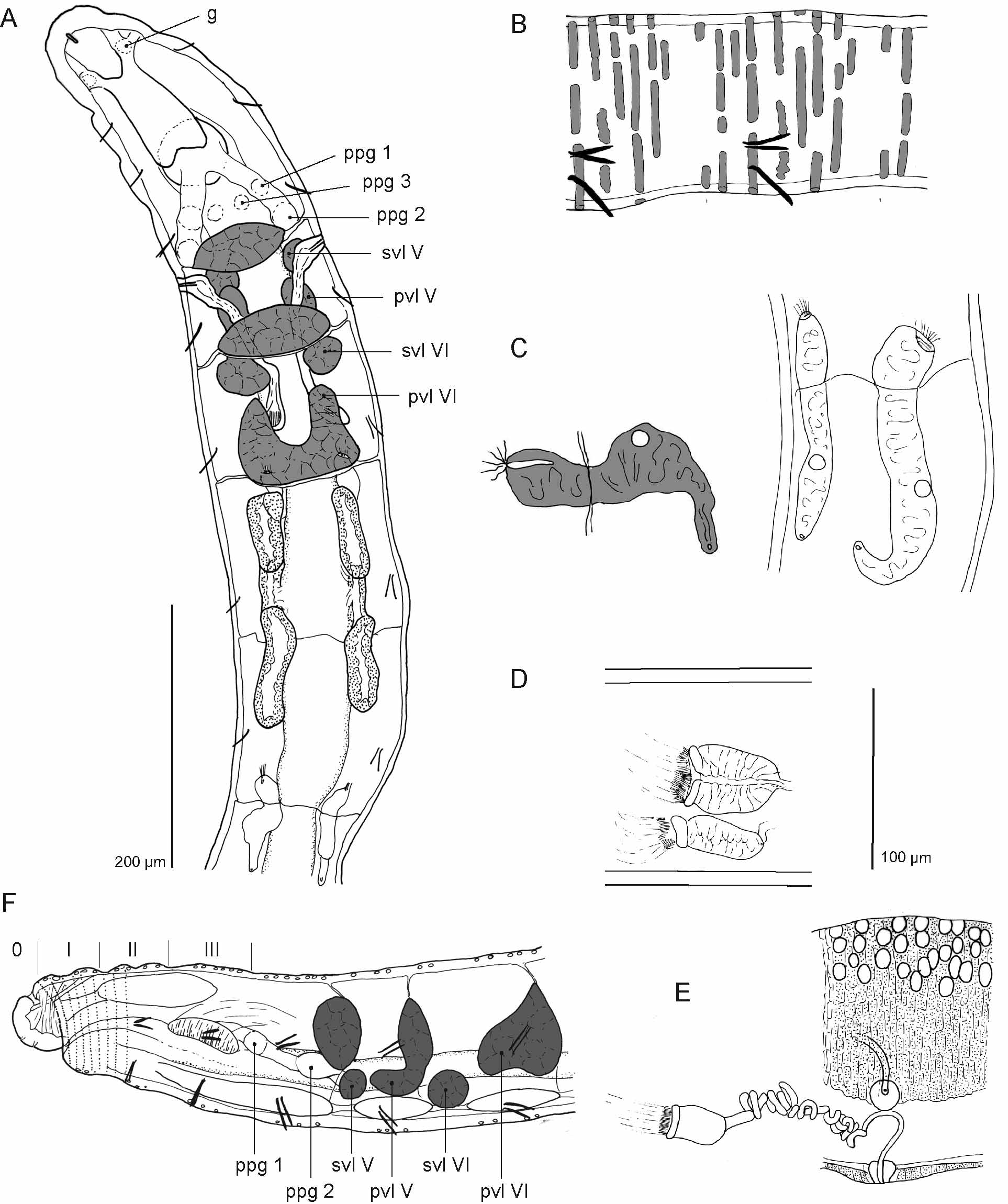
Description. Body dimensions. Live length 4–7 mm, diameter 0.1 mm, up to 0.125 mm at XII. Thin worms (length/width ratio up to 70) with strong coiling body movements. Segment number 35–40. Chaetae straight distally, in whole mounts sometimes bent in same direction as ental hook (fixation artefact?). Terminal chaetae not or only slightly longer than anterior chaetae, but distinctly thicker. e.g., 38: 2.5 µm anteriorly, 40: 3.5 µm posteriorly. Chaetae from XIII on much smaller, e.g. 15: 1.3 µm; increasing in size caudad. Epidermal gland cells ( Fig. 1 View FIGURE 1 B) conspicuous, pale or brownish, not strongly brown or yellow, transversely elongate and undulating; 7–9 rows per segment dorsally in anterior and posterior segments; fewer cells and fewer rows ventrally and in mid-body segments. Clitellum ( Fig. 1 View FIGURE 1 E) short and saddle-shaped, extension less than 1.5 segment lengths with posterior end anteriorly of level of chaetae of XIII, no clitellum ventrally; dorsally granulocytes and hyalocytes alternating, hyalocytes larger, latero-ventrally only granulocytes, here in ca. 25 regular and narrow rows (ca. 31 rows with border cells included); dorsally rows dense or indefinite. Head pore on prostomium mid-dorsally.
Prostomium without inner papillae. Body wall and cuticle thin, longitudinal muscle layer well-developed, epidermis and ring muscles forming oner layer of alternating transverse rings. Septa slightly thickened anteriorly, not or only slightly pushed backwards. Brain ( Fig. 1 View FIGURE 1 A) about 2x as long as wide, with anterior and posterior incision, sides converging anteriad. A pair of small prostomial ganglia present. Ventral nerve cord with suboesophageal ganglion in II–IV and segmental ganglia from V on. Pharyngeal glands ( Fig. 1 View FIGURE 1 A,F) in IV, V, VI: unpaired dorsal lobes; primary ventral lobes ( Fig. 1 View FIGURE 1 A, "pvl")in V and VI, largest in VI, merging into dorsal lobe; glands in VI U-shaped in dorsal view and roughly triangular in lateral view; secondary ventral lobes ( Fig. 1 View FIGURE 1 A, "svl") in V and VI, small, globular, often with slight superficial lobes. Oesophageal appendages absent. Intestinal diverticula ( Fig. 1 View FIGURE 1 A) at VII, in VII-1 / 2VIII, or intermediates, often enlarged at both ends, and folded downwards or inwards. Walls strongly textured, not hyaline. Dorsal blood vessel from X. Preclitellar nephridia at 6/7, 8/9, and 9/10, absent at 7/8. Postseptale bent upwards in the middle, with a dorsal vesicle, merging into efferent duct which is shorter than postseptale; terminal vesicle absent ( Fig. 1 View FIGURE 1 C left). Postclitellar nephridia ( Fig. 1 View FIGURE 1 C right) elongate. Coelomocytes ca. 22 µm long (ca. 20 µm in fixed specimens), oval, flattened, with fine and regular granulation, pale or with a light-brown tint; cells numerous, filling out body cavity in several segments, aggregations often opaque, brownish, but not especially dark.
Seminal vesicle present as two small sac-like dorsal invaginations of septum 10/11, directed anteriorly and containing developing sperm. Spermatozoa ca. 25 µm long, heads 13 µm long. Sperm funnel ( Fig. 1 View FIGURE 1 D,E) small, less than half as long as body diameter (ca. 40–50 µm), ca 1.5x as long as wide, barrel-shaped, with coarse and irregular granulation, flattened (e.g., 40 µm wide in top view, 20 µm wide in side view, length 50 µm), collar about as wide as funnel body. Vas deferens ( Fig. 1 View FIGURE 1 E) 5–6 µm wide, slightly wider near sperm funnel, in dense or loose irregular coils. Male copulatory organ ( Fig. 1 View FIGURE 1 E) small and inconspicuous, in line with ventral chaetae, protruded in some whole mounts as a papilla. Male pores on body surface, surrounded by small male glands (diameter ca. 15 µm) completely embedded in thickened body wall. Copulatory body muscles sparse. Spermatheca ( Fig. 1 View FIGURE 1 A) small, extending into VI, rarely VII; widened distally and proximally, narrow in the middle. One mature egg at a time, extending over 2 segments. Accessory glands absent.
Habitat. G. andreolii was found in young and medium secondary forests and in old-growth forests. It was absent in grazed and recently abandoned pastures and at agroforestry sites.
Remarks. The most diagnostic trait of G. andreolii is (1) the distribution pattern of preclitellar nephridia: present at 6/7, 8/9 and 9/10, but absent at 7/8. The same pattern is present in Achaeta neotropica Černosvitov, 1937a (Schmelz et al. 2008) but has so far not been described in Guaranidrilus . Another conspicuous trait is (2) the bending of the intestinal diverticula, present anteriorly and posteriorly. The bend may be downwards, inwards, or outwards, but it is not well-distinguished in all specimens. Further traits, diagnostic in combination: (3) about 35–40 segments, (4) chaetae not distinctly enlarged in terminal (= caudal) segments, (5) epidermal gland cells in 7–9 rows, conspicuous but not strongly brown, slightly undulating, (6) dorsal blood vessel from X, (7) clitellum saddleshaped, (8) other sexual organs small, inconspicuous. G. andreolii is similar to G. m a rq u e s i sp. nov., described in this paper, see the comparison below. Four other species of Guaranidrilus are described that lack oesophageal appendages; some of their differences with respect to G. andreolii are as follows.
G. europaeus Healy, 1979 : segment number less than 30, chaetae only 20 µm long, epidermal gland cells indistinct, in 3–5 rows per segment, dorsal blood vessel from XII or XIII.
G. f i n n i Christoffersen, 1977: segment number near 50, preclitellar nephridia 4 pairs, at 6/7 – 9/10, spermathecae and sperm funnels very large, ventral epidermal swellings ("copulatory pads") present.
G. o i e p e Righi, 1974b: First preclitellar nephridia at 8/9, intestinal diverticula only in VII, dorsal blood vessel from XII.
G. oregonensis Coates & Diaz, 1988 View in CoL : Epidermal glands in 1–4 segmental rows, preclitellar nephridia 3 pairs, 6/ 7– 8/9, clitellum girdle-shaped. The last trait needs reinvestigation (see below, G. cingulatus View in CoL , remarks).
Guaranidrilus marquesi View in CoL sp. nov. ( Figs 2 View FIGURE 2 , 8 View FIGURE 8 B)
Holotype. MZUSP 1221, adult specimen, stained whole mount, Antonina, Cachoeira , 25°19'27"S, 48°39'18"W, ca. 90 m a.s.l., old-growth forest on Cambisol [site 15], litter / top soil (0–5 cm), Oct 2004, leg. R.M. Schmelz. Paratypes. MZUSP, 27 specimens, Antonina, Cachoeira , stained whole mounts:
MZUSP 1222, 7 specimens, 25°14'38''19'21"S, 48°40'10''–42'08"W, 10–140 m a.s.l., forests of different stages on Cambisol and Gleysol [sites 7, 13, 28], May 2003, leg. R.M. Schmelz, J. Römbke.
MZUSP 1223, 3 specimens, Antonina, Cachoeira , 25°14'58''S, 48°40'12.5''W, ca. 120 m a.s.l., old-growth forest on Cambisol [site 18], Mar 2004, leg. B. Förster, R.M. Schmelz.
MZUSP 1224, 17 specimens, Antonina, Cachoeira , 25°18'32"–19'41"S, 48°39'18"– 48°40'36"W, 30–120 m a.s.l., pasture (partly abandoned) and forest on Cambisol [sites 3, 6, 12, 15], litter / top soil (0–5 cm), Oct 2004, leg. B. Förster, R.M. Schmelz.
UFPR, 28 specimens, Guarequeçaba, Itaqui, stained whole mounts:
UFPR OL-5, 4 specimens, 25°18'49.5''S, 48°27'10.4''W, 13 m a.s.l., abandoned pasture on Cambisol [site 31], Sep 2007, leg. P. Heine, R.M. Schmelz.
UFPR OL-6, 6 specimens, 25°19'7.9''S, 48°27'45.2''W, and 25°15'43.8''S, 48°29'14.8''W, 20–26 m a.s.l., young secondary forest on Cambisol [sites 34, 36], Sep 2007, leg. P. Heine, R.M. Schmelz.
UFPR OL-7, 5 specimens, Guarequeçaba, Itaqui, 25°14'45.3''–18'31.8''S, 48°27'2.3''–30'19.4''W, 8–28 m a.s.l., medium old secondary forest (35–50 ys old) [sites 37–39], Sep 2007, leg. P. Heine, R.M. Schmelz.
UFPR OL-8, 11 specimens, Guarequeçaba, Itaqui, 25°15'32.7''–18'31.8''S, 48°27'54.4''– 48°30'31.9''W, 20–93 m a.s.l., old-growth forest on Cambisol [sites 40–42], Oct 2007, leg. P. Heine, R.M. Schmelz.
UFPR OL-9, 2 specimens, 25°14'45.3''S, 48°30'19.4''W, 27 m a.s.l. medium old secondary forest on Cambisol [site 39], Jan 2008, leg. R. M. Schmelz, J. Römbke.
Additional material. Sixteen specimens from various sites in Itaqui and Cachoeira, examined in vivo, not preserved.
Etymology. Named in honour of Renato Marques, soil scientist and Brazilian coordinator of the SOLO- BIOMA project.
Description. Slow body movements. Front end and hind end opaque whitish under top light. Under the microscope, anterior and most posterior segments often brown-black due to coelomocyte aggregations. Body dimensions. Live length 8 mm, diameter ca. 0.17 mm. Segment number (34)-41-45. Chaetae in caudal segments ca. 1.7x as long as anterior chaetae, 50–60 µm long and 4–5 µm thick. Chaetae in anterior segments gradually increasing in size from ca. 20 µm in II to 25–35 µm in VIII. Chaetae in mid-body-segments small and slender, ca. 22 µm long. In juveniles, no or only slight length difference between anterior and caudal chaetae, anterior chaetae usually thicker. Epidermal gland cells ( Fig. 2 View FIGURE 2 B) elongate, dorsally distinct, ventrally indistinct, in 11 almost continuous, almost aequidistant rows, cells composed of blurred vesicles, somewhat hyaline to crystalline-granular. Clitellum over 1.5 segment lengths, beginning at half the distance between chaetae of XI and male pores, ending at level of chaetae in XIII (comp. Fig. 5 View FIGURE 5 C); saddle-shaped, i.e. not developed ventrally. Cells in dense rows; in adults, rows often indefinite dorsally. Dorsally hyalocytes and granulocytes alternating, both types rectangular, arranged in chequered pattern, hyalocytes larger (live diameter 11 µm) than granulocytes (8 µm). Below longitudinal line of lateral chaetae only granulocytes. Clitellum not elevated but conspicuous because of dark granulocytes. Ventrally some transverse striation present (cell nuclei), especially between male copulatory organs, but no distinct gland cells present. Head pore on prostomium mid-dorsally.
Prostomium without inner papillae. Body wall variable, up to 20–30 µm thick. Cuticle variable, up to 1.5 µm thick. Septa 4/5 – 6/7 slightly thickened. Brain ca. 1.5x as long as wide, ca. 80 µm long (fix), strongly incised posteriorly, sides converging anteriad. Ventral nerve cord with suboesophageal ganglion in II–IV and segmental ganglia from V on. Oesophageal appendages present in VI, ovoid bodies in dorso-lateral position of intestine ( Fig. 2 View FIGURE 2 C). Pharyngeal glands ( Fig. 2 View FIGURE 2 A,C) as unpaired dorsal lobes in IV–VI, small spherical primary and secondary ventral lobes in V and VI, primary lobes larger than secondary lobes. Intestinal diverticula ( Fig. 2 View FIGURE 2 C) at VII-2 / 3VIII or VII–VIII, lumen large and "empty", walls thin. Dorsal vessel from X in mature specimens. Preclitellar nephridia 4 pairs, from 6/7 – 9/10 ( Fig. 2 View FIGURE 2 F); postclitellar nephridia similar to preclitellar nephridia ( Fig. 2 View FIGURE 2 G). Coelomocytes ( Fig. 2 View FIGURE 2 D) grey-brownish, aggregates dark; cells ca. 25 µm long, circular, or 1.5x as long as wide and slightly longer than 25 µm, densely filled with spherical, conspicuous, non-refractile granules/vesicles, nucleus invisible even in preserved material; dense aggregations of cells in several body regions: often in V and VI, obscuring pharyngeal glands, oesophageal appendages, spermathecae; in subadults cells also present in gonadal segments (XI and XII); cells always present in caudal segments.
Seminal vesicle absent. Spermatozoa ca. 74 µm long, heads 20 µm (60 µm and 8 µm in fixed specimens, respectively). Sperm funnel ( Fig. 2 View FIGURE 2 E) less than half as long as body diameter, about 1.5x as long as wide (e.g., 55: 37 µm, fix); collar slightly narrower than funnel body; funnel body tapering distad. Male copulatory organ ( Fig. 2 View FIGURE 2 E) small, inconspicuous. Male pores on body surface, surrounding male glands slightly lobed, extending slightly into body cavity. Transverse copulatory muscles well-developed, especially posterior to male pore. Accessory glands none. Spermatheca ( Fig. 2 View FIGURE 2 A) small and inconspicuous, extending into VI, usually hidden among dense packages of coelomocytes; ectal duct ca. 20 µm long, ectal dilatation of ampulla slightly wider than ectal duct, subsequent connecting tube narrower than ectal duct, widening into ental reservoir. Habitat. G. marquesi was found in recently abandoned pastures and in young, medium and old-growth forest. It was absent at agroforestry sites and almost absent (1 specimen found) in pastures.
Remarks. Guaranidrilus marquesi is most easily recognized by (1) the strongly granulated, grey-brownish coelomocytes. The granules are spherical, instantly distinguished at x200 magnification, but not crystally refractile. Further distinguishing traits are common in the genus but diagnostic in combination: (2) posterior chaetae almost twice as long as anterior chaetae; (3) oesophageal appendages present, (4) intestinal diverticula at VII–VIII, (5) four pairs of preclitellar nephridia, from 6/7 to 9/10, (6) clitellum saddle-shaped, (7) other sexual organs inconspicuous, little sperm present, accessory glands (copulatory papillae) absent. Traits (2), (3), and (5) serve to distinguish G. m a rq u e s i from the otherwise quite similar G. andreolii .
The coelomocytes of G. marquesi , distinct in living animals, show several peculiarities after fixation: (1) Vesicles are present even in the middle of the cell, obscuring the nucleus. (2) The darkish tint of the cells is preserved in fixed material. (3) In the paracarmine-stained whole mounts the granules are intensely red. Usually live colours of cells are not preserved in fixed enchytraeids (chloragocytes excepted): In stained whole mounts, the coelomocyte cytoplasma is usually pale, and only the nucleus is stained. Furthermore, the strongly refractile vesicles or granules, present in a number of other enchytraeid species, disappear completely after passing through xylol (but see X. pitucus below). Preservation of spherical granules in coelomocytes of whole-mounted G. marquesi suggests that the substance is not lipophilic, and its stainability by paracarmine suggests that it is acidic. (4) In Bouin-fixed specimens cells are coagulated into compact masses, which suggests that the vesicles have a strong protein content.
Coagulated masses of coelomocytes obscure spermathecae and oesophageal appendages in most of the whole mounts. In living specimens, the epidermal gland cells are an additional optical barrier, but slight movements of the worm, which cause a back and forth floating of the coelomocytes, may help to distinguish the inner structures.


| MZUSP |
Museu de Zoologia da Universidade de Sao Paulo |
No known copyright restrictions apply. See Agosti, D., Egloff, W., 2009. Taxonomic information exchange and copyright: the Plazi approach. BMC Research Notes 2009, 2:53 for further explanation.
|
Kingdom |
|
|
Phylum |
|
|
Class |
|
|
Order |
|
|
Family |
|
|
Genus |
Guaranidrilus andreolii
| Schmelz, Rüdiger M., Collado, Rut & Römbke, Jörg 2011 |
G. oregonensis
| Coates & Diaz 1988 |
G. europaeus
| Healy 1979 |