
Cornopsylla
|
publication ID |
https://doi.org/10.11646/zootaxa.3646.2.2 |
|
publication LSID |
lsid:zoobank.org:pub:E28E6352-2AD5-432E-BC58-B3A345E266EA |
|
DOI |
https://doi.org/10.5281/zenodo.6159906 |
|
persistent identifier |
https://treatment.plazi.org/id/733487C2-FFD5-FF94-4AEF-0A7E65CDF856 |
|
treatment provided by |
Plazi |
|
scientific name |
Cornopsylla |
| status |
|
Cornopsylla View in CoL View at ENA Li
Cornopsylla Li, 1994: 177; 2011: 555. Type species: Cornopsylla zanthoxylae Li by original designation.
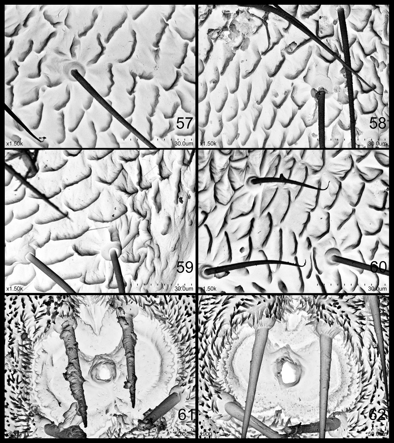
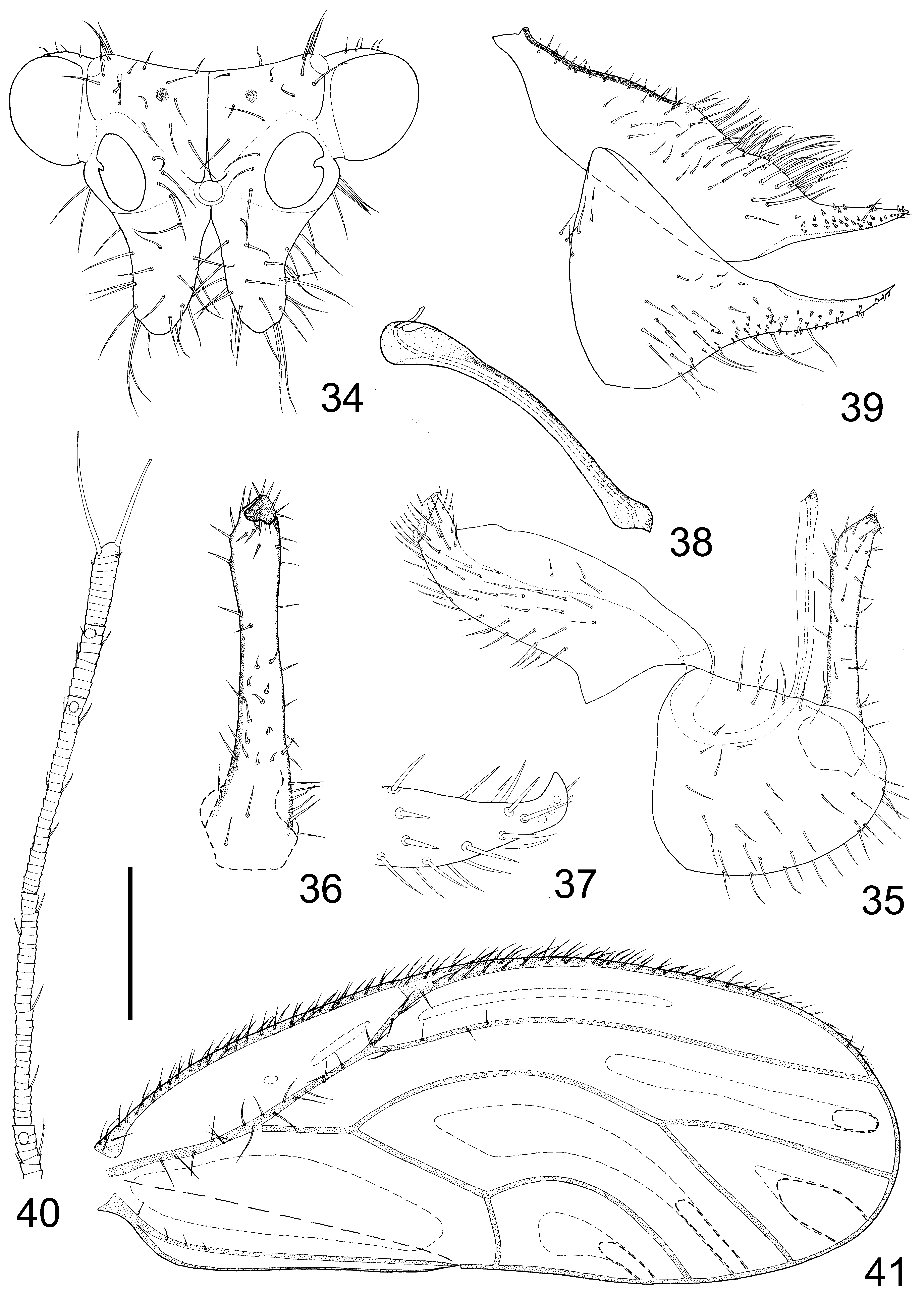
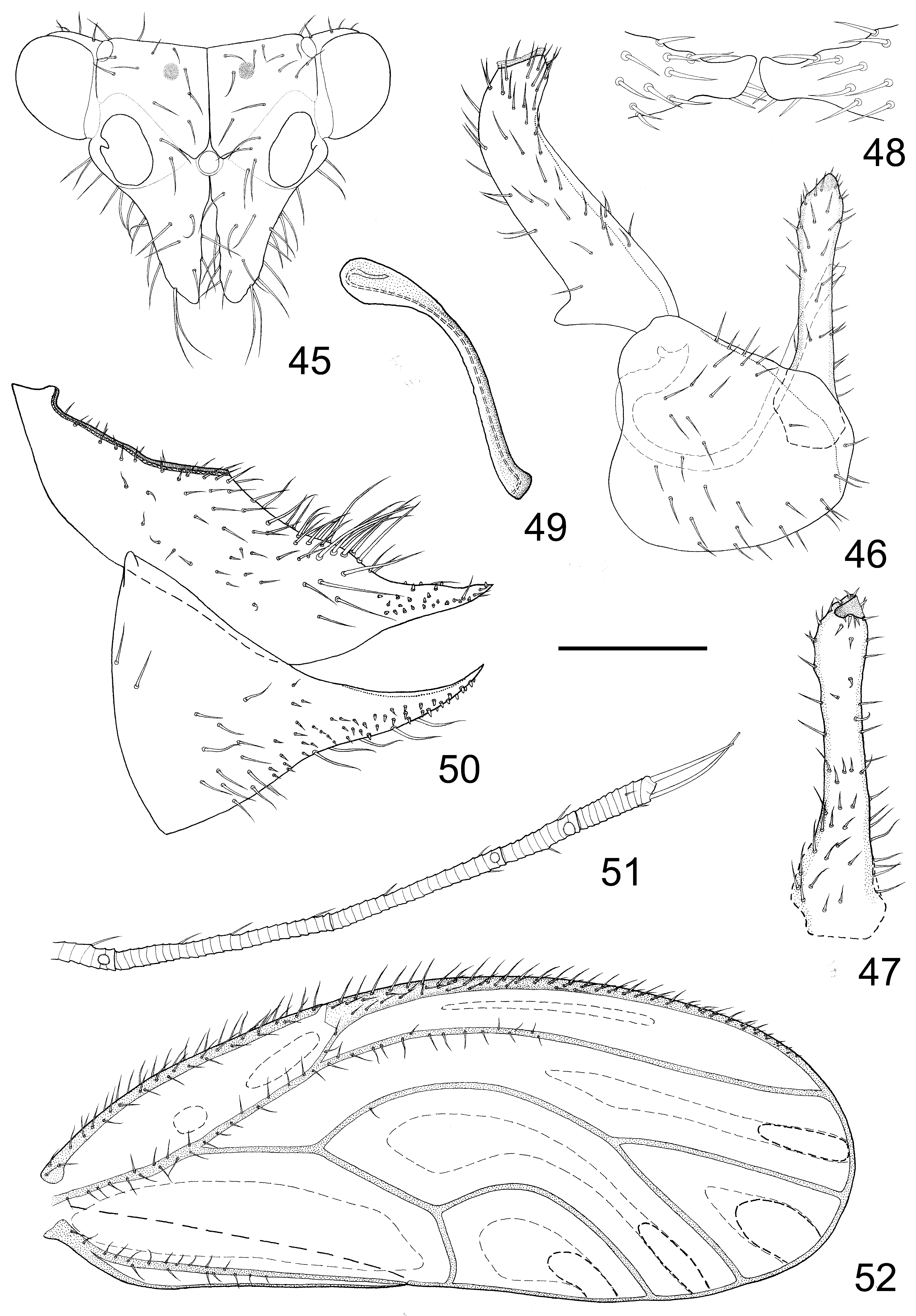
Redescription. Adult: Body medium to large sized ( Table 1), usually green in general color with orange patterns/ stripes in thoracic dorsal surface. Head strongly inclined from longitudinal body axis by about 60°-70°. Vertex concave, area surrounding lateral ocelli moderately convex, median suture complete; surface of vertex finely sculptured with scaly microstructures with margin moderately raised; microscopic setae absent, extraordinary long setae present instead ( Figs 57–60 View FIGURES 57 – 62 ). Frons mostly covered by well developed genal processes; median ocellus completely exposed, not partly covered by anterior angles of vertex. Vertex, antennal base, orbital sclerite and genal process clearly demarcated from each other by shallow sutures and differentiated superficial structures.
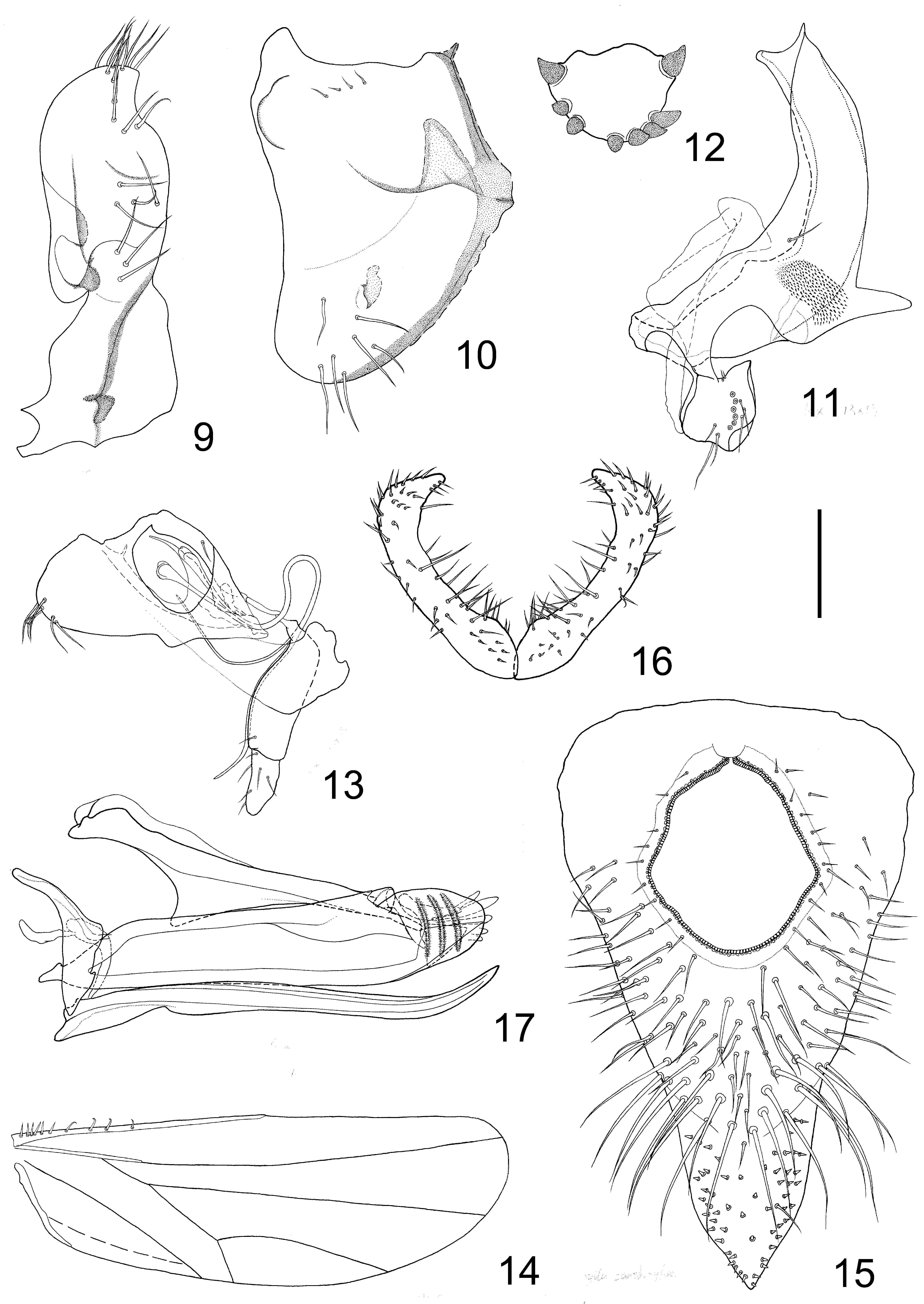
Genal processes long, conical, often longer than vertex along median suture. Antenna 10-segmented, long and slender, often over three times as long as head width ( Table 1); segments III-X often blackened to various extent; one single rhinarium present on apices of segments IV, VI, VIII and IX; terminal setae rather long, both longer than segment X; whole antenna covered with stout setae except for segment X, setae on ventral surface relatively stouter and forming a single longitudinal line, among which the one near each rhinarium is especially stout ( Figs 7 View FIGURES 1 – 8 , 29 View FIGURES 23 – 30 , 40 View FIGURES 34 – 41 & 51 View FIGURES 45 – 52 ). Clypeus ( Fig. 13 View FIGURES 9 – 17 ) well developed, stretching forward as far as anterior margin of antennal base, with one projection on ventral surface; apex with several long setae. Rostrum ( Fig. 13 View FIGURES 9 – 17 ) two-segmented, terminal segment slightly longer than half of basal segment ( Table 1); terminal segment and apex of basal segment with moderately long setae. Dorsal aspect of thorax covered with extraordinary long setae except for metapostnotum. Pronotum smoothly transiting into proepisternum, demarcated with a shallow gap; propleural sulcus reaching posterior margin of pronotum ( Fig. 9 View FIGURES 9 – 17 ). Mesopleural suture near horizontal; meso-anapleural cleft appearing as one wide gap, stretching obliquely downward; fossa of trochantinal apodeme present in central portion of mesokatepisternum ( Fig. 10 View FIGURES 9 – 17 ). Mesokatepisternum and mesoepisternum with several long setae (sometimes absent), dorsal submargins of mesopleurite and metaepisternum with several short setae. Metacoxa ( Fig. 11 View FIGURES 9 – 17 ) with well developed horn-shaped meracanthus; thin outer wall with one single seta, and a small field of micro spinules near meracanthus. Legs relatively long and slender. Metatibia without basal spine, apex with 6-9 highly sclerotised apical spurs ( Fig. 12 View FIGURES 9 – 17 ), usually randomly grouped except for the inner-most one (“thumb”) and the outer most one (“little finger”); metabasitarsus with 2 black spurs. Fore wing oblong oval, membrane hyaline; pterostigma long and narrow; C+Sc, R1, R+M+Cu1, R, base of Rs and A1+2 with extraordinary long setae, gradually turning into normal micro pterogostic setae apically; vein Cu+M about as long as vein Cu. Hind wing ( Fig. 14 View FIGURES 9 – 17 ) hyaline, shorter than fore wing, completely covered with evenly spaced surface spinules that are longer and sharper than in fore wing. Abdominal sterna densely covered with rather long setae. Male proctiger tubular and unipartite; paramere long and slender, apex strongly curved inward ( Fig. 16 View FIGURES 9 – 17 ), subapex more or less widened; aedeagus 2-segmented, simple. Female proctiger with a bulging portion before apical process; a blunt tooth-shaped lamellar process ( Fig. 15 View FIGURES 9 – 17 ) present before circumanal ring, pressing the basal margin of the latter more or less raised; subgenital plate wide and sub globular in basal half, strongly or gently narrowed in the middle, then gradually attenuated apically; membranous part anterior to base of subgenital plate with numerous long setae; valvula lateralis ( Fig. 17 View FIGURES 9 – 17 ) with three transverse ridges on outer surface, inner lube as several finger-shaped projections, projecting beyond margin; valvula dorsalis relatively slender and sclerotised basally, growing broader and less sclerotised apically, with apex near truncate; valvula ventralis ( Fig. 17 View FIGURES 9 – 17 ) rather broad.
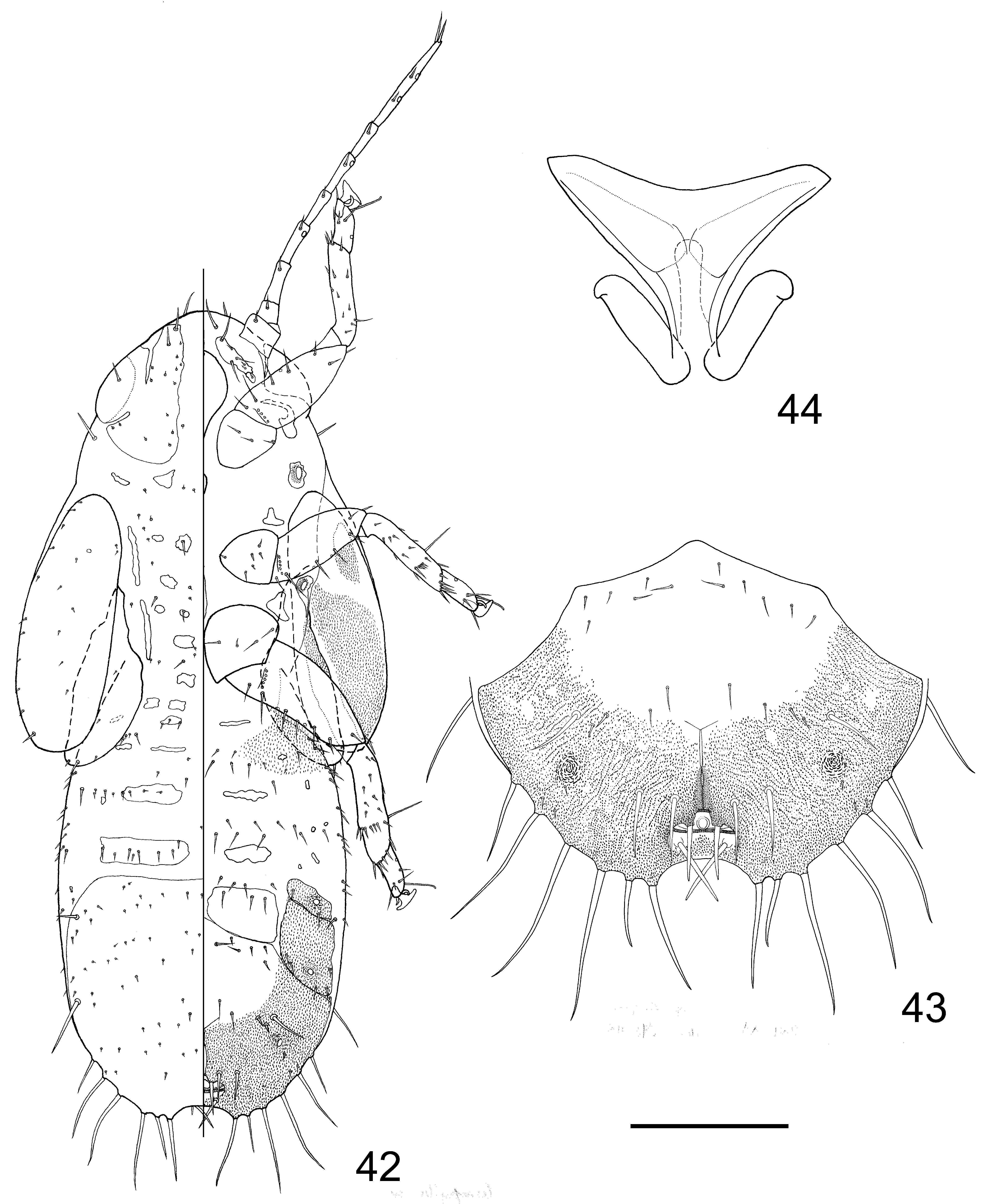
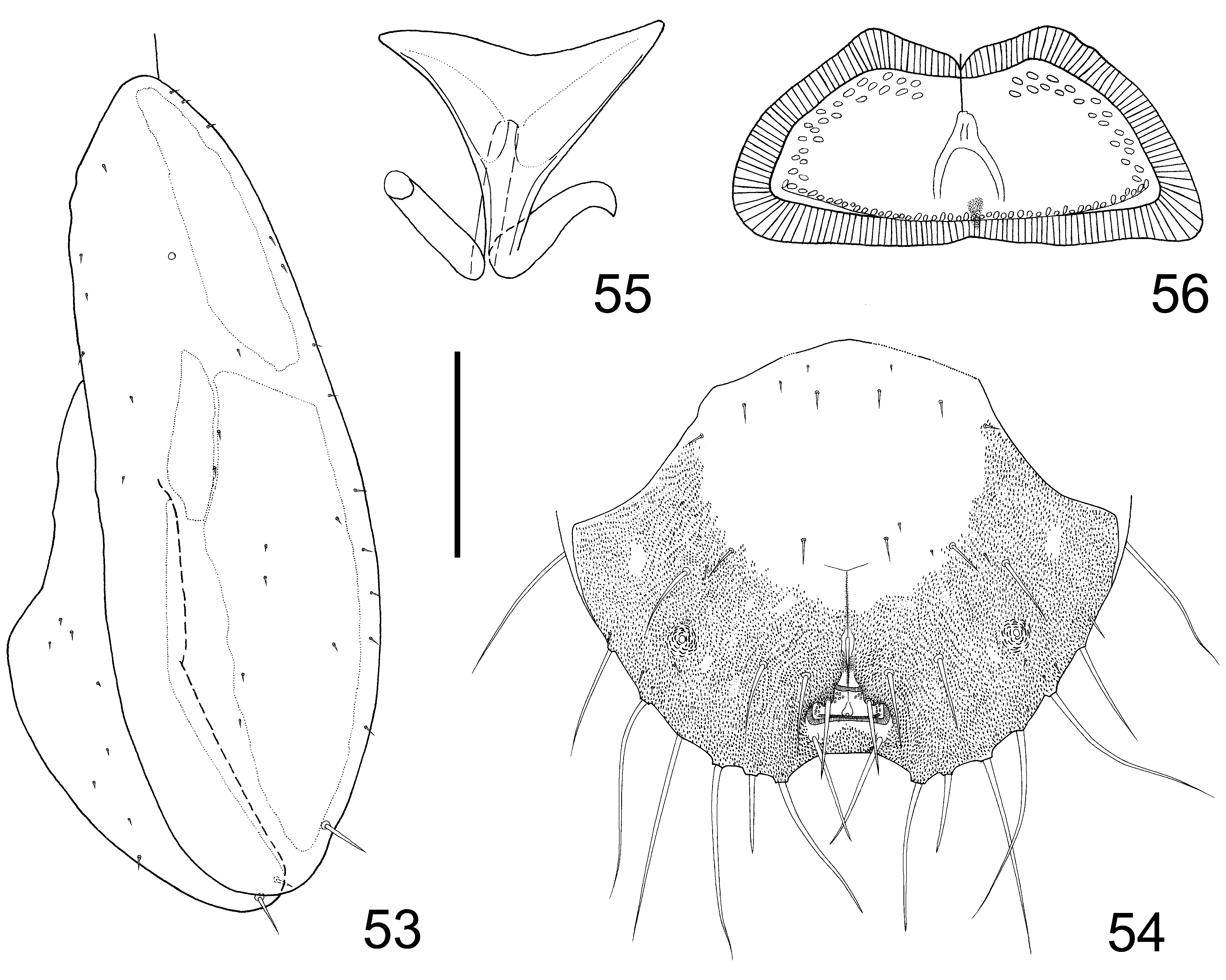
Fifth instar nymph ( Figs 20 View FIGURES 20 – 22 , 31 View FIGURES 31 – 33 & 42 View FIGURES 42 – 44 ): Body long and narrow. Head near semicircular; anterior margin of dorsal surface with 1+1 long setae and 1+1 short setae; ventral surface with 1+1 long and narrow oblique sclerites with several normal setae of various lengths; clypeus with 1+1 long normal setae in anterior submargin; dorsal sclerite (cephaloprothorax) partly fused with sclerite surrounding compound eye, with scattered minute setae and short normal setae; compound eye obscurely demarcated from sclerite surrounding it; ocular seta rooted slightly anterior to centre of dorsal surface of compound eye, projecting slightly beyond margin of the latter when pressed to be procumbent. Antenna 8-segmented, with one single rhinarium on apices of segments 4 and 6, and two rhinaria on segment 8; segments 3-7 with one strong seta each on submargin ventrally, segment 8 with one strong seta near each rhinarium ventrally. One long seta present in dorsal submargin behind compound eye. Prothorax dorsal sclerites partly fused with head, with 1+1 long and narrow sclerites and 1+1 trapezoidal free sclerites. Dorsal sclerites of thorax scattered, with lateral 2+2 long, narrow and longitudinal. Scattered short and minute normal setae present in dorsal surface of thorax. Fore wing pad oblong oval and without humeral lobe, subacute anteriorly and slightly constricted in middle, apical angle rather blunt; dorsal surface with minute setae, one pore present in middle of apical 1/5; one long seta present in outer margin slightly anterior to apical angle; ventral surface with micro spinules arranged in blocks. Hind wing pad blade-shaped, with minute setae in dorsal surface; ventral surface with micro spinules arranged in narrow bands along outer margin; apical angle with one long and one short setae. In ventral surface of thorax, one free sclerite present anterior to each coxa. One sclerite with a spiracle present anterior to meso- and metacoxa each. Each leg with one pore in dorsal surface of tarsus; one rather long normal setae present in dorsal surface of meso- and metatibiotarsus each, right on subdividing edge. Tarsal arolium fan-shaped, with well developed unguitractor and moderately long petiole; apical margin more or less depressed. Apex of abdomen inwardly emarginated as a semicircular indentation, about 1.5 times as wide as outer circum anal pore ring. Dorsal surface of abdomen with scattered sclerites, 3+3 transverse free sclerites and transverse bands of moderately long setae present anterior to caudal plate. Dorsal surface of caudal plate with transverse bands of minute setae. 1+1 short and thick setae present in posterior part of dorsal surface of caudal plate. Ventral surface of abdomen with 1+1 triangular fields of long and unicuspid micro spinules in lateral part of base, each surrounding one small sclerite within and with one small protruding pore near lateral margin. Scattered small sclerites and 2+2 large free sclerites with one spiracle each present in ventral surface of abdomen laterally. 4+4 median free sclerites present in ventral surface of abdomen anterior to caudal plate, with posterior pair more or less fused with it. Transverse bands of setae present in ventral surface. Caudal plate often dark brown or black. Micro spinules present on 2+2 lateral free sclerites with spiracle and marginal area of caudal plate in ventral surface of abdomen, in caudal plate arranged in lines ( Figs 21 View FIGURES 20 – 22 , 32 View FIGURES 31 – 33 , 43 View FIGURES 42 – 44 & 54 View FIGURES 53 – 56 ). Ventral surface of caudal plate with 1+1 spiracles in lateral part. Anus ventral. Field anterior to circum anal pore ring rising upward and strongly extending caudad ( Figs 21 View FIGURES 20 – 22 , 32 View FIGURES 31 – 33 , 43 View FIGURES 42 – 44 & 54 View FIGURES 53 – 56 ), covering part of the pore ring. Outer circum anal pore ring ( Figs 56 View FIGURES 53 – 56 , 61 & 62 View FIGURES 57 – 62 ) complete, consisting of long-narrow slit-shaped pores. Inner circum anal pore ring ( Figs 56 View FIGURES 53 – 56 , 61 & 62 View FIGURES 57 – 62 ) consisting of small ellipse pores; posterior margin complete, anterior margin broken in middle. 1+1 strong setae present on edge of extending lobe. 1+1 strong setae present behind outer circum anal pore ring, usually across each other. 2+2 strong setae present anterolaterally to anal pore ring. Margin of caudal plate with 8+8 strong setae ( Figs 21 View FIGURES 20 – 22 , 32 View FIGURES 31 – 33 , 43 View FIGURES 42 – 44 & 54 View FIGURES 53 – 56 ), lateral-most 2+2 rooted in submargin of dorsal surface, lateral 6+6 gradually tapering apically and median-most 2+2 with coneshaped apex.
C. zanthoxylae C. magna C. rotundiconis C. trichotoma
Male (n=5) Female (n=5) Male (n=5) Female (n=5) Male (n=1) Female (n=1) Male (n=5) Female (n=4) BL 3.66±0.09 4.17±0.16 3.70±0.14 4.55±0.19 3.59 4.39 3.40±0.11 4.03±0.22 HW 0.75±0.02 0.83±0.02 0.73±0.04 0.86±0.03 0.70 0.86 0.72±0.01 0.80±0.03 AL 2.26±0.03 2.60±0.10 2.28±0.15 2.74±0.12 2.38 2.59 2.16±0.11 2.59±0.25 T1 0.134±0.004 0.150±0.006 0.135±0.011 0.154±0.006 0.163 0.165 0.142±0.003 0.161±0.003 T2 0.111±0.003 0.118±0.001 0.110±0.008 0.124±0.004 0.115 0.114 0.115±0.004 0.119±0.002 WL 2.85±0.05 3.29±0.17 2.99±0.08 3.69±0.11 2.86 3.60 2.78±0.06 3.28±0.17 TL 0.70±0.02 0.76±0.04 0.71±0.05 0.86±0.04 0.68 0.84 0.69±0.02 0.77±0.04 AL/HW 3.07±0.12 3.15±0.23 3.20±0.28 3.08±0.21
T1/T2 1.23±0.04 1.24±0.03 1.43±0.03 1.29±0.06
R1/R2 1.83±0.10 1.92±0.17 1.93 1.94±0.02 (n=2)
fifth instar nymph (n=3) fifth instar nymph (n=5) fifth instar nymph (n=2) fifth instar nymph (n=2) BL 2.11±0.16 2.56±0.07 2.40±0.16 2.27±0.30
HW 0.55±0.03 0.65±0.05 0.63±0.01 0.54±0.02
FL 0.79±0.06 0.76±0.03 0.80±0.09 0.74
HL 0.56±0.005 0.54±0.03 0.54±0.06 0.57
AW 0.128±0.009 0.120±0.04 0.116±0.016 0.140
Remarks. Adults of species of this genus are very hard to distinguish by none-genital characters, especially the venation of wings. Although fields of surface spinules on the fore wing could be used, the intraspecific sexual difference should be noticed. Morphologically, the emblematical “extraordinary long setae” in many body parts are considered to be specified microscopic/micropterogostic setae, as the latter are not found wherever the former occur. For 5th instar nymphs, the “cave” covering the circum anal pore ring gives the plane of the latter an oblique position. Because of this, and the exact point of vision affected by raising or declining of the abdomen, the shape of circum anal pore rings is hard to tell from ventral view, thus becoming difficult to use in specific diagnosis. SEM has to be introduced into the study of the circum anal pore fields of nymphs of this genus.


No known copyright restrictions apply. See Agosti, D., Egloff, W., 2009. Taxonomic information exchange and copyright: the Plazi approach. BMC Research Notes 2009, 2:53 for further explanation.