
Sphaerephesia
|
publication ID |
https://doi.org/ 10.11646/zootaxa.4000.2.3 |
|
publication LSID |
lsid:zoobank.org:pub:7EDEDAEE-642C-4F9D-A04D-141815D73343 |
|
DOI |
https://doi.org/10.5281/zenodo.5667459 |
|
persistent identifier |
https://treatment.plazi.org/id/81007D79-8D43-2575-FF0F-FADFFE37197F |
|
treatment provided by |
Plazi |
|
scientific name |
Sphaerephesia |
| status |
|
Figs 4 View FIGURE 4 E–F, 5F, 7
Material examined. East of Malabar, Sydney, New South Wales, Australia, AM W.42720 (1 spec.), 1 km south of ocean outfall, 33° 59' 00" S, 151° 17' 33" E, 79.4m, 20 Jun 1996.
Comparative material. Sphaerephesia mamalaensis Magalhães et al., 2011 , holotype, NMNH 1154142; paratypes, NMNH 1154143.
Diagnosis. Body strongly flattened dorso-ventrally, with four longitudinal rows of sessile, macrotubercles, pear-shaped or with distal short papilla, and minute spherical papillae arranged in four transversal rows per segment. Distance between dorsal macrotubercles exceeding distance between those and lateral ones. Parapodia with ventral cirri as long as acicular lobe and apparently lacking papillae. Six to eight chaetae per parapodium, with shaft distally enlarged and blades 1–3 times as long as wide. Male ‘copulatory organs’ present as an elongated conical papilla between ventral cirri of chaetigers 7 and 8.
Table 2. Description and position of the structures referred to as copulatory organs in the literature and the present study. These structures are provided, in all cases, with obvious pores. Unless stated, as in the case of the inflated ventral cirri, these structures are present in addition to normal ventral cirri present in other parapodia. Abbreviations: ch, chaetiger; btw, between.
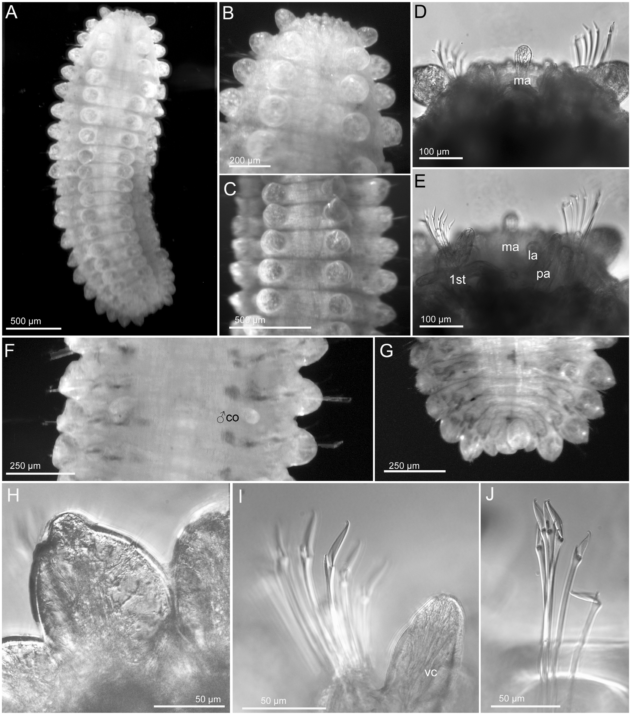
Description. Measurements and general morphology. Male, 3.2 mm long, 1 mm wide, 20 chaetigers. Body ellipsoid ( Fig. 7 View FIGURE 7 A), strongly flattened dorsoventrally. Dorsum slightly sub-trapezoidal, flattened ventrum. Segmentation conspicuous ( Fig. 7 View FIGURE 7 A). Preserved specimen lacking pigmentation. Anterior end bluntly rounded ( Fig. 7 View FIGURE 7 A–B, D–E).
Head. Prostomium with five digitiform appendages ( Fig. 7 View FIGURE 7 D–E), including a pair of palps, in ventral most position; a pair of lateral antenna, similar in shape and size to palps; and a median antenna, shorter (two thirds) than lateral antennae and with a rounded distal end. Antenniform papillae not observed ( Fig. 7 View FIGURE 7 D–E). A pair of tentacular cirri similar in shape and size to lateral antennae and palps. Head with several rounded and hemispherical papillae, difficult to count due to retraction of prostomium ( Fig. 7 View FIGURE 7 B, D–E).
Tubercles. First chaetiger with two macrotubercles, sessile, pear-shaped. Rest of chaetigers with four macrotubercles each, arranged in four longitudinal rows along dorsum, similar in shape to first ones but larger ( Fig. 7 View FIGURE 7 A–C), or with short terminal papillae ( Fig. 7 View FIGURE 7 G–H). Distance between mid-rows is larger than between these and lateral macrotubercles ( Figs 4 View FIGURE 4 E, 7A–C). Additional minute hemispherical papillae are present over dorsum, arranged in four transversal rows per segment ( Fig. 4 View FIGURE 4 E). Ventral surface with small spherical papillae, arranged in 3–4 transversal rows ( Fig. 4 View FIGURE 4 F).
Parapodia. Parapodia sub-conical, as long as wide, wrinkled. Acicular lobe oval, projecting distally. Ventral cirri oval, slightly longer than acicular lobe in anterior segments and similar in length in the rest. Mid-body parapodia without papillae; two or three small spherical papillae located ventral to parapodial base, not considered herein parapodial ( Fig. 5 View FIGURE 5 F). Dorsal surface of parapodia not clearly visible due to proximity and large size of macrotubercles.
Chaetae. Compound chaetae present in all chaetigers, arranged in a curved transverse row around acicular lobe and numbering 6–8 per fascicle ( Figs 5 View FIGURE 5 F, 7D–E, I–J). Shaft with slight widened distal end with delicate almost inconspicuous spinulation (under compound microscope). Blades ranging between 2–4 times longer than wide, those from mid-fascicle longer, with fine and short spinulation along superior edge and a distal recurved tip ( Fig. 7 View FIGURE 7 I, J).
Pygidium . Pygidium terminal, with mid-ventral spherical anal cirrus and a pair of dorsal anal cirri, similar in shape to macrotubercles but slightly smaller ( Fig. 7 View FIGURE 7 G).
Internal features. Muscular pharynx not visible though body wall. Eyes not seen.
Reproductive features. Male ‘copulatory organs’ present as an elongated conical papilla between ventral cirri of chaetiger 7 and 8 ( Figs 4 View FIGURE 4 F, 7F).
Remarks. Of the eight currently accepted species of Sphaerephesia , only S. mamalaensis has four longitudinal rows of macrotubercles and chaetae with short blades (less than five times longer than wide). Sphaerephesia mamalaensis shares several diagnostic features with the Australian specimen: median antenna is slightly shorter than lateral, macrotubercles are not clearly papillated but more pear shaped, there are 3–4 transversal rows of papillae per segment on both dorsal and ventral surfaces, chaetae have blades 2–4 times longer than wide showing intra-fascicle variation ( Magalhães et al. 2011; per. obs.). The specimen herein described differs from S. mamalaensis in the conspicuous delineation of segments, the absence of parapodial papillae and the different shape of the pygidial ventral cirrus, spherical (generally digitiform), but this could be an artefact of this specimen. This specimen could belong to an undescribed species, but features such as the shape of the pygidial cirrus and the absence of parapodial papillae should be confirmed with more specimens.
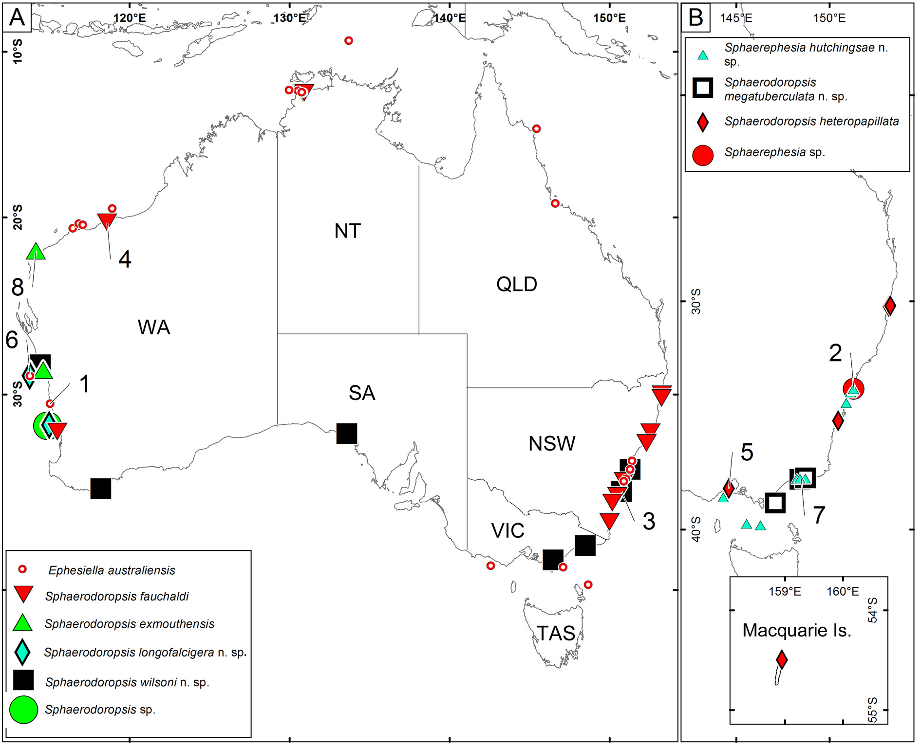
Distribution. Sydney, New South Wales ( Fig. 15 View FIGURE 15 ).
Habitat. Sand, 80 m depth.


| NMNH |
Smithsonian Institution, National Museum of Natural History |
No known copyright restrictions apply. See Agosti, D., Egloff, W., 2009. Taxonomic information exchange and copyright: the Plazi approach. BMC Research Notes 2009, 2:53 for further explanation.