
Sphaerodoropsis exmouthensis Hartmann-Schröder, 1981
|
publication ID |
https://doi.org/ 10.11646/zootaxa.4000.2.3 |
|
publication LSID |
lsid:zoobank.org:pub:7EDEDAEE-642C-4F9D-A04D-141815D73343 |
|
DOI |
https://doi.org/10.5281/zenodo.5667463 |
|
persistent identifier |
https://treatment.plazi.org/id/81007D79-8D5F-2570-FF0F-FFABFC411C8A |
|
treatment provided by |
Plazi |
|
scientific name |
Sphaerodoropsis exmouthensis Hartmann-Schröder, 1981 |
| status |
|
Sphaerodoropsis exmouthensis Hartmann-Schröder, 1981 View in CoL
Figs 1 View FIGURE 1 D–I, 4G–H, 5G, 8
Sphaerodoropsis exmouthensis Hartmann-Schröder, 1981: 44 View in CoL , Figs 94–99; 1984: 31; 1986: 51.
Material examined. Holotype ZMH P.16493: Western Australia, Exmouth, Tantabiggy Creek, in sand on the reef plateau, 11 Oct. 1975. Paratype MZH P.16494 (1 spec.) same sample. Additional material. ZMH P.16995 (1 spec. for SEM), Drummonds core, Geraldton, Western Australia, Australia; in fine sand with seagrass, 22 Oct 1975.
Diagnosis. Ellipsoid body with strongly convex dorsum. Four longitudinal rows of sessile and spherical (sometimes pear-shaped) dorsal macrotubercles and three transversal rows (total up to 11) of dorsal spherical papillae per segment. Distance between dorsal-most macrotubercles exceeding distance between those and lateral ones. Parapodia with long digitiform acicular lobe, shorter ventral cirrus and a single rounded papilla on anterior distal end. Four or five chaetae per parapodium, with thin shafts slightly enlarged distally and lacking spinulation, and short blades (5–7 times longer than wide), serrated and with smooth distal end. Males with ‘copulatory organ’ as conical papillae ventral to ventral cirri between chaetiger 6 and 7.
Re-description. Measurements and general morphology. Male, body ellipsoid, measuring 1.3 mm long and 0.42 mm wide, with 19 chaetigers. Tegument with transverse wrinkles, segmentation inconspicuous. Dorsum convex and ventrum flattened. Preserved specimen lacking pigmentation.
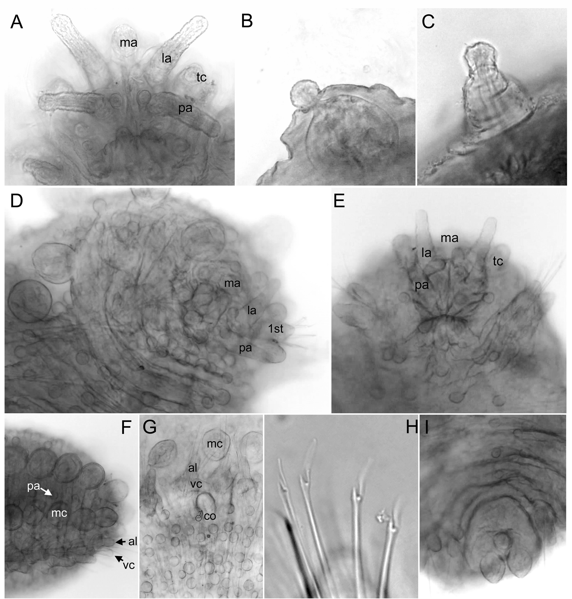
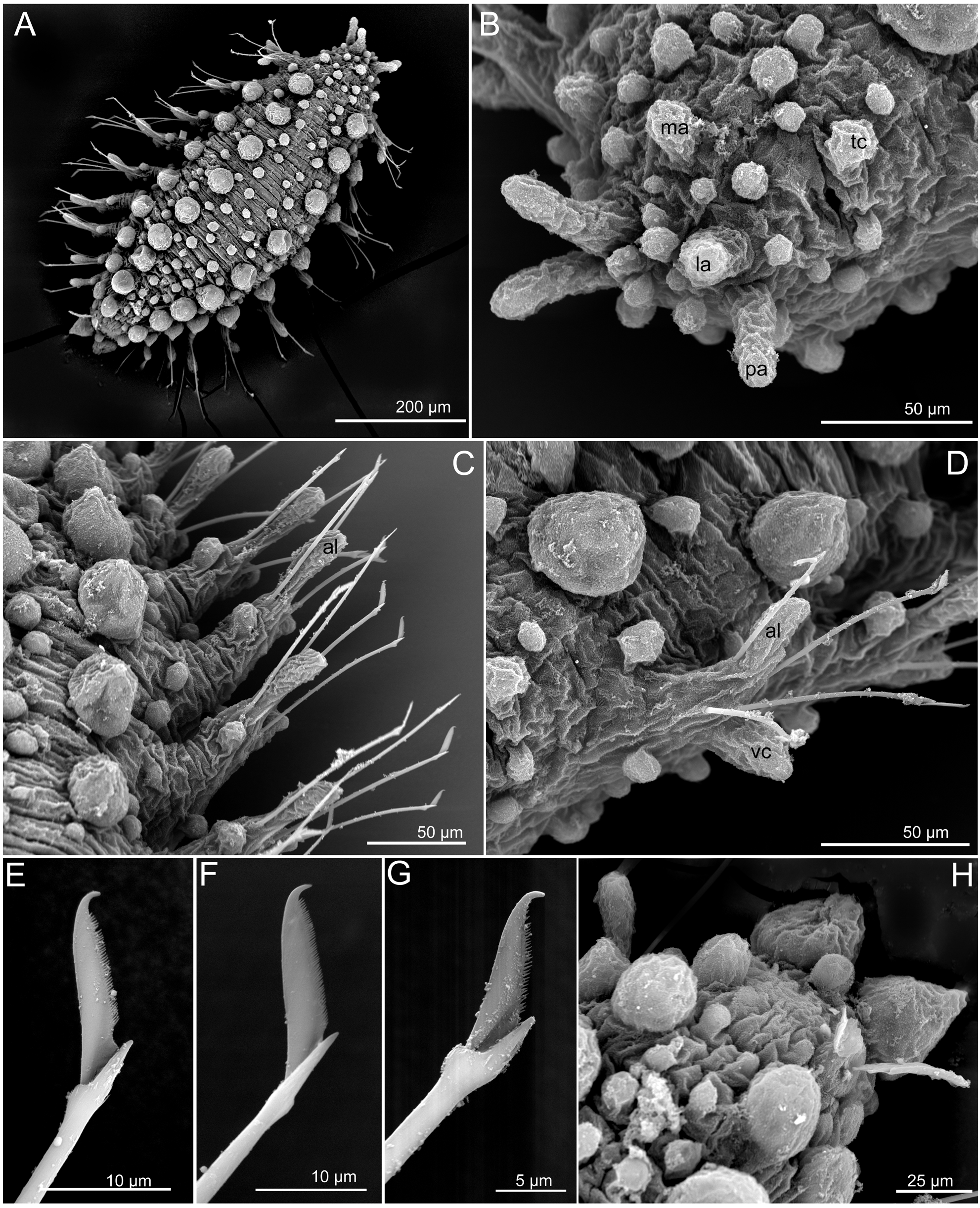
Head. Anterior end bluntly rounded. Prostomium with five longer appendages, including a pair of digitiform palps, in ventral-most position; a pair of lateral antennae similar in size and shape to palps; and a median antenna, ellipsoid, about one half of the size of lateral antennae ( Figs 1 View FIGURE 1 D–E, 8A–B). Antenniform papillae absent ( Figs 1 View FIGURE 1 D– E, 8A–B). Up to 10 spherical papillae confined by prostomial appendages and the mouth ( Figs 1 View FIGURE 1 D–E, 8A–B). A pair of tentacular cirri similar in shape and size to median antenna and palps and several scattered papillae similar to prostomial ( Fig. 8 View FIGURE 8 A–B).
Tubercles. First chaetiger with two macrotubercles, slightly smaller than following, spherical and sessile ( Figs 1 View FIGURE 1 D, 8A). Rest of chaetigers with four macrotubercles each, arranged in four longitudinal rows along dorsum ( Figs 1 View FIGURE 1 F, 4G, 8A). Distance between mid-dorsal rows is similar or slightly larger than between these and lateral macrotubercles ( Figs 1 View FIGURE 1 A, 4G). Lateral macrotubercles are slightly smaller than dorsal ( Figs 1 View FIGURE 1 F, 8A). Spherical papillae are present over dorsum, arranged in three transversal rows per segment ( Figs 4 View FIGURE 4 G, 8A). Around 6–8 papillae between mid-macrotubercles and around 2–3 between these and the lateral ones in mid-segments, papillae closer to macrotubercles slightly larger than the rest ( Figs 4 View FIGURE 4 G, 8A, D). None to two papillae between lateral macrotubercles and parapodia ( Fig. 8 View FIGURE 8 D). Ventral surface with spherical papillae, arranged in 2–3 transversal rows, with a total of 9–13 papillae in mid-body segments; numbers decreasing towards posterior end ( Figs 1 View FIGURE 1 G, 4H), with a group of three near the base of parapodia and a midline group of 5–7 papillae, being the anterior larger. Body epithelium with ellipsoid granules.
Parapodia. Parapodia sub-conical, about 1–2 times longer than wide. First chaetiger with a long (as long as parapodia), digitiform acicular lobe projecting distally; ventral cirri shorter and wider ( Fig. 8 View FIGURE 8 D), parapodial papillae absent. Second and following chaetigers similar or with longer acicular lobes, and one dorso-anterior spherical papillae ( Figs 5 View FIGURE 5 G, 8C).
Chaetae. Compound chaetae present in all chaetigers, arranged in a curved transverse row around acicular lobe, numbering 3–5 per fascicle ( Figs 5 View FIGURE 5 G, 8C–D). Shaft with slightly widened distal end and delicate, almost inconspicuous, spinulation. Blades similar in length along fascicles (5–7 times longer than maximum width), with fine and short spinulation along superior edge and a distal recurved tip ( Figs 1 View FIGURE 1 H, 8E–G).
Pygidium . Pygidium terminal, with distal dorsal spherical papillae, a mid-ventral digitiform anal cirrus and a pair of pear-shaped dorsal anal cirri, slightly larger than macrotubercles ( Figs 1 View FIGURE 1 I, 8H).
Internal features. Eyes not observed, muscular pharynx inconspicuous.
Reproductive features. Holotype, male, with ‘copulatory organs’ as conical papillae ventral to ventral cirri between chaetiger 6 and 7 ( Figs 1 View FIGURE 1 G, 5H). Females have not been examined herein but there is a record of a gravid female with 13 chaetigers ( Hartmann-Schröder 1981).
Variation. Specimens measure 0.5–1.3 mm long and 0.25–0.42 mm width, in all cases body is about 4–5 times longer than wide. Paratype with eight chaetigers. Median antenna is in the three specimens examined shorter than lateral and no antenniform papillae have been observed in specimens examined. Prostomial papillae confined by antennae and mouth are less than 10. Macrotubercles are, in the specimen studied under SEM, not completely rounded and somehow pear-shaped ( Fig. 8 View FIGURE 8 C–D). Number of epithelial papillae, including parapodial with a single papilla in all except first chaetiger, and arrangement seem to be constant. Parapodia do not exceed twice the length of the width. Number of chaetae range between 4–5 per parapodium. Length of blades shows a slight variation in number of chaetae and the relative length of blades. Examined specimens are not transparent and muscular pharynx was not conspicuous. Eyes have not been observed. ‘Copulatory organs’ only observed in holotype.
Remarks. In the original description there were some details lacking and others that have been interpreted differently herein. The palps and lateral antennae (referred to as outer and inner antennae respectively in original description) were described as being shorter and oval and digitiform to filiform but are considered here to be similar in shape and size. The arrangement of epithelial papillae both on dorsal and ventral body surface seems not random ( Fig. 8 View FIGURE 8 A). Parapodia were described with a rounded papilla on superior margin that has here interpreted as anterior; chaetae were considered as smooth but fine dentation is observed under SEM. Pygidial macrotubercles as pear shaped (as drawn in original description, Hartmann-Schröder 1981).
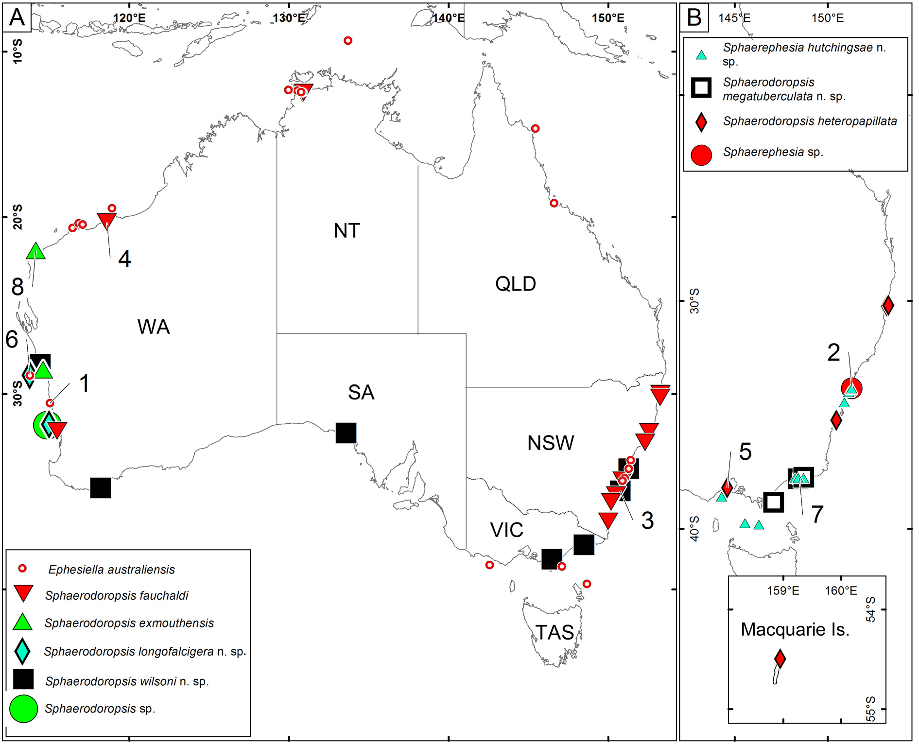
Type loc al ity. Tantabiddy Creek, Exmouth, Western Australia ( Fig. 15 View FIGURE 15 ).
Distribution. Western Australia (Exmouth and Geraldton; Fig. 15 View FIGURE 15 ),?South Australia (Ceduna and Kangaroo Island) ( Hartmann-Schröder 1981, 1984, 1986). South Australian material needs confirmation; they could belong to Sphaerodoropsis wilsoni n. sp. described below.
Habitat. Sand near reef plateau or sea-grasses, shallow depths.


No known copyright restrictions apply. See Agosti, D., Egloff, W., 2009. Taxonomic information exchange and copyright: the Plazi approach. BMC Research Notes 2009, 2:53 for further explanation.
|
Kingdom |
|
|
Phylum |
|
|
Class |
|
|
Order |
|
|
Family |
|
|
Genus |
Sphaerodoropsis exmouthensis Hartmann-Schröder, 1981
| Capa, Maria & Bakken, Torkild 2015 |
Sphaerodoropsis exmouthensis Hartmann-Schröder, 1981 : 44
| Hartmann-Schroder 1981: 44 |